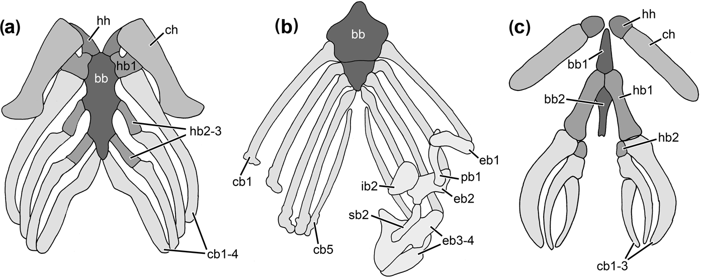
The hyobranchial or visceral skeleton of gnathostomes plays a fundamental role in breathing and feeding. In gnathostome fishes and aquatic amphibians, movements of the apparatus assist in opening the jaws and expanding the bucco-pharyngeal cavity, thus generating negative pressure within the mouth cavity and initiating a rapid inflow of water together with the prey, a mechanism that is referred to as suction feeding (e.g., Lauder & Reilly Reference Lauder, Reilly, Bels, Chardon and Vandewalle1994). The hyobranchial skeleton shows a wide variety of morphologies and characteristics among the different lineages and taxa of gnathostome fishes and tetrapods. The ‘generalised’ or plesiomorphic configuration of the hyobranchial skeleton is a complex set of paired segmented arches, consisting of the mandibular arch (i.e., the jaws), followed by the hyoid arch and the subsequent branchial (or gill) arches, which are linked ventrally to the median basibranchial series (Nelson Reference Nelson1969; Janvier Reference Janvier1996). The anteriormost element of the basibranchial series is usually referred to as basihyal and is connected with the hyoid arch; the posteriorly-following elements of the series are the basibranchials which are associated with the branchial arches. Whereas the mandibular arch consists of only two segments (the palatoquadrate dorsally and the Meckelian cartilage ventrally), the hyoid arch can be subdivided into the hypohyal, the ceratohyal and the hyomandibula (from ventral to dorsal). One or two small bones might be interconnected between the ceratohyal and the hyomandibular in osteichthyan fishes, the symplectic and the interhyal (Janvier Reference Janvier1996). The posteriorly-following branchial arches are plesiomorphically five in number, but may vary from one to six in gnathostome fishes and tetrapods. The following segments can be distinguished per branchial arch (from ventral to dorsal): hypo-, cerato-, epi- and pharyngobranchial (divided into infra- and suprapharyngeal) (Fig. 1). The gills (either external or internal gills or both) are attached to the branchial arches. During the fish–to–tetrapod transition and the subsequent conquest of land by vertebrates in the Late Palaeozoic, the hyobranchial apparatus of fishes had to be remodelled during the change from an aquatic to a rather terrestrial existence, in order to perform terrestrial feeding. Furthermore, the hyobranchial skeleton had to facilitate an increasing amount of aerial respiration, with a corresponding decrease in the importance of gill breathing. Better knowledge of the transformation of the hyobranchial apparatus during the fish–to–tetrapod transition and its further evolution in basal tetrapods is thus crucial for understanding early tetrapod history and the adaptations to life on land.
Hyobranchial skeletons of extant actinopterygians and sarcopterygians: (a) the actinopterygian Polypterus; distal arch elements are omitted (redrawn from Nelson Reference Nelson1969); (b) the coelacanth Latimeria; distal arch elements are omitted on the left side (redrawn from Forey Reference Forey1998); (c) the paedomorphic salamander Necturus (redrawn from Stadtmüller Reference Stadtmüller, Bolk, Göppert, Kallius and Lubosch1936). Abbreviations: bb=basibranchial; cb=ceratobranchial; ch=ceratohyal; eb=epibranchial; hb=hypobranchial; hh=hypohyal; ib=infrapharyngobranchial; pb=pharyngobranchial; sb=suprapharyngobranchial.

Coates & Clack (Reference Coates and Clack1991) and Clack et al. (Reference Clack, Ahlberg, Finney, Dominguez Alonso, Robinson and Ketcham2003) showed that the Late Devonian stem-tetrapods Acanthostega and Ichthyostega have a hyobranchial skeleton that closely resembles those of bony fishes. They discovered osteological correlates of internal gills, such as three or four pairs of ossified ceratobranchials with grooves for gill arch arteries, and a cleithrum (or clavicle) that bears a medially directed postbranchial lamina. In osteichthyan fishes, this lamina forms the posterior wall of the opercular chamber (Coates & Clack Reference Coates and Clack1991). A hyobranchial skeleton with similar three or four pairs of ceratobranchials has been demonstrated in a variety of adult aquatic basal tetrapods from the Permo-Carboniferous and the Triassic (Bystrow Reference Bystrow1938; Nilsson Reference Nilsson1946; Wellstead Reference Wellstead1991; Andrews & Carroll Reference Andrews and Carroll1991; Schoch Reference Schoch2002; Hellrung Reference Hellrung2003; Jenkins et al. Reference Jenkins, Shubin, Gatesy and Warren2008). Schoch & Witzmann (Reference Schoch and Witzmann2011) showed that most of these aquatic forms possessed the osteological correlates of internal gills similar to the Devonian forms, whereas their larvae bore external gills resembling those of extant salamander larvae. The adult hyobranchial apparatus of more terrestrial basal tetrapods such as eryopid, zatracheid and dissorophoid temnospondyls or “microsaurs”, on the other hand, is poorly known, and only a few descriptions of rather fragmentary elements exist (Romer Reference Romer1969; Boy Reference Boy1985; Witzmann & Schoch Reference Witzmann and Schoch2006; Clack & Milner Reference Clack and Milner2010). In contrast, the larval hyobranchium of many of these forms is well known based on the presence of ossified or cartilaginous branchial arches often associated with branchial dentition and external gills (Boy Reference Boy1974; Boy & Sues Reference Boy, Sues, Heatwole and Carroll2000; Witzmann Reference Witzmann2004; Milner Reference Milner2007; Schoch & Milner Reference Schoch and Milner2008).
Although a large number of thorough osteological descriptions of basal tetrapods do exist, the majority of these studies are concentrated on the skull and on the postcranium, but deal only superficially with the morphology and the phylogenetic alteration of the hyobranchial skeleton. This may be due to the fact that the sometimes tiny and feebly ossified hyobranchial skeletal elements of early tetrapods are often poorly preserved and are thus difficult to interpret. However, numerous new finds of hyobranchial elements in recent years allow for a detailed investigation of these bones. In the following study, the hyobranchial morphologies of early tetrapods are reviewed, based primarily on first-hand examination and supplemented by published descriptions. This study will focus on those parts of the hyobranchial apparatus that can anatomically be compared between taxa. The following questions will be settled. What is the plesiomorphic condition of the hyobranchial apparatus in tetrapods, and which phylogenetic signals can be recovered in hyobranchial morphologies of the different tetrapod lineages? Can “terrestrial” and “aquatic” hyobranchial apparatus be distinguished in early tetrapods, and in which taxa can internal gills be assumed, based on the osteological correlates used by Coates & Clack (Reference Coates and Clack1991) and Schoch & Witzmann (Reference Schoch and Witzmann2011)? This study may further serve as a basis for later functional analyses of breathing and feeding in early tetrapods based on hyobranchial morphology.
1. Phylogenetic framework and general comments on hyobranchial morphology
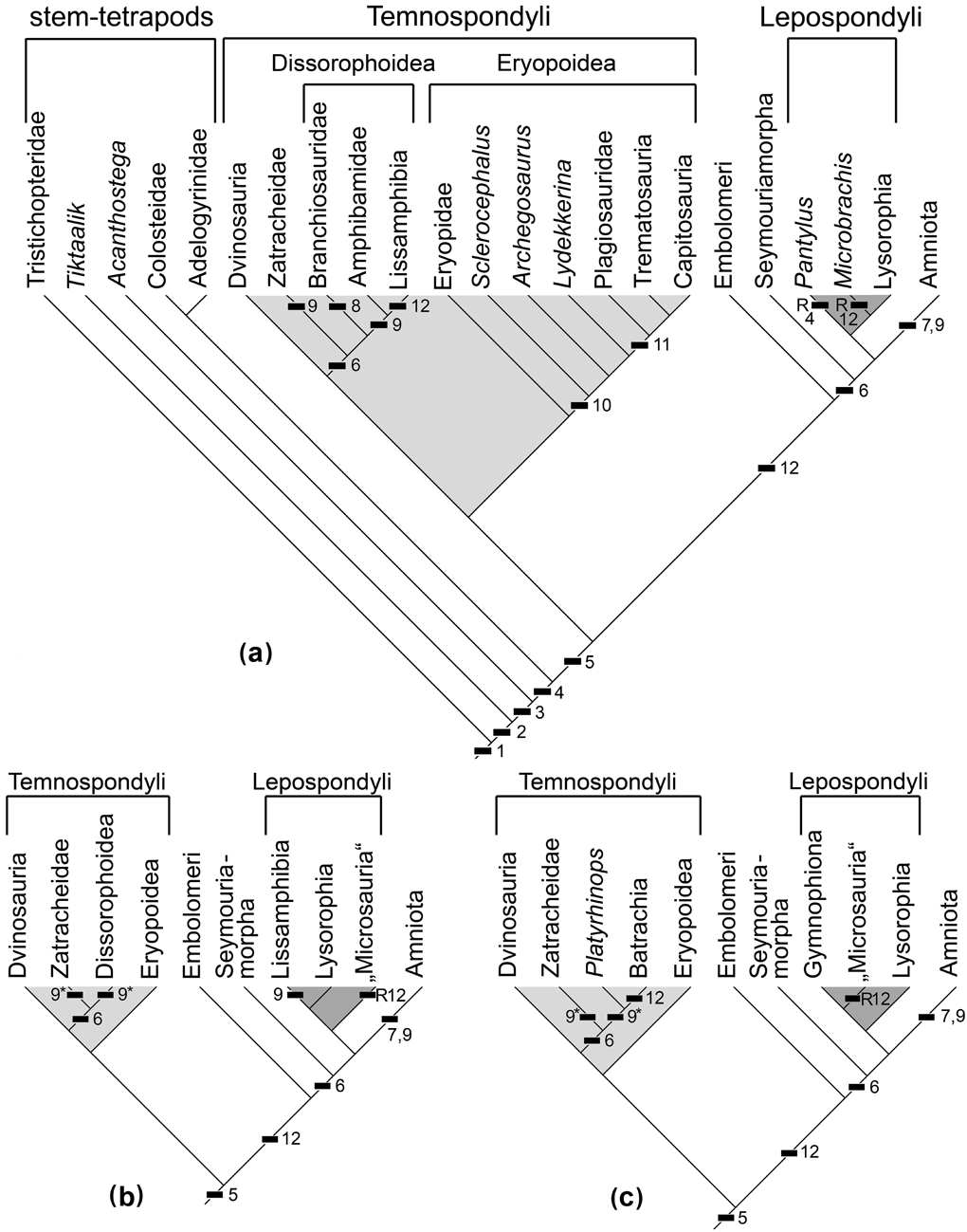
In the present study, the phylogenetic results of Ruta & Coates (Reference Ruta and Coates2007) and Schoch (Reference Schoch2013) are taken as phylogenetic framework of early tetrapod interrelationships. The term ‘basal tetrapod’ is accordingly used for non-amniote crown-group tetrapods of the Palaeozoic and Mesozoic. Among them, temnospondyls are regarded as the clade that includes all groups of lissamphibians, and “anthracosaurs” (including seymouriamorphs) and lepospondyls are regarded as stem-amniotes. Devonian forms, as well as colosteids, adelogyrinids and baphetids, are referred to as stem-tetrapods. Basal tetrapods and stem-tetrapods are informally designated here as “early tetrapods”. For an alternative view of early tetrapod relationships, see Marjanović & Laurin (Reference Marjanović and Laurin2008, and references therein).
“Proximal” is used here to refer to the portion of a hyobranchial element that is close to the basibranchial (i.e. to the ventral midline), whereas “distal” refers to the opposite direction. This appears to be clearer than to use “ventral” and “dorsal”, respectively, as is sometimes done, since the distal ends of certain hyobranchial elements are often located laterally rather than dorsally with respect to the proximal parts.
Identification of hyobranchial elements is often difficult in fossil specimens because of the frequently poor and incomplete state of preservation. Many excellently preserved specimens of stem-tetrapods and basal tetrapods, however, such as Acanthostega, dvinosaurians, plagiosaurids, branchiosaurids, adelogyrinids, etc., have the hyobranchial apparatus almost completely ossified and articulated. The knowledge of the morphology and configuration of the hyobranchial apparatus in these forms enables comparison with fragmentary hyobranchia, or even isolated elements in other taxa, and their identification. In contrast to most gnathostome fishes that possess a number of basibranchial elements in the ventral midline of the hyobranchium (Nelson Reference Nelson1969; Janvier Reference Janvier1996), only one unpaired basibranchial bone is known in basal tetrapods (sometimes also called the copula, Boy Reference Boy1974), which is located normally ventral to the parasphenoid. Elements of the hyoid arch can be distinguished from those of the branchial arches as follows. Hypohyals are usually rather small, (elongate) rectangular and located anterolateral to the anterior tip of the basibranchial. The ceratohyals are located distal to the hypohyals. When fully ossified, they are normally the longest elements of the hyobranchial skeleton. Generally, they have a flattened and often blade-like morphology. The hyomandibula of bony fishes has already transformed to the stapes in stem-tetrapods; although initially part of the hyobranchial skeleton, the stapes will not be considered here, since it became independent from the hyoid arch in tetrapods and attained a completely new functional role (Clack Reference Clack, Webster, Fay and Popper1992). Hypobranchials form the proximal part of the branchial arches and are rod-like bones located lateral and posterolateral to the basibranchial. They are mostly straight and rather short. Distal to them are the much longer ceratobranchials that are most often slightly curved. Posterolateral grooves for the branchial arteries on the ceratobranchials may indicate the presence of fish-like internal gills (Coates & Clack Reference Coates and Clack1991; Schoch & Witzmann Reference Schoch and Witzmann2011), whereas ceratobranchials without grooves may have borne external gills or no gills at all. Hyobranchial skeletal elements in early tetrapods always have unfinished ends, i.e. they were usually continued in cartilage to a certain degree.
Reilly & Lauder (Reference Reilly and Lauder1988) demonstrated on the basis of comparative osteology and myology that it is most parsimonious to designate the proximal-most elements in the salamander branchial arches as hypobranchials and the distally following segments as ceratobranchials. This interpretation is followed here in the description of basal tetrapod hyobranchia, whose basic configuration of skeletal elements corresponds to that in larval salamanders. This view is in accordance with most authors who studied fossil tetrapod hyobranchia, e.g. Bystrow (Reference Bystrow1938), Boy (Reference Boy1974), Andrews & Carroll (Reference Andrews and Carroll1991), Coates & Clack (Reference Coates and Clack1991), Witzmann (Reference Witzmann2004) and Schoch & Witzmann (Reference Schoch and Witzmann2011); but in contrast to Sushkin (Reference Sushkin1936), Romer (Reference Romer1969), Carroll & Gaskill (Reference Carroll and Gaskill1978) and Wellstead (Reference Wellstead1991), who preferred the usage of the terms ceratobranchial and epibranchial for the two proximal-most segments of branchial arches.
If one or more particular hyobranchial elements are not preserved in any known specimen of a certain early tetrapod taxon, it might be assumed that the respective element(s) were cartilaginous in life. A fundamental problem in analysing the hyobranchial skeleton of early tetrapods, however, is how the failure of a cartilaginous element to fossilise is distinguished from the evolutionary loss or absence of the element in question. This problem becomes even worse if one considers, for example, the extensive variation in ossification patterns of the hyobranchium in extant salamanders. This problem will be addressed below in the discussion paragraph. A further problem is that many of the hyobranchial elements of early tetrapods are very small and might easily get lost after the death of the animal and are therefore simply not preserved.
Early growth stages (or larvae) are distinguished from adult stem- and basal tetrapods by the presence of external gills, which are a larval characteristic at least in temnospondyls (and extant amphibians) and seymouriamorphs (Witzmann Reference Witzmann2004). However, since direct preservation of gills is rather exceptional, the existence of dentigerous branchial platelets, which in life would be attached to the branchial arches and indicate opened gill clefts, plus an overall poor degree of ossification of the endocranial and postcranial skeleton (especially vertebral centra, pelvis, and carpals and tarsals), are considered to indicate a larval form and distinguish it from an adult one.
2. Material studied
The following specimens were investigated for the present study:
Tetrapodomorph fishes. Eusthenopteron foordi (Late Devonian), NRM PZ 2609.
Stem-tetrapods. Acanthostega gunnari (Late Devonian), MGUH f.n.1227, f.n.1300; Greererpeton burkemorani (Middle and Late Carboniferous), CMNH 11090, 11130, 11219, 11132, 11073, 11320; Adelogyrinidae (Early Carboniferous), Adelogyrinus simorhynchus: NMS.G.1889.101.17; Adelospondylus watsoni, NMS.G.1885.57.51.
Temnospondyli. Dvinosaurus (Late Permian): D. primus, PIN 2005/39, Am/40, Am/41; D. campbelli, PIN 4818/410; PIN uncatalogued (several isolated ceratobranchials); Trimerorhachis insignis (Early Permian), UCMP 105175, 154434, 105146, 105221, 105163, 142027; Tabanchuia oomie (Early Triassic), UCMP 42777, 42781, 42780; Isodectes obtusus (Early Permian), AMNH 11037; Branchierpeton amblystomum (Early Permian), MMG SaP 179, MNC-F 10368; Apateon gracilis (Early Permian), MB.Am.406; Micropholis stowi (Early Triassic), BSM 1934 VIII 43; Platyrhinops lyelli (Late Carboniferous), MB.Am.23, 331; Iberospondylus schultzei (Late Carboniferous), PU-ANF 14; Sclerocephalus (Permo-Carboniferous), S. haeuseri: BSM AS I 575, SMNS 81791, 91800, MB.Am.1309, UHC-P 0489; S. nobilis, NHMM-PW 2005/2; Glanochthon (Early Permian), G. latrirostris: MB.Am.211; G. angusta, GPIT.Am.34; Archegosaurus decheni (Early Permian), MB.Am.983, IGS U II 1/2; Lydekkerina huxleyi (Early Triassic), BSM 1934 VIII 44; Stanocephalosaurus birdi (Wellesaurus peabodyi), UCMP 56098, 36040; Trematolestes hagdorni (Middle Triassic), SMNS 81790; Callistomordax kugleri (Middle Triassic), SMNS 90700; Metoposaurus diagnosticus (Middle Triassic), SMNS 5143; Gerrothorax pulcherrimus (Middle Triassic), SMNS 83866, 83382, MGUH 28919, NRM-PZ B.18b; Plagiosuchus pustuliferus (Middle Triassic), SMNS 84794.
Lepospondyli. Microbrachis pelikani (Late Carboniferous), AMNH 2557.
Seymouriamorpha. Ariekanerpeton sigalovi (Early Permian), PIN 2079/35, 41, 53, 117, 450, 563, 566, 601, 630, 631, 645, 679, 719, 755, 815.
Institutional abbreviations. AMNH, American Museum of Natural History, New York, USA; BSM, Bayerische Staatssammlung für Paläontologie und historische Geologie, Munich, Germany; CMNH, Cleveland Museum of Natural History, Cleveland / Ohio, USA; GPIT, Institut und Museum für Geologie und Paläontologie Tübingen, Germany; IGS, Institut de Géologie Strasbourg, Université Louis Pasteur, France; MB, Museum für Naturkunde Berlin, Germany; MGUH, Geological Museum, University of Copenhagen, Denmark; MMG SaP, Museum für Mineralogie und Geologie zu Dresden, Germany, Sachsen-Perm-Sammlung; MNC, Museum für Naturkunde Chemnitz, Germany; NHMM, Naturhistorisches Museum Mainz, Germany; NMS, National Museums of Scotland, Edinburgh, UK; NRM, Naturhistoriska riksmuseet Stockholm, Sweden; PIN, Paleontological Institute and Museum of the Russian Academy of Science, Moscow, Russia; PU-ANF, Departamento de Paleontología, Universidad Complutense, Madrid, Spain; SMNS, Staatliches Museum für Naturkunde, Stuttgart, Germany; UCMP, Museum of Paleontology of the University of California, Berkeley, USA; UHC-P, Ulrich Heidtke Collection, Paläontologische Sammlung, Niederkirchen, Germany.
3. Description
3.1. Stem-tetrapods
3.1.1. Acanthostega
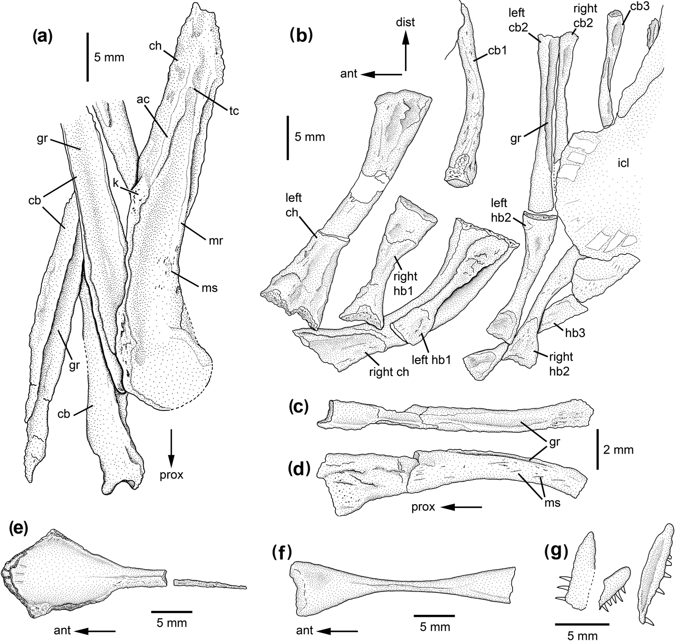
The largest preserved hyobranchial elements in Acanthostega gunnari are the ceratohyals (Fig. 2a). The left ceratohyal is well preserved in MGUH f.n.1227 and is exposed in ventro-lateral view. Coates & Clack (Reference Coates and Clack1991) suggested that it had rotated around 180° compared to its original orientation and swapped sides. However, the shape of the ceratohyal in Tiktaalik (Downs et al. Reference Downs, Daeschler, Jenkins and Shubin2008, fig. 6) and in Trimerorhachis (see below) suggests that the preserved orientation is correct. The ceratohyal is approximately 1.2 times longer than the ceratobranchials. Its proximal end is rounded and flattened, whereas the distal end is tapering. At mid-length, the anterodorsal margin bears a knob-like expansion with a distal “notch”, from which an anterodorsal crest runs towards the distal tip of the ceratohyal. A conspicuous ridge (medial ridge sensu Coates & Clack Reference Coates and Clack1991) extends parallel to the posteroventral margin from the expanded proximal blade in a distal direction. It exhibits fan-shaped striae in its proximal region that can be interpreted as muscle scars (Fig. 2a). This ridge is connected to the smaller anterodorsal crest via a short, transversally aligned crest.
Hyobranchial skeleton of stem-tetrapods: (a) Acanthostega gunnari MGUH f.n.1227, ceratohyal and ceratobranchials; (b) Adelogyrinus simorhynchus NMS.G.1889.101.17, hyobranchial skeleton and anterior part of interclavicle; (c–d) Adelospondylus watsoni NMS.G.1885.57.51, isolated ceratobranchial: (c) posterolateral view; (d) anterior view; (e–g) Greererpeton burkemorani: (e) CMNH 11090, basibranchial in ventral view; (f) CMNH 11130, basibranchial in ventral view; (g) CMNH 11219, dentigerous branchial platelets. Abbreviations: ac=anterodorsal crest; ant=anterior; cb=ceratobranchial; ch=ceratohyal; dist=distal; gr=groove; hb=hypobranchial; icl=interclavicle; k=knob; mr=medial ridge; ms=muscle scars; prox=proximal; tc=transversally aligned crest.

Anteromedial to the ceratohyals is a pair of small, squarish bones in MGUH f.n.1227. Their margins are ill-defined and the elements seem to be crushed, thus indicating a poor degree of ossification in the living animal. Their small size and anteromedial location indicate that these elements are hypohyals (see also Coates & Clack Reference Coates and Clack1991).
Coates & Clack (Reference Coates and Clack1991) did not comment on the number of ceratobranchials in Acanthostega, whereas Clack & Coates (Reference Clack, Coates, Hoch and Brantsen1993) restored its hyobranchial skeleton with three ceratobranchials per side. This was reproduced in Clack (Reference Clack2012). Re-examination of the gill skeleton showed that four pairs of ossified ceratobranchials are present in Acanthostega. As discovered by Coates & Clack (Reference Coates and Clack1991), their posterolateral surface bears a deep groove whose width corresponds almost to the width of the ceratobranchial and is visible along the whole length of the element (Fig. 2a). At their proximal end, the ceratobranchials bear two blunt processes and are slightly wider than at the distal tip. Distal (dorsal) to left ceratobranchial 4 in MGUH f.n.1227 is a short rod-like element that can be interpreted as epibranchial (see also Clack & Coates Reference Clack, Coates, Hoch and Brantsen1993 and Clack Reference Clack2012). Clack et al. (Reference Clack, Ahlberg, Finney, Dominguez Alonso, Robinson and Ketcham2003) discovered similar grooved branchial arches in the Devonian stem-tetrapod Ichthyostega.
3.1.2. Adelospondyli: Adelogyrinus
The ceratohyals are the largest and broadest elements of the hyobranchium (Fig. 2b) in the only known specimen of Adelogyrinus simorhynchus (NMS.G.1889.101.17). Their proximal and distal ends are broadened, and the shaft is curved and laterally convex and bears no processes. Five further elements can be interpreted as hypobranchials according to their location and orientation (Fig. 2b); thus, at least three pairs of hypobranchials were present in the living animal, as already reconstructed by Andrews & Carroll (Reference Andrews and Carroll1991). The first pair is the stoutest, with the distal end being broader than the proximal one, and the posterior edge being more concave than the anterior one. The second and third pairs are more slender, but have otherwise similar proportions.
Four or five long, slender ceratobranchials can be recognised distal to the hypobranchials (Fig. 2b). One element of the first pair is preserved dorsal to the first hypobranchials. The bone is bent and slightly broader proximally than distally. Crests or grooves are not visible. The second pair of ceratobranchials is preserved dorsal to the second pair of hypobranchials. These bones are straight, and the left one has a posterolateral groove that extends along almost its entire length; near the proximal end of the bone, the groove curves posteriorly and medially and becomes untraceable. One ceratobranchial of the third pair is preserved dorsal to the third pair of hypobranchials. The proximal portion of the element is concealed by the interclavicle. The posterolateral surface of the distal portion is slightly concave, and a distinct crest is visible laterally on the shaft that delimits a posterior groove on the ceratobranchial. Posterior to this element, a small part of a bone is visible, most of which is overlain by the interclavicle. It might represent the second element of the third pair of ceratobranchials. According to this description, Adelogyrinus has three pairs of ossified ceratobranchials (contra two pairs sensu Andrew & Carroll Reference Andrews and Carroll1991), and at least the second and third pairs of them bear posterolateral grooves.
3.1.3. Adelospondyli: Adelospondylus
An isolated, curved bone that is associated with Adelospondylus watsoni, specimen NMS.G.1885.57.51, is broadened at one end and tapers continuously towards the other end (Fig. 2c, d). It is ovate in cross-section. The surface of the more slender portion bears numerous striae that might represent muscle scars. The convex edge of this curved bone bears a groove that diminishes towards the broadened end. This bone, although conspicuously smaller, closely resembles the ceratobranchials in Dvinosaurus (see below). If the interpretation as a ceratobranchial is correct, then the broadened part represents the proximal end of the bone.
3.1.4. Colosteidae: Greererpeton
In Greererpeton burkemorani (CMNH 11090), the anterior part of the rod-like basibranchial attains more than five times the width of the slender shaft and is diamond-shaped (Fig. 2e). The ventral surface of the broadening is slightly convex, with a narrow concave part lateral to it. The basibranchial is not bent in lateral view. The shaft bears a ventral keel or crest, which diminishes anteriorly on the broadened portion. In CMNH 11130, it is clearly visible that the basibranchial is broadened also in its posterior part, attaining approximately two thirds the width of the anterior portion (Fig. 2f). In CMNH 11219, two or three short, stout rod-like bones are present posterolateral to the basibranchial and might represent remains of hypobranchials.
Dentigerous branchial platelets that were attached to the cartilaginous ceratobranchials in the living animal can be found in two adult skulls and a juvenile (CMNH 11219, 11320, 11132). These platelets are elongated, ovate in outline and bear one row of approximately 20 teeth at the bulge-like, presumably posterior margin (Fig. 2g). The internal surface of the platelets is concave. Due to incomplete preservation, it cannot be ascertained how many rows of platelets were present.
3.1.5. Colosteidae: Colosteus
Hook (Reference Hook1983, fig. 7a) figured short, rod-like elements in Colosteus scutellatus with slightly expanded ends that can be interpreted as hypobranchials. Furthermore, Hook (Reference Hook1983, fig. 12a, b) described elongated dentigerous branchial platelets that closely resemble those of Greererpeton, but are proportionally longer.
3.1.6. Baphetidae
Milner & Lindsay (Reference Milner, Milner and Walsh1998) provisionally designated a stout bone between the mandibular rami of Baphetes cf. B. kirkbyi as a ceratobranchial. However, since this bone is comparatively short and straight, this interpretation is doubtful. Clack (Reference Clack2003) tentatively described a possible ceratohyal in Kyrinion martilli. The bone has a slightly expanded proximal end, a short shaft and a blade-like expanded distal portion. Apart from articulatory facets at the distal and proximal ends, a further facet is present at the mid length of the bone.
3.2. Temnospondyli
3.2.1. Dvinosauria: Dvinosaurus
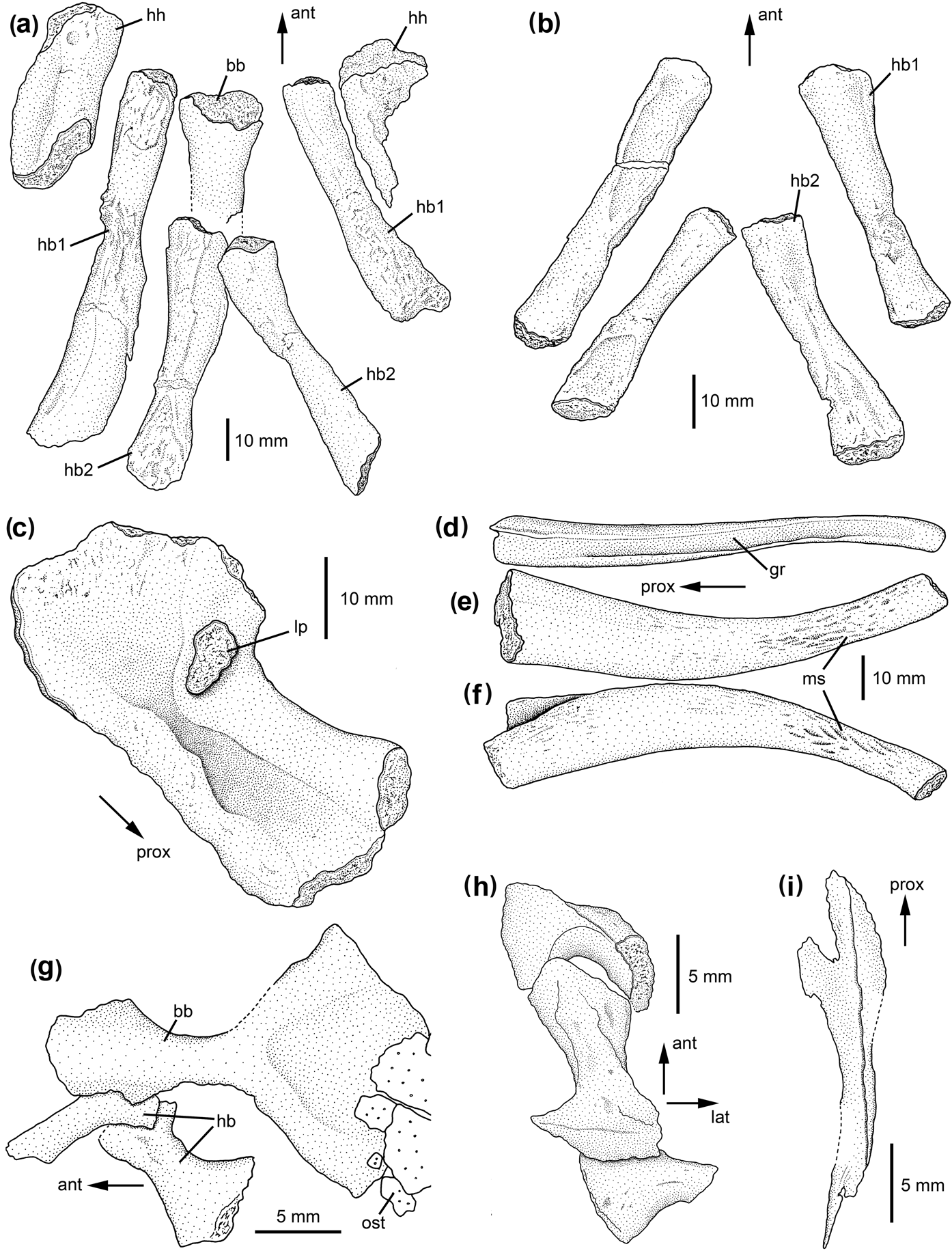
The basibranchial of D. primus is preserved in one specimen, PIN Am/40, and only its anterior part is exposed. It is a robust, rod-like bone and rounded-oval in cross section (Fig. 3a). The slightly expanded anterior portion is straight and becomes deeper than the shaft. In the same specimen, the first pair of hypobranchials is almost one third longer than the second pair (Fig. 3a). The first hypobranchials appear to be broader distally than proximally. Two pairs of ossified hypobranchials are preserved in the smaller skull of D. primus (PIN 2005/39). The first pair is slightly longer than the second pair (Fig. 3b). The bones are robust, round to ovate in cross section and slightly expanded at the ends. One pair of hypohyals is present in PIN Am/41, the right one being well preserved (Fig. 3a) and reaching about half the length of the first hypobranchial. It is rounded and rhombic in outline, with the medial portion being bulge-like, thickened and medially concave, whereas the lateral portion is thinner and laterally convex. Both portions are separated by a slight longitudinal depression.
Hyobranchial skeleton of dvinosaurian temnospondyls: (a–c) Dvinosaurus primus: (a) PIN Am/40, basibranchial, hypobranchials and hypohyals in ventral view; (b) PIN 2005/39, hypobranchials in ventral view; (c) PIN 2005/39, ceratohyal in lateral view; (d–f) Dvinosaurus campbelli, PIN 4818/410, isolated ceratobranchial: (d) posterolateral view; (e) posterior view; (f) anterior view; (g–i) Trimerorhachis insignis: (g) UCMP 105146, basibranchial and two right hypobranchials in ventral view; (h) UCMP 105175, three left hypobranchials in ventral view; (i) UCMP 105221, ceratohyal. Abbreviations: ant=anterior; bb=basibranchial; gr=groove; hb=hypobranchial; hh=hypohyal; lat=lateral; lp=lateral process; ms=muscle scars; ost=osteoderm; prox= proximal.

The ossified part of the right ceratohyal is well preserved in lateral and dorsal view in PIN 2005/39, whereas the left element has been removed since the descriptions of Sushkin (Reference Sushkin1936) and Bystrow (Reference Bystrow1938). It is a broad, stout bone with expanded proximal and distal ends (Fig. 3c). Its distal end is blade-like, rounded and laterally concave; this is the broadest part of the element. The concavity narrows and extends towards the proximal end. It is framed anterodorsally by a strong, bulge-like thickening, whereas the posteroventral margin is thinner. The distal end of the bulge-like thickening forms a lateral process (tuberculum ceratohyalis sensu Bystrow Reference Bystrow1938 and Shishkin Reference Shishkin1973) with unfinished surface. From the base of this process, a thinner process extends anterodorsally and forms the anterior edge of the posterior blade; dorsally (distally), it forms an elongate, unfinished surface.
The description of the four pairs of ceratobranchials by Sushkin (Reference Sushkin1936) and Bystrow (Reference Bystrow1938) was based on the skull PIN 2005/39. Since then, the left ceratobranchials have been removed, and the right ceratobranchials are in a very poor state of preservation. It is clearly evident that ceratobranchial 1 is the most robust branchial arch, but it cannot be determined if two grooves are present on it, as stated by Bystrow (Reference Bystrow1938). In PIN Am/41, three ceratobranchials are located on the left and at least two on the right side behind the skull, but they are very poorly preserved. The following description is based on well-preserved, isolated ceratobranchials of D. campbelli (mainly PIN 4818/410). The ceratobranchials are robust, curved elements that are well ossified (Fig. 3d–f). The proximal half is broad and anteroposteriorly flattened. The element tapers continuously towards its distal tip, with the proximal end attaining almost three times the width of the distal end. The posterolateral groove for the branchial artery (Sushkin Reference Sushkin1936; Bystrow Reference Bystrow1938; Schoch & Witzmann Reference Schoch and Witzmann2011) is clearly visible (Fig. 3d), but appears proportionally shallower and narrower than that of Acanthostega. The groove becomes shallower distally and disappears before reaching the distal tip. Proximally, the groove expands into a broad depression. The distal third of the anterior and posterior face possesses distinct muscle scars consisting of ridges and depressions (Fig. 3e, f), closely resembling those of Adelospondylus (Fig. 2d).
3.2.2. Dvinosauria: Trimerorhachis
The basibranchial of Trimerorhachis insignis consists of a stout, short shaft and is broadened anteriorly and posteriorly (Fig. 3g). The ventral surface of the anterior broadening is convex and becomes slightly concave towards the lateral ends where the bone gets thinner. The shaft is dorsoventrally flattened in cross-section and bears no ventral crest. The posterior part of the basibranchial is extremely expanded, forming a triangular blade which is approximately double the width of the anterior broadening. The ventral surface of the posterior blade is concave. The posterior expansion is almost half the length of the complete basibranchial and is dorsoventrally thinner than the anterior portion. The basibranchial is not bent in a ventral or dorsal direction.
Three pairs of short hypobranchials are present, whose morphology is best visible in UCMP 105146 and 105175 (Fig. 3g, h). The anterior two pairs have hook-like proximal and distal expansions, and the third pair is smaller and only proximally broadened.
In UCMP 105221 is an ossified element that is approximately the same length as the basibranchial and is located anterior to the ceratobranchials. The bone is flattened, rod-like, slightly bent, and is expanded proximally into a blade that is more than double the width of the shaft (Fig. 3i). The distal end appears to taper in an acute apex. A sharp crest is aligned longitudinally along shaft and blade. This bone most probably represents a ceratohyal.
Ceratobranchials in Trimerorhachis were described by Olson (Reference Olson1979), Witzmann (Reference Witzmann2004) and Schoch & Witzmann (Reference Schoch and Witzmann2011). Olson (Reference Olson1979) suspected five pairs of ceratobranchials in Trimerorhachis, whereas Boy & Sues (Reference Boy, Sues, Heatwole and Carroll2000) regarded the first ceratobranchial in Olson's (Reference Olson1979, fig. 7) figure as a ceratohyal. According to Witzmann (Reference Witzmann2004) and Schoch & Witzmann (Reference Schoch and Witzmann2011), four pairs of ceratobranchials are present, each of which is a slender, slightly curved element with widened proximal end. A posterolateral groove runs from the proximal to the distal end. The grooves are broad, proportionally comparable to those of Acanthostega, but they are not as deep. Branchial platelets with three or four teeth at the posterior edge have been found associated with the ceratobranchials (Witzmann Reference Witzmann2004).
3.2.3. Dvinosauria: Thabanchuia
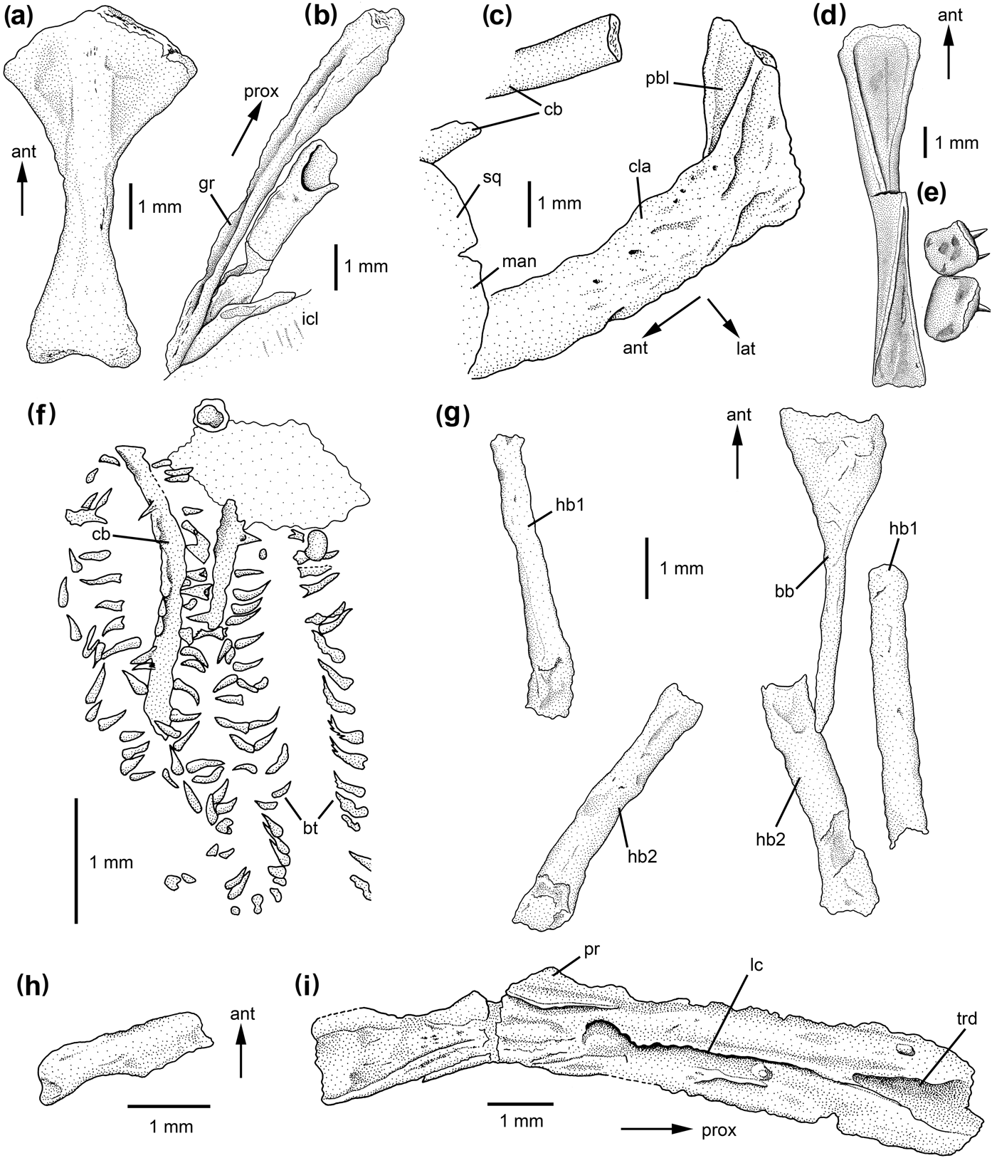
The basibranchial of Thabanchuia oomie consists of a short, stout shaft and is anteriorly and posteriorly distinctly expanded (Fig. 4a). The shaft is dorsoventrally flattened in cross-section and bears no ventral crest. The anterior expansion is bulge-like convex ventrally in its median part (as the anterior continuation of the shaft), but flattens increasingly in an anterior direction. It becomes dorsoventrally thinner laterally where it has a ventrally concave surface. The posterior expansion is dorsoventrally thinner with respect to the shaft, faintly concave ventrally and has a bifurcated posterior end with two small, blunt processes. The basibranchial is neither curved dorsally nor ventrally.
Hyobranchial skeleton of dvinosaurian and dissorophoid temnospondyls: (a–c) Thabanchuia oomie: (a) UCMP 42780 (holotype), basibranchial in ventral view; (b) UCMP 42781, ceratobranchials; (c) UCMP 42780 (holotype), clavicle in anterolateral view showing postbranchial lamina; (d–e) Isodectes obtusus, AMNH 11037: (d) basibranchial in ventral view; (e) dentigerous branchial platelets; (f) Apateon gracilis, MB.Am.406, ossified ceratobranchials with branchial teeth; (g–h) Micropholis stowi: (g) BSM 1934 VIII 43 (specimen B sensu Broili & Schröder, Reference Broili and Schröder1937), basibranchial and hypobranchials in ventral view; (h) BSM 1934 VIII 43 (specimen A sensu Broili & Schröder, Reference Broili and Schröder1937), ceratohyal; (i) Platyrhinops lyelli, MB.Am.23, ceratohyal of a large individual. Abbreviations: ant=anterior; bb=basibranchial; bt=branchial teeth; cb=ceratobranchial; cla=clavicle; gr=groove; hb=hypobranchial; icl=interclavicle; lat=lateral; lc=longitudinal crest; man=mandible; pbl=postbranchial lamina; pr=process; prox=proximal; sq=squamosal; trd=triangular depression.

At least three pairs of slender, slightly curved ceratobranchials are present (Fig. 4b). As stated by Warren (Reference Warren1999), the ceratobranchials appear hollow inside, indicating perichondral ossification of the elements, whereas the inner parts were still cartilaginous. They are somewhat expanded proximally and may possess a slightly bifurcated proximal end, reminiscent of the ceratobranchials of Acanthostega. Posterolateral grooves extend almost the entire length of the ceratobranchials, but diminish in depth proximally and distally.
The ascending process of the clavicle is clearly visible in UCMP 42781 and 42780. This process bears a broad lamina which extends medially and is anteriorly concave (Fig. 4c). This lamina can be interpreted as a postbranchial lamina.
3.2.4. Dvinosauria: Isodectes
In one specimen of Isodectes obtusus (AMNH 11037), the basibranchial is preserved as a slender, rod-like element with a ventrally concave anterior expansion that is framed by elevated lateral margins (Fig. 4d). A sagittal crest divides the anterior expansion into two equally sized areas. The basibranchial gets conspicuously deeper towards the shaft, where it is much deeper than wide and tapers ventrally to a thin, sharp crest. The posterior portion widens again and attains two thirds of the width of the anterior portion.
The dentigerous branchial platelets that were attached to the cartilaginous ceratobranchials in life are quadrangular, or only slightly longer than wide (Fig. 4e). One edge is bulge-like thickened and bears 5–6 teeth. Because of incomplete preservation, it cannot be ascertained how many rows of branchial platelets were present in Isodectes. Milner (Reference Milner1982) described a small larva of Isodectes with a rod-like basibranchial that has faintly expanded anterior and posterior ends, dentigerous branchial platelets and three pairs of external gills.
3.2.5. Further dvinosaurians
Kourerpeton bradyi is represented by a single specimen for which locality and geological age are unknown. It has four pairs of ceratobranchials preserved (Olson & Lammers Reference Olson, Lammers and Churcher1976, fig. 1). A revision of this taxon is currently being undertaken by Marcello Ruta and Andrew R. Milner. Berman (Reference Berman1973) described the only known specimen of Lafonius lehmani, which might be a larval or juvenile specimen. From the hyobranchial apparatus, Berman (Reference Berman1973) identified elongate dentigerous branchial platelets with space for approximately ten teeth at one margin (Berman Reference Berman1973, fig. 5e).
3.2.6. Zatracheidae: Acanthostomatops
Three or four pairs of ossified curved ceratobranchials are present in larval Acanthostomatops vorax that lack grooves and branchial teeth (Steen Reference Steen1937; Witzmann & Schoch Reference Witzmann and Schoch2006). A basibranchial and hypobranchials have not been found. In an adult specimen, Witzmann & Schoch (Reference Witzmann and Schoch2006) noted the absence of ossified ceratobranchials; instead, they identified at least four pairs of slender, linear bones ventral to the basal plate of the parasphenoid and interpreted them as part of a tongue supporting the hyobranchium.
3.2.7. Dissorophoidea: Micromelerpetontidae
From the larval hyobranchial apparatus of Micromelerpeton credneri, branchial platelets that are rounded rectangular and have up to six teeth are preserved (Boy Reference Boy1995). The platelets are strongly concave on the internal side that was attached to the four pairs of cartilaginous ceratobranchials. In a few individuals, two pairs of feebly ossified hypobranchials and ceratohyals (?hypohyals), but no basibranchials, are preserved (Boy Reference Boy1995, Reference Boy2003; Boy & Sues Reference Boy, Sues, Heatwole and Carroll2000). The hyobranchium of the adult, land-dwelling individuals is not known (Boy Reference Boy1995).
In contrast to Micromelerpeton, Branchierpeton amblystomum is known only from larval specimens. The basibranchial is a tiny rod-like element which is anteriorly and posteriorly slightly expanded (Werneburg Reference Werneburg1991, fig. 13d; pers. obs. MMG SaP179). The morphology of the branchial platelets corresponds to those of Micromelerpeton. One specimen of Branchierpeton (MNC-F 10368) has four pairs of ossified ceratobranchials preserved (described by Werneburg Reference Werneburg1998 as a larval Acanthostomatops vorax, but see assignment to B. amblystomum in Witzmann & Schoch Reference Witzmann and Schoch2006). The ceratobranchials are long, slender elements whose proximal ends are expanded (approximately twice the width of the shaft), whereas the distal ends are tapering and hook-like. The original bone surface is not preserved, but in places, the denser perichondral bone (i.e. the cortex) can be distinguished from the spongy endochondral bone.
3.2.8. Dissorophoidea: Branchiosauridae and Trematopidae
Branchiosaurids have a highly derived larval hyobranchial apparatus adapted to filter feeding on planktonic prey (Boy & Sues Reference Boy, Sues, Heatwole and Carroll2000; Schoch & Milner Reference Schoch and Milner2008). The often multi-ended branchial teeth are not associated with bony platelets, but are isolated and directly attached to the four pairs of (mostly) cartilaginous ceratobranchials. Six rows of teeth are present and face towards each other, forming a zipper-like structure in the three pairs of gill clefts (Boy & Sues Reference Boy, Sues, Heatwole and Carroll2000). Ossified elements of the branchiosaurid hyobranchium comprise a tiny, rod-like basibranchial, hypobranchials and ceratohyals (or hypohyals), similar to those of micromelerpetontids (see above) (Werneburg Reference Werneburg1991; Boy & Sues Reference Boy, Sues, Heatwole and Carroll2000). Few specimens of branchiosaurids with ossified ceratobranchials are known (Werneburg Reference Werneburg1991). In the specimen of Apateon gracilis (MB.Am.406), the slightly curved ceratobranchials are long, slender and not expanded at their ends (Fig. 4f). The larvae of trematopids also have a hyobranchial apparatus that closely resembles that of branchiosaurids and was adapted for filter feeding (Milner Reference Milner2007; Schoch Reference Schoch2009). The hyobranchium of metamorphosed branchiosaurids and trematopids is unknown.
3.2.9. Dissorophoidea: Amphibamidae
The basibranchial of Micropholis stowi is clearly evident in ventral exposure in individual B of specimen BSM 1934 VIII 43 (sensu Broili & Schröder Reference Broili and Schröder1937). It consists of a deep, narrow shaft that shows no posterior expansion, whereas the anterior portion is conspicuously expanded and attains more than five times the width of the shaft (Fig. 4g). The ventral surface of the anterior broadening is smooth. The basibranchial shows no ventral or dorsal curvature. Two pairs of robust hypobranchials are visible in individual B. They are about twice as broad as the basibranchial shaft and roundish in cross-section, and attain about 90% the length of the basibranchial. An ossified, short element anterolateral to the basibranchial fragment in individual A was interpreted as a hypohyal or a ceratohyal by Boy (Reference Boy1985). In the present study, this rod-like bone is interpreted as ossified part of a ceratohyal due to its elongate shape (Fig. 4h).
Pasawioops mayi has a very slender basibranchial whose anterior end is expanded and flattened (Fröbisch & Reisz Reference Fröbisch and Reisz2008). These authors further described one pair of hypobranchials, which are rod-like elements and are longer and more robust than the basibranchial.
Sigurdsen & Bolt (Reference Sigurdsen and Bolt2010, fig. 3b) reported feebly ossified, slender hyobranchial elements in a few specimens of Doleserpeton annectens. They consist of a short basibranchial bone (“basihyal”) and a pair of rod-like ceratohyals and one hypobranchial.
Clack & Milner (Reference Clack and Milner2010) detected an elongated, flattened bone in the anterior region of the palate of a small and a larger specimen of Platyrhinops lyelli and interpreted the bones as ceratohyals. The small specimen (MB.Am.331) bears two rod-like bones ventral to the vomers. As Clack & Milner (Reference Clack and Milner2010) pointed out, it cannot be decided if the two elements represented one element that is broken, or two separate bones. The bones are slender and show no differentiation, except for the fact that the anatomical right element bears a wide longitudinal groove all along its length. In the larger specimen (MB.Am.23), the element found by Clack & Milner (Reference Clack and Milner2010) is broader and flattened and is slightly curved (Fig. 4i). It bears a longitudinal, sharp crest on its (presumed) ventrolateral side, dividing the surface into equal dorsal and ventral parts. At the (presumed) proximal portion, the crest is divided and frames a notch-like, triangular depression. In the (presumed) distal third of the bone, the medial crest has diminished. There is a pointed elevation or process in the distal third of the (presumed) anterior margin. At least the bone in the larger skull (MB.Am.23) can be interpreted as ceratohyal with some confidence, because of the flattened, albeit slender morphology and its anterolateral location ventral to the skull.
Bourget & Anderson (Reference Bourget and Anderson2011) detected a pair of poorly preserved, flattened elements in Rubeostratilia texensis that resemble the described possible ceratohyal in the small specimen of Platyrhinops. However, their orientation and location ventral to the parasphenoid might indicate that they represent hypobranchials.
3.2.10. Eryopidae: Onchiodon
In larvae of Onchiodon labyrinthicus, Boy (Reference Boy1990) and Witzmann (Reference Witzmann2005) described the rod-like basibranchial that has flattened anterior and posterior ends. The anterior end is more expanded than the posterior one. The basibranchial is unknown in adult O. labyrinthicus and O. thuringiensis (see Werneburg Reference Werneburg2007), but Werneburg (Reference Werneburg1997, figs 1d, 4b) described and illustrated the basibranchial in adult specimens of O. frossardi. In this form, the expanded anterior end of the basibranchial has approximately four times the width of the rather short shaft, and the posterior end attains almost the width of the anterior end. There is no evidence of downwards curvature of the anterior portion of the bone. The dentigerous branchial platelets of Onchiodon larvae correspond in morphology and arrangement to those of micromelerpetontids (Boy Reference Boy1990). Four pairs of slender ceratobranchials without grooves are ossified in a number of specimens of O. labyrinthicus, consisting of spongy bone, to which the branchial platelets are attached (Witzmann Reference Witzmann2005).
3.2.11. Temnospondyli incertae sedis: Iberospondylus
The basibranchial of Iberospondylus schultzei (PU-ANF 14) is a robust, rod like element with a distinctly expanded, flattened anterior end (Fig. 5a, b). This anterior portion is curved ventrally and has two concave areas on its ventral surface, separated in the midline by a shallow longitudinal crest. The slender shaft is deeper than wide in cross section. The posterior end is flattened again and is expanded, but attains less than half the width of the anterior portion.
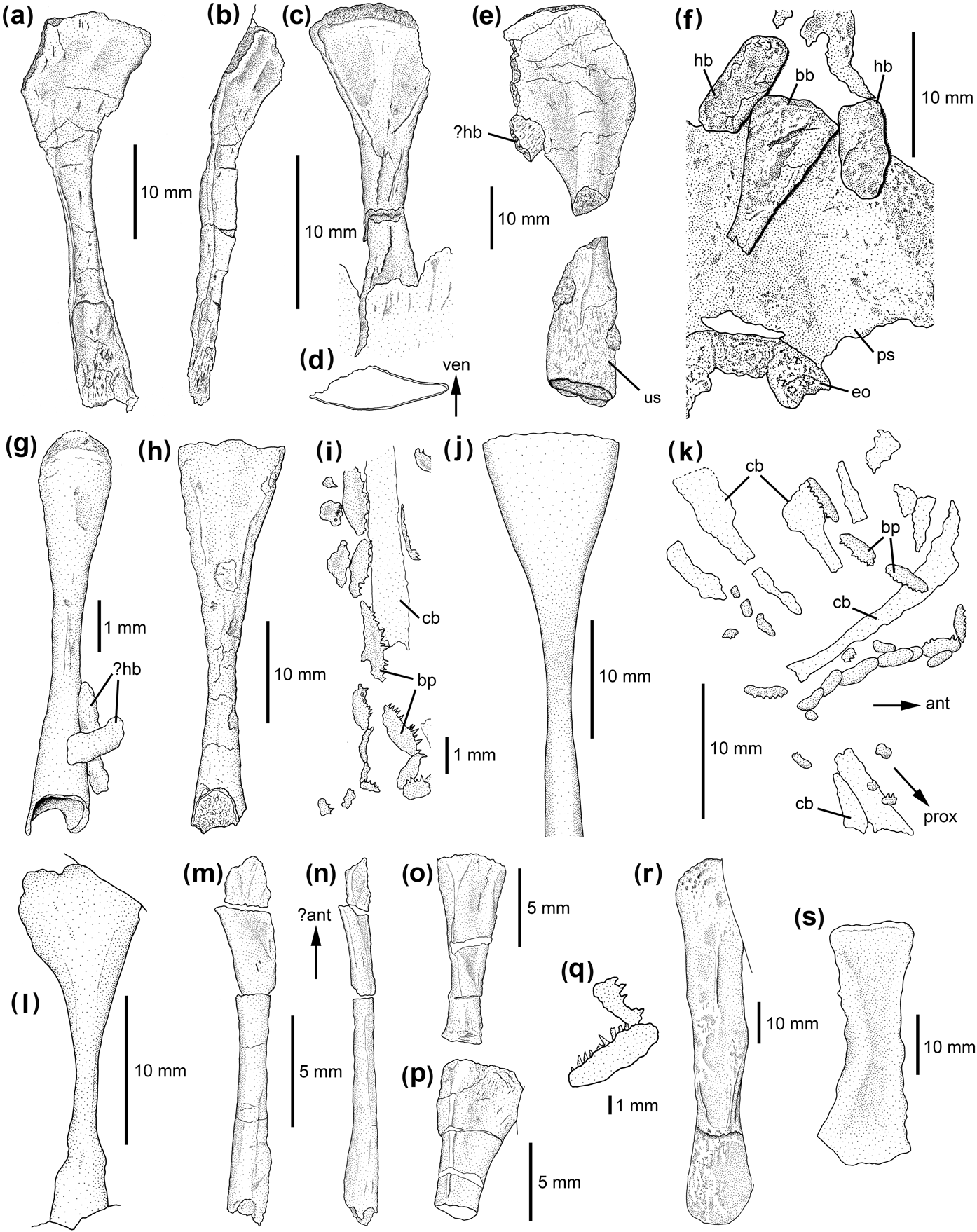
Hyobranchial skeleton of Iberospondylus and stereospondylomorph temnospondyls: (a–b) Iberospondylus schultzei, PU-ANF 14: (a) basibranchial in ventral view; (b) basibranchial in ventrolateral view; (c) Sclerocephalus haeuseri, SMNS 81791, basibranchial in ventral view; (d) cross-section of the anterior portion in (c); (e) Sclerocephalus nobilis, NHMM-PW 2005/2, basibranchial with possible hypobranchial fragment in ventral view; (f) Sclerocephalus haeuseri, UHC-P 0489, parasphenoid with exoccipitals, basibranchial and hypobranchials in ventral view; (g) Glanochthon latirostris, MB.Am.211, basibranchial with possible hypobranchial fragments in ventral view; (h) Glanochthon angusta, GPIT Am 34, basibranchial in ventral view; (i) Glanochthon latirostris, MB.Am.224, imprint of cartilaginous ceratobranchial with ossified dentigerous branchial platelets; (j–k) Archegosaurus decheni: (j) MB.Am.953, basibranchial in ventral view; (k) IGS U II 1/2, imprints of cartilaginous ceratobranchials with ossified dentigerous branchial platelets; (l) Lydekkerina huxleyi, BSM 1934 VII 44, basibranchial in ?ventral view; (m–p) Trematolestes hagdorni, SMNS 81790: (m) basibranchial in ventral view; (n) basibranchial in lateral view; (o–p) possible hypobranchials; (q) Callistomordax kugleri, SMNS 90700, dentigerous branchial platelets; (r) Metoposaurus diagnosticus SMNS 5143, basibranchial in ventral view; (s) Stanocephalosaurus birdi (Wellesaurus peabodyi), UCMP 36040, basibranchial in ventral view. When not otherwise indicated, anterior is to the top. Abbreviations: ant=anterior; bb=basibranchial; bp=branchial platelets; cb=ceratobranchial; eo=exoccipital; hb=hypobranchial; prox=proximal; ps=parasphenoid; us=unfinished surface.

3.2.12. Stereospondylomorpha: Sclerocephalus
The ontogeny of the basibranchial in Sclerocephalus haeuseri was described by Boy (Reference Boy1972, Reference Boy1974, Reference Boy1988) and Schoch (Reference Schoch2003), highlighting the proportional broadening of the anterior end during growth. In adult specimens, the basibranchial morphology resembles closely that of Iberospondylus, as described above (Fig. 5c, d). The ventral curvature of the expanded anterior portion and the two concave areas, however, are not apparent in larvae and juveniles. The only known specimen of S. nobilis (NHMM-PW 2005/2) has a large and massive basibranchial element (Fig. 5e). Its anterior expansion is further ossified anteriorly; thus, the ventral curvature is more pronounced and the bone has a shovel-like appearance. The shaft is stout and ventrally convex, and bears a sharp crest more posteriorly. Interestingly, the flattened and slightly expanded posterior portion of the basibranchial has an unfinished surface not only at its posterior edge, but at least also on the complete ventral surface (the lateral and dorsal sides are concealed). Apart from S. nobilis, this structure is also evident in specimen BSM AS I 575 of S. haeuseri.
In one specimen of S. haeuseri (UHC-P 0489), one pair of short, rod-like bones is preserved anterolateral to the basibranchial ventral to the base of the cultriform process (Fig. 5f). They are interpreted here as the anterior pair of hypobranchials, but might alternatively represent ossified hypohyals. In S. nobilis, a rod-like bone articulates with the right posterolateral edge of the expanded anterior portion of the basibranchial and might represent a part of a hypobranchial (Fig. 5e).
Larvae of Sclerocephalus haeuseri possess three or four rows of dentigerous branchial platelets that are more elongate than those of micromelerpetontids and bear between five and ten teeth (Boy Reference Boy1988; Schoch Reference Schoch2003). These platelets are not confined to larval specimens, but occur also in at least some adults (Schoch & Witzmann Reference Schoch and Witzmann2009a).
3.2.13. Stereospondylomorpha: Glanochthon
In Glanochthon latrirostris, the basibranchial has not been found in adults (Boy Reference Boy1993; Schoch & Witzmann Reference Schoch and Witzmann2009b), but is clearly visible in the juvenile MB.Am.211 (Fig. 5g). Its anterior portion is deeper and about 2.5 times wider than the shaft and slightly curved ventrally. The shaft is round in cross-section and bears a faint ventral ridge. The posterior portion is again deeper and expanded, attaining approximately 80% of the width of the anterior portion. Posterolateral to the basibranchial are two short, poorly ossified rods that might represent hypobranchials. The basibranchial is preserved in an adult specimen of G. angusta (GPIT.Am.34) and is more slender and less robust than that of Sclerocephalus (Fig. 5h). The anterior expansion is flattened and is about 3.5 times wider than the shaft, whereas the posterior extension attains only twice the width of the shaft. A ventral curvature is not determinable. Two anterior concave areas on the ventral surface are faintly indicated.
Four rows of dentigerous branchial platelets (sometimes associated with imprints of cartilaginous ceratobranchials) are preserved in small specimens and are retained at least in medium-sized skulls (Schoch & Witzmann Reference Schoch and Witzmann2009b). They are elongate and have space for more than 20 teeth (Fig. 5i).
3.2.14. Stereospondylomorpha: Archegosaurus
The ontogeny of the basibranchial in Archegosaurus decheni was documented by Hofker (Reference Hofker1926) and Witzmann (Reference Witzmann2006) and resembles that of Sclerocephalus. In adults, the expanded anterior portion is six times wider than the slender shaft and is curved ventrally (Fig. 5j). The posterior portion is only slightly expanded (1.6 times the width of the shaft), but has become conspicuously deeper.
The imprints of cartilaginous ceratobranchials have distally and proximally expanded ends (Fig. 5k). Imprints of two short rod-like bones posterior to the parasphenoid might represent hypobranchials 1 and 2, and a short element anterolateral to them might be a ceratohyal (Hofker Reference Hofker1926, fig. 8). The morphology of the denticulate branchial platelets and their arrangement correspond to those of Glanochthon and are present in larvae and retained at least in middle-sized specimens. Some of the platelets may bear an ornamentation of concentric rings which may represent growth rings (Witzmann Reference Witzmann2006).
3.2.15. Stereospondylomorpha: basal stereospondyls
Van Hoepen (Reference Van Hoepen1915) and Schoch (Reference Schoch2002) described elongate branchial platelets in Uranocentrodon senekalensis that were arranged in at least three rows. Each platelet has space for more than 12 teeth. A rod-like bone that is located next to the cultriform process of the parasphenoid in Lydekkerina huxleyi (BSM 1934 VIII 44) has an anterior expanded portion that attains more than five times the width of the slender shaft (Fig. 5l). The surface of the anterior expanded portion is slightly convex. Posteriorly, the bone widens again and attains three times the width of the shaft. Because of its location and morphology, the element can be interpreted as the basibranchial.
3.2.16. Stereospondylomorpha: Lapillopsidae
Among this presumably semi-terrestrial group of stereospondyls, a specimen of Lapillopsis nana is preserved with a slender, rod-like bone ventral to the parasphenoid (Yates Reference Yates1999, fig. 7). Yates (Reference Yates1999) suggested that it might represent a part of the hyobranchium and could be a possible hypobranchial (probably because of its oblique orientation with respect to the longitudinal axis of the skull). However, due to its isolated nature ventral to the parasphenoid, it might also represent a slightly rotated basibranchial.
3.2.17. Stereospondylomorpha: Trematosauroidea
In the type specimen of Trematolestes hagdorni (SMNS 81790), a long, very slender bone is located approximately in the midline of the skull between the orbits (Fig. 5m, n). Its shaft is round-ovate in cross-section and the caudally oriented end is flattened and slightly expanded with a concave surface. The opposite end is less expanded, and has become deeper rather than flattened. Because of its morphology and position, Schoch (Reference Schoch2006) regarded this element as the basibranchial, and this interpretation is followed here. The element has probably rotated around 180° and the broader portion represents the anterior end. Posterior to the basibranchial is a pair of short bones (Fig. 5o, p). Because of their position and length, these bones might be hypobranchials. More posterior, lateral to the left clavicle, is a bone fragment that might represent the expanded proximal end of a ceratobranchial.
In the stem-metoposaurid Callistomordax kugleri, several long-ovate branchial platelets with space for approximately 15 teeth at one edge are preserved (Fig. 5q). Additionally, a slightly curved bone fragment with a longitudinal groove is present that probably represents a part of a ceratobranchial.
In specimen SMNS 5143 of Metoposaurus diagnosticus, a rod-like bone is preserved ventral to the cultriform process (Fig. 5r). Its shaft is stout, dorsoventrally flattened, and the anterior and posterior ends are slightly broadened and rounded. The anterior end is somewhat deeper than the shaft and bears a pitted surface. A longitudinal crest is visible in the anterior half of the bone. In the posterior half, two crests converge towards the posterior end. Morphology and location of the bone strongly suggest the interpretation as a basibranchial.
3.2.18. Stereospondylomorpha: Capitosauroidea
In two specimens of Stanocephalosaurus birdi (Wellesaurus peabodyi; UCMP 56098, 36040), a stout linear bone is visible that is located next to the cultriform process of the parasphenoid and oriented parallel to it. It is rather undifferentiated and slightly expanded anteriorly and posteriorly (Fig. 5s) and probably represents the basibranchial. Warren & Hutchinson (Reference Warren and Hutchinson1988, fig. 6) described a basibranchial in Rewanobatrachus (Parotosuchus) aliciae. This bone has a long, slender shaft and is slightly expanded anteriorly and posteriorly. In Parotosuchus wadei, Damiani & Warren (Reference Damiani and Warren1997, fig. 2) found thin rod-like bones between the clavicles and the posterior end of the mandibles. They tentatively interpreted these bones as ribs, but did not rule out the possibility that they might represent ossified ceratobranchials.
3.2.19. Stereospondylomorpha: Brachyopoidea
The small and only known specimen of the brachyopoid Platycepsion wilkinsoni has four ossified ceratobranchials and an ossified part of a possible ceratohyal is present anterior to them (Watson Reference Watson1956; Warren & Marsicano Reference Warren and Marsicano1998). Warren et al. (Reference Warren, Rozefelds and Bull2011) found fragments of ossified, grooved ceratobranchials in a specimen of Bothriceps australis.
3.2.20. Stereospondylomorpha: Gerrothorax (Plagiosauridae)
The view of Jenkins et al. (Reference Jenkins, Shubin, Gatesy and Warren2008) is followed here that other Gerrothorax species cannot be differentiated from G. pulcherrimus. Therefore, G. “rhaeticus” from Sweden (Nilsson Reference Nilsson1946) is treated here as the same taxon as G. pulcherrimus from Greenland (Jenkins et al. Reference Jenkins, Shubin, Gatesy and Warren2008) and Germany (Hellrung Reference Hellrung2003). In the following, the partially articulated hyobranchial apparatus SMNS 83866, MGUH 28919 and NRM-PZ B.18b will be described separately.
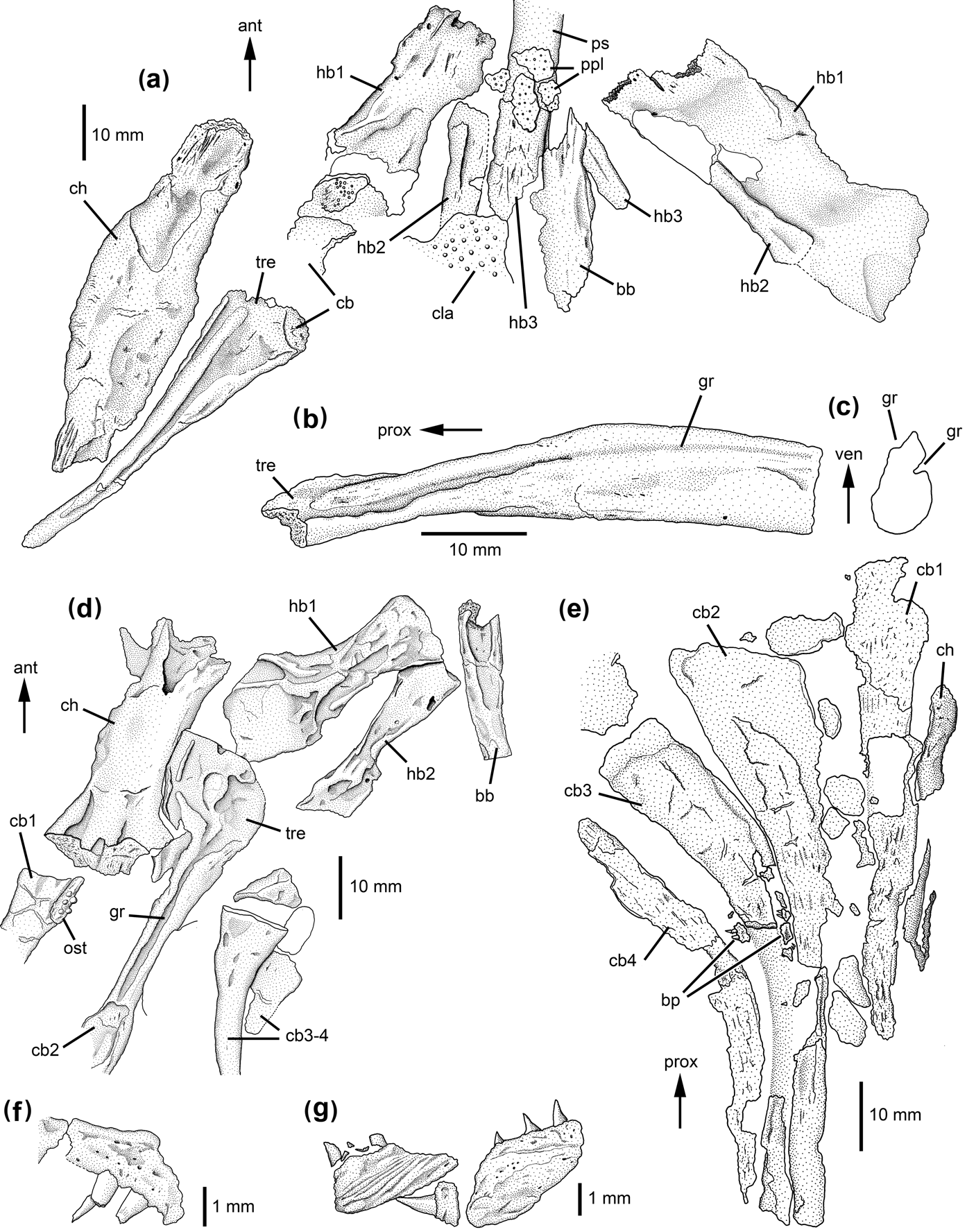
The Stuttgart specimen SMNS 83866 is an almost complete skeleton of Gerrothorax, in which large parts of the hyobranchial apparatus are preserved in ventral view (Fig. 6a). The large paired elements in the anterior part of the hyobranchium are elongated and rectangular in outline. The anatomical right element appears to have a concave posteromedial edge, whereas this region is obscured in the opposing element. Poor preservation precludes recognition of more anatomical details. In contrast to Hellrung (Reference Hellrung2003), who reconstructed these elements as hypohyals, they are interpreted here as the first pair of hypobranchials for the following reasons. First, the ceratobranchials and not the ceratohyals are located distal to these elements; second, comparison with tetrapodomorph fishes (Jarvik Reference Jarvik1954, Reference Jarvik1963; Downs et al. Reference Downs, Daeschler, Jenkins and Shubin2008), Acanthostega (Coates & Clack Reference Coates and Clack1991; this study), other temnospondyls (Bystrow Reference Bystrow1938; this study) and extant salamanders (e.g. Stadtmüller Reference Stadtmüller, Bolk, Göppert, Kallius and Lubosch1936; Deban & Wake Reference Deban, Wake and Schwenk2000) shows the hypohyals to be mostly rather small elements proximal to the ceratohyals. If this interpretation is correct, then three ossified pairs of hypobranchialia are present in this specimen, the posterior two pairs being more slender and rod-like. Additionally, a fragment of the basibranchial can be identified between the hypobranchial elements (contra Hellrung Reference Hellrung2003). It appears to be rod-like and slender. Two ceratobranchials can definitely be identified posterior to the hypobranchials (see also Hellrung Reference Hellrung2003), and a bone fragment directly posterior to the first hypobranchial probably represents a further ceratobranchial. Thus, probably at least three ossified ceratobranchials are present in this specimen. Their following morphological description is supplemented by the isolated finds SMNS 83370 and 83382. The ceratobranchials are widened proximally in a triangular extension, and taper continuously in a distal direction (Fig. 6a–c). The shaft is slender and may be slightly curved, and broadens again somewhat at its distal end. With the exception of short striae and tiny ridges, the dorsomedial surface of the ceratobranchials is smooth. In contrast, the ventrolateral surface is rather complex. A broad, shallow ridge extends from the proximal end distally. It divides the proximal triangular expansion into two concave fields. The broad ridge increases distinctly in height at the transition from the triangular expansion to the shaft and tapers into a rather sharp ventral crest, so that the shaft becomes deeper than wide. Two grooves extend along the lateral face of the shaft, a posteroventral and an anteroventral one (Fig. 6c). The posterior of these furrows represents the distal continuation of the posterior concave field; it is rather shallow and located immediately posterior to the apex of the ridge. Anterior to the crest is a deeper furrow that represents the distal continuation of the anterior concave field. Branchial platelets and teeth are not visible. Each ceratohyal has an elongate, oval shape and is the longest preserved element of the hyobranchium. The anterolateral edge is slightly convex, whereas the posteromedial edge appears slightly concave. Approximately the proximal half of the ventrolateral surface consists of a distinct depression with longitudinal striae at the proximal end. The distal end of the element bears similar longitudinal striae, but no depression is visible.
Hyobranchial skeleton of plagiosaurid stereospondyls: (a–f) Gerrothorax pulcherrimus: (a) SMNS 83866, partial hyobranchial apparatus in ventral view; (b) SMNS 83382, isolated ceratobranchial, lateral view; (c) SMNS 83382, isolated ceratobranchial, cross-section of distal end; (d) MGUH 28919: partial hyobranchial apparatus in ventral view; (e) NRM-PZ B.18, four ceratobranchials and fragmentary ceratohyal; (f) NRM-PZ B.18, dentigerous branchial platelets; (g) Plagiosuchus pustuliferus, SMNS 84794, dentigerous branchial platelets. Abbreviations: ant=anterior; bb=basibranchial; bp=branchial platelets; cb=ceratobranchial; ch=ceratohyal; cla=clavicle; gr=groove; hb=hypobranchial; ost=osteoderm; ppl=palatal platelets; prox=proximal; ps=parasphenoid; tre=triangular expansion; ven=ventral.

The Copenhagen specimen MGUH 28919 is a rather small, apparently juvenile specimen from Greenland. Several ossified elements of the hyobranchium are articulated and exposed in ventral view (Fig. 6d). The large paired elements regarded as hypohyals by Jenkins et al. (Reference Jenkins, Shubin, Gatesy and Warren2008) are interpreted here as hypobranchials 1 for the same reasons as the hypohyals sensu Hellrung (Reference Hellrung2003) in SMNS 83866 (see above). Consequently, the posterior following, much narrower element can be regarded as second hypobranchial, and the third hypobranchial is not preserved. The ventral surface of the first hypobranchial bears a ridge along the shaft and bifurcates at the distal broadening of the bone. The anterolateral edge of the bone bears a notch-like depression. If the designation as first and second pair of hypobranchials is correct, then the median, unpaired element can be interpreted as basibranchial rather than a basihyal sensu Jenkins et al. (Reference Jenkins, Shubin, Gatesy and Warren2008). It is quite short and only slightly broader anteriorly than posteriorly. It is dorsoventrally flattened in cross-section. At least three, but probably four ceratobranchials are preserved which correspond in outline to the ceratobranchials described in the Stuttgart material. On the anatomical right side, ceratobranchial 1 is preserved only in its proximal part. Ceratobranchial 2 is straight, grooved and has a trough in its proximal end (Fig. 6d). Ceratobranchials 3 and 4 are only fragmentarily preserved, but at least ceratobranchial 3 appears to be slightly curved. On the anatomical left side, three grooved ceratobranchials are preserved (probably ceratobranchials 1–3, not shown in the Figure). Whereas the anteriormost element is rather straight, the posteriorly following ones are increasingly curved and are becoming shorter, similar to NRM-PZ B.18b (see below). The ceratohyals are the broadest elements, but in contrast to SMNS 83866, they are shorter than the ceratobranchials, probably because of incomplete ossification. They have a slightly convex lateral and a concave medial margin. The medial margin is distinctly thickened, and the proximal portion bears a notch-like depression whose length accounts for approximately one quarter of the length of the ceratohyal. In its distal part, the bone bears smaller, paired depressions.
The Stockholm specimen NRM-PZ B.18b is very poorly preserved, and only the outline of the hyobranchial elements, not their surface structure, can be seen (Fig. 6e). The first ceratobranchial is rather straight, whereas the posteriorly following three elements get increasingly curved. The length of the first ceratobranchial cannot be ascertained, but ceratobranchials 2–3 get increasingly shorter. Associated with ceratobranchials 2 and 3 are ossified branchial platelets of rectangular outline, with space for 3–4 posterior teeth (Fig. 6f). Judging from its position lateral to ceratobranchial 1, an elongate, very poorly preserved element can best be interpreted as ceratohyal (see also Nilsson Reference Nilsson1946). The elements interpreted by Nilsson (Reference Nilsson1946) as three hypobranchials are too fragmentarily preserved to draw any conclusions.
3.2.21. Stereospondylomorpha: Plagiosuchus (Plagiosauridae)
Next to the clavicular stem in an articulated specimen of Plagiosuchus pustuliferus (SMNS 84794) are several branchial platelets bearing curved teeth at one edge (Fig. 6g). Because of poor preservation, they are of ill-defined outline and it cannot be ascertained if they are quadrangular or rather elongate. Interestingly, they bear an ornament of fine, curved ridges and furrows. Four pairs of grooved ceratobranchials are ossified in Plagiosuchus (Schoch & Witzmann Reference Schoch and Witzmann2011, fig. 4g, h). Due to poor preservation, it cannot be determined if the ceratobranchials have the widened proximal ends that are visible in Gerrothorax.
3.3. Lepospondyli
3.3.1. Lysorophia
The extensive hyobranchial apparatus of lysorophians was described in detail by Wellstead (Reference Wellstead1991). Hypohyals and ceratohyals are more flattened than the rather cylindrical, stout four pairs of hypobranchials and the four pairs of ceratobranchials. Some specimens of Brachydectes elongates bear “accidental bones” in the first branchial arch between the hypobranchials and ceratobranchials, the homology of which is uncertain (Wellstead Reference Wellstead1991). The ceratobranchials bear no grooves.
3.3.2. “Microsauria”
“Microsaurs” are probably not a monophyletic assemblage (Marjanović & Laurin Reference Marjanović and Laurin2013, and references therein) and are therefore written in quotation marks. Microbrachis pelikani is the only lepospondyl in which branchial platelets were described (Carroll & Gaskill Reference Carroll and Gaskill1978; Olori Reference Olori2011). According to Olori (Reference Olori2011), the platelets are round or ovate and may bear three denticles. In some platelets, the bone surface is concave, probably representing the side that was attached to the ceratobranchials as in colosteids and temnospondyls (see above). In specimen AMNH 2557, posterolateral to two preserved rows of branchial platelets is a wrinkled structure that extends mediolaterally. Because of its location and alignment, this structure might represent preservation of external gills proper.
The hyobranchial apparatus of Pantylus cordatus is well ossified and was described by Romer (Reference Romer1969, fig. 16) and Carroll & Gaskill (Reference Carroll and Gaskill1978) in a specimen which has been sectioned since then. Three pairs of elongate bones are present that are blade-like, expanded in their proximal portion and continue distally into a narrower stem. At about midlength, these bones curve in a dorsomedial direction. The anteriormost of these bones appears to bifurcate distally. Distal to them are located three pairs of stout, broadened bones. Because the proximal bones are proportionally very long and curved and the distal ones are rather short, the hyobranchial elements in Pantylus can best be designated as ceratobranchials and epibranchials sensu Reilly & Lauder (Reference Reilly and Lauder1988). The basibranchial or parts of the hyoid arch are not preserved.
Further microsaurs with hyobranchial remains are Pelodosotis elongatum, Tambaroter carolli and Hapsidopareion lepton. Narrow, rod-like hyobranchial elements of uncertain affinities (? ceratohyal and hypobranchials) are present in Pelodosotis and Tambaroter (Carroll & Gaskill Reference Carroll and Gaskill1978; Henrici et al. Reference Henrici, Martens, Berman and Sumida2011), whereas in Hapsidopareion, a short, robust bone posterior to the articular and stapes was interpreted as a probable hyoid element by Carroll & Gaskill (Reference Carroll and Gaskill1978).
3.3.3. Nectridea, Aïstopoda and Acherontiscidae
No unambiguous remains of hyobranchial elements have been demonstrated so far in nectrideans (Bossy & Milner Reference Bossy, Milner and Wellnhofer1998) or in aïstopods (Carroll Reference Carroll and Wellnhofer1998; Anderson et al. Reference Anderson, Carroll and Rowe2003). Carroll (Reference Carroll1969) interpreted four disarticulated, flattened bones of rectangular outline just behind the skull of Acherontiscus caledoniae as elements of the hyobranchial apparatus, but a more precise assignment is not possible.
3.4. “Anthracosauria”
No cartilaginous or ossified hyobranchial skeletal elements and no branchial teeth have ever been found in the paraphyletic “anthracosaurs” (embolomeres, gephyrostegids and seymouriamorphs), despite the fact that numerous specimens of larval seymouriamorphs preserve three pairs of pinnate external gills (Ivakhnenko Reference Ivakhnenko1987; Klembara Reference Klembara1995; Bulanov Reference Bulanov2003; pers. obs.). Pawley (Reference Pawley2006) described a postbranchial lamina on the cleithrum of the embolomere Archeria.
3.5. Reconstructions of hyobranchial apparatus
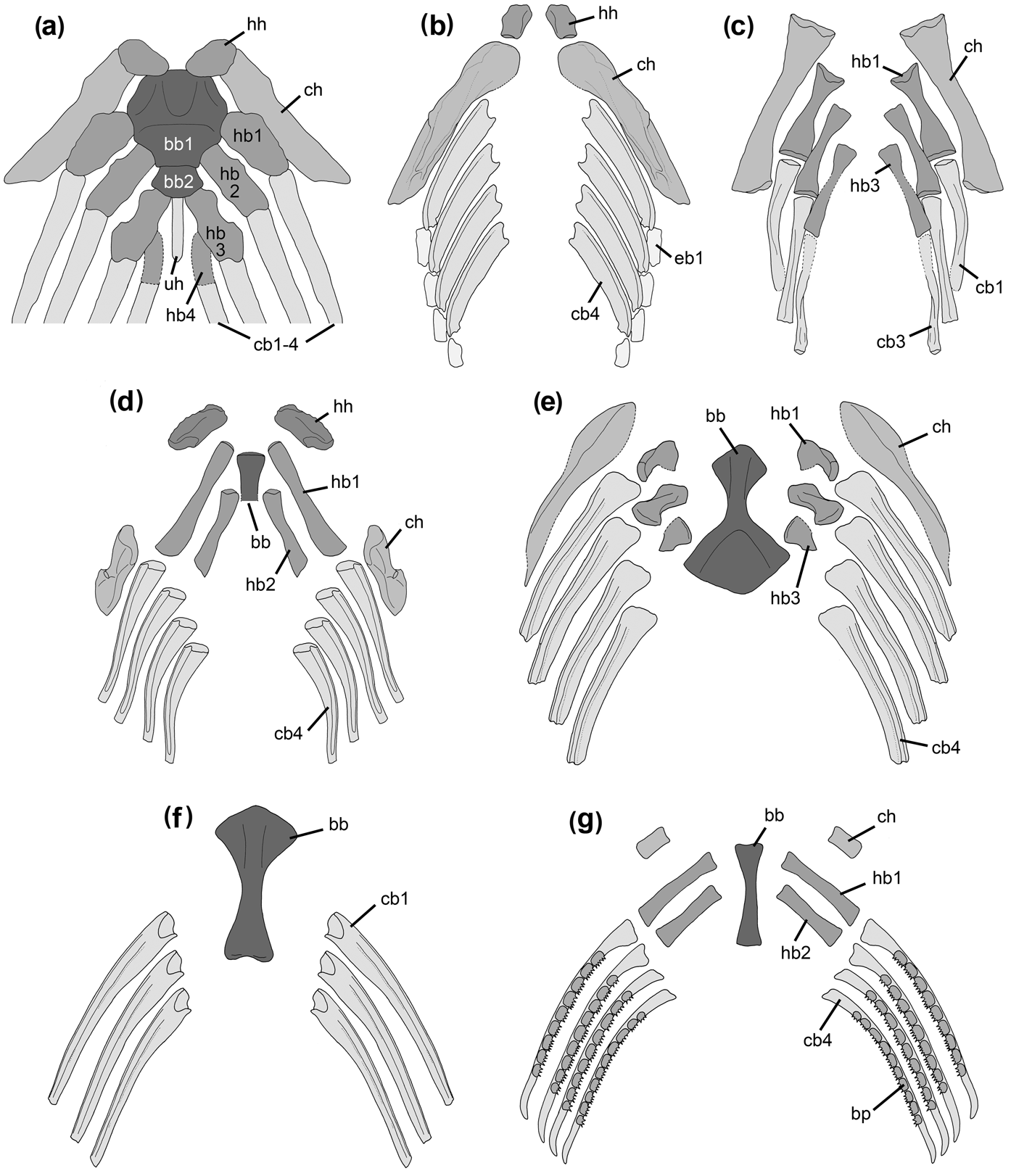
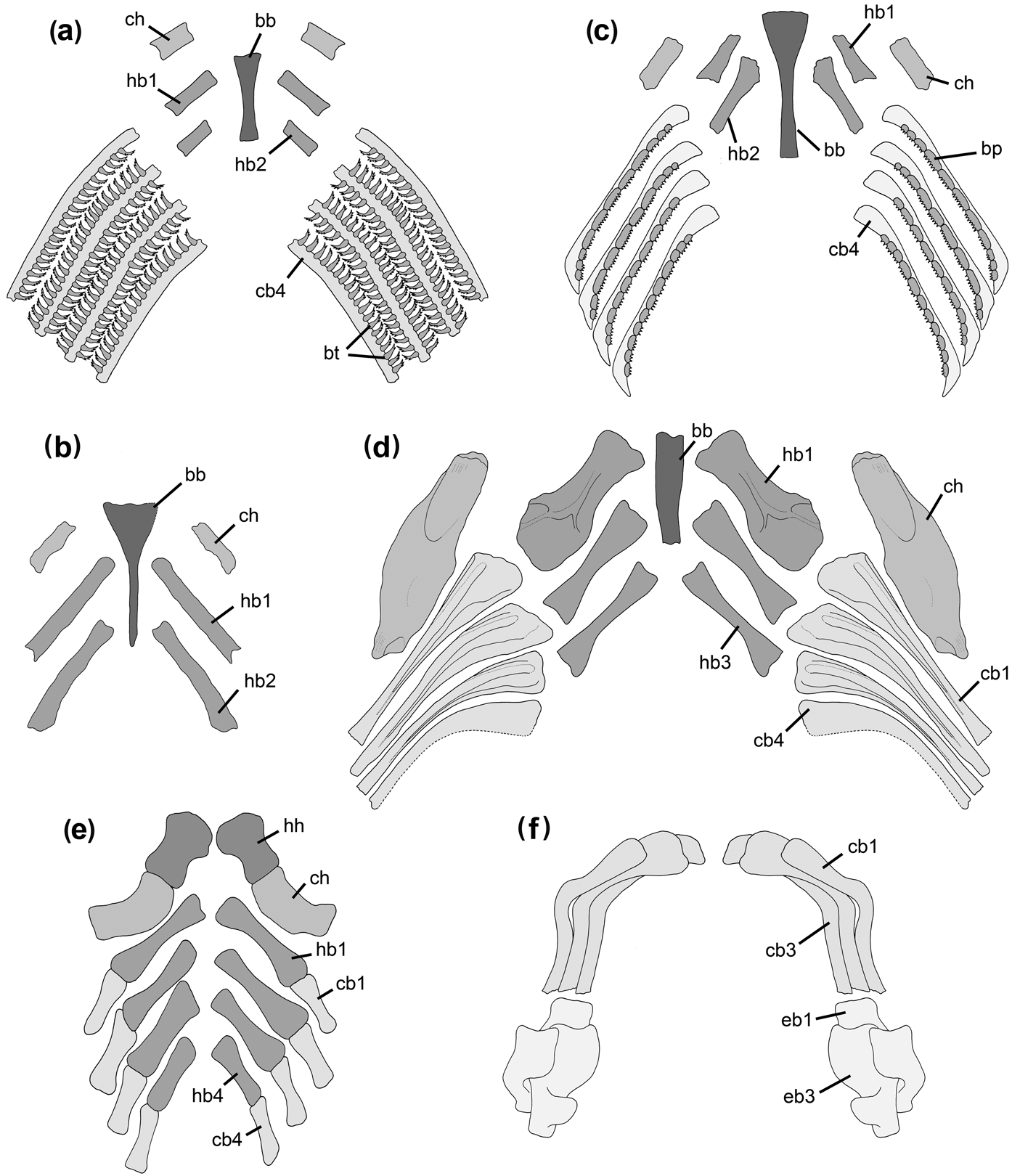
The hyobranchial apparatus of different basal tetrapods are reconstructed in Figures 7 and 8 and are compared with that of the tetrapodomorph fish Tiktaalik. Due to the often fragmentary preservation and the poor degree of ossification, these reconstructions must in part be tentative.
Reconstructions of hyobranchial apparatus in Tiktaalik and different basal tetrapods: (a) Tiktaalik roseae, redrawn after Downs et al. (Reference Downs, Daeschler, Jenkins and Shubin2008); (b) Acanthostega gunnari, based on MGUH f.n.1227 and 1300; (c) Adelogyrinus simorhynchus, based on NMS.G.1889.101.17; (d) Dvinosaurus sp., based on PIN 2005/39 and 4818/410; (e) Trimerorhachis insignis, based on UCMP 105146, 105175, 105221 and 154434; (f) Thabanchuia oomie, based on UCMP 42780 and 42781; (g) larval micromelerpetontid, based on MNC-F 179 and MMG SaP 10368 (Branchierpeton amblystomum), ceratohyals and hypobranchials are supplemented after Boy & Sues (Reference Boy, Sues, Heatwole and Carroll2000, Micromelerpeton credneri). Abbreviations: bb=basibranchial; bp=branchial platelets; cb=ceratobranchial; ch=ceratohyal; eb=epibranchial; hb=hypobranchial; hh=hypohyal; uh=urohyal.

Reconstructions of hyobranchial apparatus in different basal tetrapods: (a) Apateon sp., ossified ceratobranchials and branchial teeth based on MB.Am.406, basibranchial, ceratohyal and hypobranchials supplemented after Boy & Sues (Reference Boy, Sues, Heatwole and Carroll2000); (b) Micropholis stowi, based on BSM 1934 VIII 43; (c) Archegosaurus decheni, based on IGS U II 1/2 and MB.Am.983, ceratohyals and hypobranchials supplemented after Hofker (Reference Hofker1926); (d) Gerrothorax pulcherrimus, based on SMNS 83866, MGUH 28919 and NRM-PZ B.18; (e) Brachydectes elongatus, redrawn after Wellstead (Reference Wellstead1991), “accessory bones” omitted; (f) Pantylus cordatus, modified after Romer (Reference Romer1969). Abbreviations: bb=basibranchial; bp=branchial platelets; bt=branchial teeth; cb=ceratobranchial; ch=ceratohyal; eb=epibranchial; hb=hypobranchial; hh=hypohyal.

4. Discussion
4.1. Phylogenetic patterns of the hyobranchium in basal tetrapods
4.1.1. Basibranchial skeleton
In osteichthyan fishes, the basibranchial skeleton might be composed of a series of cartilaginous and/or ossified basibranchial elements, or the basibranchial can be developed as a single element (Nelson Reference Nelson1969). The basibranchial series is most often subdivided into several ossification centres, including an anterior basihyal in actinopterygians. However, the number of elements varies between taxa, and the basal actinopterygian Polypterus has only a single basibranchial ossification (Fig. 1a). Among extant sarcopterygian fishes, Latimeria has also a single basibranchial (Fig. 1b), whereas in the extant dipnoan Neoceratodus, the basibranchial series comprises a basihyal and two small basibranchials. Ventral to the basibranchial series, an unpaired bone referred to as urohyal may be present in certain osteichthyans. In actinopterygians, this bone is either a tendon bone, as in Polypterus, or a dermal bone, as in teleosts. In sarcopterygian fishes, the element is an endoskeletal bone (Arratia & Schultze Reference Arratia and Schultze1990).
In tetrapodomorph fishes, the basibranchial series comprises two bones, an anterior basibranchial 1 and a posterior basibranchial 2 (Jarvik Reference Jarvik1954; Lebedev Reference Lebedev1995; Johanson & Ahlberg Reference Johanson and Ahlberg1997; Downs et al. Reference Downs, Daeschler, Jenkins and Shubin2008). Basibranchial 1 is a massive bone of octagonal shape in Tiktaalik (Fig. 7a), Medoevia and Gogonasus, or it is elongate rectangular as in Eusthenopteron and Mandageria. It has extensive articulation facets for the hypohyals and the first two pairs of hypobranchials. Posteriorly, the bone is connected with basibranchial 2, which is distinctly smaller and usually articulates with the third pair of hypobranchials. The vertically oriented, blade-like urohyal is connected to the ventral surface of the basibranchial series. A sublingual rod (regarded as the basihyal by Nelson Reference Nelson1969) may extend from basibranchial 1 towards the mandibular symphysis in Eusthenopteron (Jarvik Reference Jarvik1954, Reference Jarvik1963) and Mandageria (Johanson & Ahlberg Reference Johanson and Ahlberg1997). It is difficult to assess to which element of the basibranchial series the single ossified basibranchial element in basal tetrapods is homologous. According to the relative position of the hypobranchials, it might represent the first basibranchial of tetrapodomorph fishes, or it could likewise represent a fusion between basibranchials 1 and 2. Bystrow (Reference Bystrow1938) regarded the basibranchial in Dvinosaurus as the second basibranchial, but gave no reasons for this assignment. The basibranchial skeleton in Acanthostega is not preserved. Given the excellent preservation of the partially articulated hyobranchium in this Devonian stem-tetrapod, one can assume that the basibranchial skeleton was cartilaginous in the living animal and is thus not preserved. The colosteid Greererpeton is the earliest and basalmost tetrapod in which the basibranchial is known; therefore, basibranchial morphology of this form can be regarded as the plesiomorphic state for basal tetrapods: a slender, rod-like shaft bearing a ventral crest, a broadened, flat anterior portion, and a posterior end that is broadened to a lesser degree than the anterior one. The ventral surface of the anterior broadening bears narrow concave portions next to the lateral margins. Anterior and posterior extensions in cartilage were present, and no articulation facets for hypohyals and hypobranchials are visible.
In further basal tetrapod evolution, an ossified basibranchial is only known in temnospondyls where it retained its rod-like morphology; however, some modifications are observable in different temnospondyl lineages. Within dvinosaurians, the ventral crest is absent, and the bone is rather stout with conspicuously expanded anterior and posterior ends in Trimerorhachis and Thabanchuia. In Dvinosaurus, in contrast, the anterior portion of the basibranchial is only slightly expanded and deeper than the shaft. This morphology might be regarded as a paedomorphic trait, since the basibranchials of larval and juvenile stereospondylomorphs (Boy Reference Boy1974; Schoch Reference Schoch2003; Witzmann Reference Witzmann2006; this study) have a very similar morphology. The dvinosaurian Isodectes has a similarly slender basibranchial, but it differs from other dvinosaurians by a sharp ventral crest formed by the shaft and the distinct ventral concavity of the anterior broadened portion. In the amphibamid dissorophoids Micropholis and Pasawioops, the basibranchial is slender and anteriorly expanded, as in Greererpeton, but has no ossified posterior expansion. In basal stereospondylomorphs, the basibranchial has retained the slender outline as in colosteids, but the anterior expansion is curved ventrally, and the lateral concavities on the ventral surface of the expansion, as present in Greererpeton, have become two distinct concave areas. The temnospondyl Iberospondylus, whose phylogenetic affinities are still not clear (Schoch Reference Schoch2013), has exactly the same basibranchial morphology as basal stereospondylomorphs. In the basal stereospondyl Lydekkerina, the basibranchial is similar to that of Micropholis, whereas in more derived stereospondyls, the element seems to ossify rarely and is only known in Stanocephalosaurus, Rewanobatrachus, Trematolestes and Metoposaurus, where it is a rather undifferentiated rod. The fact that, from the hyobranchial apparatus, only the basibranchial was ossified in several taxa of early tetrapods might be connected with the functional role of this element as the point of insertion of the rectus cervicis muscle. This muscle aids in depression of the mandible when pulled posteroventrally in larval salamanders, a mechanism that is probably plesiomorphic for gnathostomes (Lauder & Reilly Reference Lauder, Reilly, Bels, Chardon and Vandewalle1994). Additionally, the rigid basibranchial bone served for pressing the prey against the toothed palate to seize and to manipulate it. In particular, the elaborated, downturned anterior extension of the basibranchial in basal stereospondylomorphs may have served for the insertion of a powerful rectus cervicis muscle. In more derived stereospondylomorphs, the stereospondyls, a retroarticular process is often well developed on the mandibles and thus a strong depressor mandibulae muscle is able to insert on the lower jaw (Schoch & Milner Reference Schoch, Milner and Wellnhofer2000). This development in stereospondyls might be correlated with a morphological simplification (and obviously frequent non-ossification in many forms) of the basibranchial.
Although the general configuration, especially of the larval (or paedomorphic) hyobranchial apparatus of extant salamanders (Fig. 1c) and caecilians, resembles that of early tetrapods (anurans are highly derived because of the extreme feeding specialisations of their larvae, Lauder & Reilly Reference Lauder, Reilly, Bels, Chardon and Vandewalle1994), homologisation of the basibranchial elements in extant amphibians and early tetrapods has proven to be difficult. In extant larval salamanders, the basibranchial series consists of a first basibranchial (or anterior copula) and a second basibranchial (posterior copula or urohyal), whereas adult forms have a first basibranchial that is followed posteriorly by a median bone, the os thyroideum (Rose Reference Rose, Heatwole and Davies2003). A basibranchial series of basihyal and posteriorly following basibranchials 1–4 were described in larvae of the caecilian Epicrionops bicolor (Wake Reference Wake1989), whereas in the adult form, only one basibranchial is present (the reformed basibranchial 1). At present, it cannot be said which of the basibranchial elements in salamanders and caecilians corresponds to the basibranchial ossification in basal tetrapods. There might have been two or more basibranchials elements in early tetrapods, from which only one element was ossified and is preserved; alternatively, the single preserved basibranchial element might have given rise to the two or more centres, as in extant amphibians. Boy (Reference Boy1974) suspected that the two basibranchial elements in larval urodeles are homologous to the single basibranchial bone in basal tetrapods, whereas Jarvik (Reference Jarvik1954, Reference Jarvik1963) and Kanyukin (Reference Kanyukin2006) homologised basibranchial 2 in larval urodeles with the urohyal of tetrapodomorph fishes.
4.1.2. Hyoid arch
The hyoid arch in basal tetrapods is represented by hypohyals and ceratohyals, whereas the hyomandibula was modified into the stapes in stem-tetrapods (Clack Reference Clack, Webster, Fay and Popper1992). Where preserved in tetrapodomorph fishes, the small hypohyals are stout, curved elements that fit into articulation facets on the anterodorsal edge of basibranchial 1 (Jarvik Reference Jarvik1954; Johanson & Ahlberg Reference Johanson and Ahlberg1997; Long et al. Reference Long, Barwick and Campbell1997; Downs et al. Reference Downs, Daeschler, Jenkins and Shubin2008) (Fig. 7a). In basal tetrapods, these elements are very rarely preserved, but the known hypohyals of basal tetrapods remain stout and rather small, and are not received by ossified facets on the basibranchial (Fig. 7b, d). Whereas the ceratohyals of Medoevia and Eusthenopteron consist of an anterior (ceratohyal 2) and posterior (ceratohyal 1) part (Jarvik Reference Jarvik1954; Lebedev Reference Lebedev1995), the proportions of the ceratohyal in basal tetrapods resemble those of the tetrapodomorph fish Tiktaalik (Downs et al. Reference Downs, Daeschler, Jenkins and Shubin2008, fig. 6, supplementary fig. 3). In Tiktaalik, the ceratohyal is a single element that is elongate, blade-like, tapering distally, and has its widest portion in its proximal half (Fig. 7a). The proximal end forms an articulation surface with the hypohyals. In basal tetrapods, the ceratohyal is adequately preserved only in rare cases. When fully ossified, it is the longest element of the hyobranchial apparatus, as in Acanthostega (Fig. 7b), adelogyrinids (Fig. 7c), Trimerorhachis (Fig. 7e) and Gerrothorax (Fig. 8d), and appears broader and more flattened than the other paired elements. In Dvinosaurus (Fig. 7d) and the baphetid Kyrinion (Clack Reference Clack2003), the blade-like ceratohyals are proportionally much stouter elements. This can probably be attributed to incomplete ossification of the element, i.e. the distal and proximal portions were cartilaginous and only the middle part of the shaft was ossified. The amphibamid Platyrhinops has a long and slender, albeit flattened ceratohyal, whereas in other larval and adult dissorophoids in which the ceratohyal is known, it is a rather rod-like element.
4.1.3. Branchial arches
Five branchial arches are plesiomorphic for gnathostomes (Janvier Reference Janvier1996), and five arches are present in coelacanths (Forey Reference Forey1998), porolepiforms (Kanyukin Reference Kanyukin2006), and possibly originally also in dipnoans (Miles Reference Miles1977). Thus, five branchial arches are probably also plesiomorphic for sarcopterygians. Plesiomorphically, four branchial arches are present in basal tetrapods and tetrapodomorph fishes. This configuration of the hyobranchial skeleton with the retention of four branchial arches in stem-tetrapods and basal tetrapods closely resembles that of osteichthyan fishes and larval salamanders. Only in very few taxa of basal tetrapods, is there evidence of a deviation from this pattern (see below).
In tetrapodomorph fishes, at least three (Eusthenopteron, Jarvik Reference Jarvik1954, Reference Jarvik1963) or four pairs (Medoevia, Lebedev Reference Lebedev1995; Mandageria, Johanson & Ahlberg Reference Johanson and Ahlberg1997; probably Gogonasus, Long et al. Reference Long, Barwick and Campbell1997; probably Tiktaalik, Downs et al. Reference Downs, Daeschler, Jenkins and Shubin2008) of hypobranchials are reported. These bones are stout elements and differentiated with crests and furrows, and hypobranchial 4 usually has facets for articulation with hypobranchial 3 (Lebedev Reference Lebedev1995; Johanson & Ahlberg Reference Johanson and Ahlberg1997; Long et al. Reference Long, Barwick and Campbell1997). In Acanthostega, no discrete hypobranchials can be found (Fig. 7b); this taxon might be specialised in the reduction of these elements or in their fusion with the ceratobranchials. This resembles the situation in dipnoans (Miles Reference Miles1977) and coelacanths (Forey Reference Forey1998), which normally lack discrete hypobranchials, and fusion of hypobranchials and ceratobranchials has occurred frequently in several taxa of extant salamanders (e.g. Elwood & Cundall Reference Elwood and Cundall1994; Rose Reference Rose, Heatwole and Davies2003). A similar situation might be present also in Thabanchuia (Fig. 7f), in which no hypobranchials have been found and whose ceratobranchials have similar “double headed” proximal ends, possibly for articulation with the basibranchial. The “double headed” proximal ends in fact support the hypothesis that the hypobranchials were reduced or fused with the ceratobranchials; however, it cannot be ruled out that they were cartilaginous and are simply not preserved. In colosteids, hypobranchials are short and undifferentiated (Hook Reference Hook1983; this study), whereas in adelogyrinids (Fig. 7c) and lysorophians (Fig. 8e), they are long and slender. The plesiomorphic condition in temnospondyls is not clear, since among basal temnospondyls, Dvinosaurus has long, well ossified hypobranchials (Fig. 7d), whereas in Trimerorhachis, the bones are very short (Fig. 7e), similar to colosteids. In basal tetrapods, the hypobranchials are undifferentiated simple rods, as compared to tetrapodomorph fishes, with the exception of the first pair of hypobranchials in Gerrothorax (Fig. 8d) that has elaborated crests and depressions. The plesiomorphic number of hypobranchials in tetrapods is four pairs (as apparent in lysorophians; Wellstead Reference Wellstead1991), whereas adelogyrinids (Fig. 7c) possess three pairs. Most temnospondyls have reduced the number of these bones to two pairs, with the exception of Trimerorhachis (Fig. 7e) and Gerrothorax (Fig. 8d). In extant larval or neotenic salamanders, a third and even a fourth pair of rudimentary hypobranchials may occur (Drüner Reference Drüner1902, Reference Drüner1904), and larvae of caecilians possess three pairs of hypobranchials (Stadtmüller Reference Stadtmüller, Bolk, Göppert, Kallius and Lubosch1936; Boy Reference Boy1974). Thus, albeit there is a trend to reduce the hypobranchials to two pairs, their number is subject to variation within the different groups of basal tetrapods.
Jarvik (Reference Jarvik1954) and Downs et al. (Reference Downs, Daeschler, Jenkins and Shubin2008) reported four pairs of ceratobranchials in Eusthenopteron and Tiktaalik (Fig. 7a), respectively, whereas the actual number of ceratobranchials is not known in Medoevia (Lebedev Reference Lebedev1995), Gogonasus (Long et al. Reference Long, Barwick and Campbell1997) and Mandageria (Johanson & Ahlberg Reference Johanson and Ahlberg1997). In tetrapods, the plesiomorphic number of ceratobranchials is likewise four pairs, as found in Acanthostega (Fig. 7b), lysorophians (Fig. 8e), Dvinosaurus (Fig. 7d), plagiosaurids (Fig. 8d), stereospondylomorphs (Fig. 8c), and at least the larvae of dissorophoids (Figs 7g, 8a), zatracheids and eryopids. The ceratobranchials of Acanthostega, with their curved shape and the broad, deep posterolateral groove, closely resemble those in tetrapodomorph fishes, Devonian lungfishes (Miles Reference Miles1977) and porolepiforms (Kanyukin Reference Kanyukin2006; Downs et al. Reference Downs, Daeschler, Jenkins and Shubin2011). This recalls also the situation in dvinosaurian temnospondyls and adelogyrinids, with the difference that the grooves appear proportionally shallower and narrower than in Acanthostega. Among plagiosaurids, ceratobranchial morphology in Gerrothorax differs from that of other basal tetrapods and their fish-like relatives, in that they have a distinctly widened, triangular proximal portion which is flattened compared to the shaft and possesses an elaborate pattern of ridges and furrows. The different morphology of these arches in Gerrothorax might be associated with the general strong dorsoventral flattening of this animal. The ossified ceratobranchials in dissorophoid, zatracheid and eryopid larvae are proportionally more slender, and possess no posterolateral grooves (Witzmann Reference Witzmann2005; Witzmann & Schoch Reference Witzmann and Schoch2006), which are also not apparent in lysorophians (Wellstead Reference Wellstead1991).
The epibranchials, which are located distal to the ceratobranchials were demonstrated in Eusthenopteron (Jarvik Reference Jarvik1954) and are probably preserved in Gogonasus as well (Long et al. Reference Long, Barwick and Campbell1997). The only early tetrapods with unequivocal epibranchials preserved are Acanthostega (Fig. 7b) (Clack & Coates Reference Clack, Coates, Hoch and Brantsen1993; Clack Reference Clack2012; this study) and probably Pantylus (Fig. 8f). Because atavistic epibranchials may occur in individuals of extant urodele larvae (Reilly & Lauder Reference Reilly and Lauder1988), and Elwood & Cundall (Reference Elwood and Cundall1994, fig. 9) illustrated a possible second epibranchial in Cryptobranchus, it can be assumed that the genetic potential to form epibranchials was obviously still present in early tetrapods.
4.1.4. Denticulated pharyngeal/branchial platelets
Teeth or denticles in the pharyngeal region are characteristic for osteichthyans (Jarvik Reference Jarvik1980). In Eusthenopteron, pharyngeal bony plates of different size are present on the basibranchials, ceratohyals, hypobranchials, ceratobranchials, epibranchials and the neurocranium (Jarvik Reference Jarvik1954, Reference Jarvik1963). Their surface is covered by a shagreen of small teeth (denticles), whereas some larger teeth may be present along the free edges (Jarvik Reference Jarvik1954; pers. obs. NRM PZ 2609). A very similar pattern of denticulated platelets was found in Mandageria (Johanson & Ahlberg Reference Johanson and Ahlberg1997), whereas corresponding structures associated with hyobranchial elements were not described in Tiktaalik and Acanthostega, and it is not clear whether they are simply not preserved or were actually absent in the living animals. In early tetrapods, denticulate platelets are restricted to the cartilaginous or ossified ceratobranchials in aquatic forms with opened gill clefts (branchial platelets) (Schoch Reference Schoch2002; Witzmann Reference Witzmann2004). In temnospondyls, denticulate ossified platelets are developed in the interpterygoid vacuities (Witzmann Reference Witzmann2006, and references therein), and this might be a novel feature of this group. In contrast to these interpterygoid platelets and the pharyngeal platelets of fishes, the shagreen of denticles on the branchial platelets is reduced and the platelets bear only larger teeth at one edge (probably homologous to the large teeth along the free edge in Eusthenopteron). The earliest and basalmost tetrapods with preserved branchial platelets are colosteids, whose platelets are elongate and bear a large number of associated teeth on one edge. This might be the plesiomorphic morphology of branchial platelets in tetrapods, which was altered in different lineages of basal tetrapods: among dvinosaurians, stereospondylomorphs (Sclerocephalus, Plagiosuchus, Gerrothorax) and many dissorophoids the platelets are quadrangular rather than elongate, and branchiosaurids and trematopids have even reduced the platelets proper and modified the branchial teeth for filter feeding (Boy & Sues Reference Boy, Sues, Heatwole and Carroll2000; Milner Reference Milner2007; Schoch & Milner Reference Schoch and Milner2008). Interestingly, no branchial teeth have ever been found in “anthracosaurs”, including seymouriamorphs, although numerous specimens of larval seymouriamorphs with external gills are known (Klembara Reference Klembara1995; Bulanov Reference Bulanov2003; Klembara & Ruta Reference Klembara and Ruta2005; pers. obs.). The lack of pharyngeal teeth is enigmatic in “anthracosaurs”, since the presence of external gills in larvae and the postbranchial lamina at least in Archeria (Pawley Reference Pawley2006) might suggest the presence of open gill clefts. Also in lepospondyls, no branchial teeth have ever been found, with the exception of the “microsaur” Microbrachis (Olori Reference Olori2011). Extant amphibians have completely reduced the teeth on the branchial arches. Larval salamanders, however, possess gill rakers consisting of cartilage or connective tissue on the ceratobranchials. These gill rakers may mineralise in rare cases, as reported in larvae of the salamander, Rhyacotriton olympicus (Worthington & Wake Reference Worthington and Wake1971). Gill rakers of adjacent branchial arches interdigitate to allow the animal to close the gill clefts, in this respect resembling the configuration of branchial teeth in branchiosaurids (Carroll Reference Carrol2004), but gill rakers must not be confused with branchial teeth (Schoch Reference Schoch2001). Among caudates, branchial teeth were reported by Gao & Shubin (Reference Gao and Shubin2012) in the Late Jurassic Beiyanerpeton and by Skutschas & Gubin (Reference Skutschas and Gubin2012) in the Paleocene–early Eocene Seminobatrachus. This indicates that among lissamphibians, at least early crown-group salamanders retained teeth on the branchial arches.
4.2. Phylogenetic implications
The retention of three or four grooved ceratobranchials and/or the presence of a postbranchial lamina on the dermal shoulder girdle in several taxa give strong evidence that internal gills were widespread in stem-tetrapods and basal tetrapods. Grooved ceratobranchials plus a postbranchial lamina have been found in Acanthostega (Coates & Clack Reference Coates and Clack1991) and among temnospondyls in the dvinosaurian Thabanchuia (this study) and the plagiosaurid Plagiosuchus (Schoch & Witzmann Reference Schoch and Witzmann2011). Grooved ceratobranchials, but no evidence of a postbranchial lamina, were demonstrated in Ichthyostega (Clack et al. Reference Clack, Ahlberg, Finney, Dominguez Alonso, Robinson and Ketcham2003), adelogyrinids (this study), the temnospondyls Dvinosaurus (Sushkin Reference Sushkin1936; Bystrow Reference Bystrow1938; this study) and Trimerorhachis (Witzmann Reference Witzmann2004; Schoch & Witzmann Reference Schoch and Witzmann2011; this study), the plagiosaurid Gerrothorax (Hellrung Reference Hellrung2003; Schoch & Witzmann Reference Schoch and Witzmann2011; this study) and the stem-metoposaurid Callistomordax kugleri (Schoch Reference Schoch2008; this study). A postbranchial lamina, but no ossified ceratobranchials, is present in Greererpeton (Lebedev & Coates Reference Lebedev and Coates1995), possibly Whatcheeria (Lombard & Bolt Reference Lombard and Bolt1995) and Baphetes (Milner et al. Reference Milner, Milner and Walsh2009), the trematosaurid Trematolestes (Schoch & Witzmann Reference Schoch and Witzmann2011) and among “anthracosaurs” probably in Archeria (Pawley Reference Pawley2006). Taking the analyses of Ruta & Coates (Reference Ruta and Coates2007) and Schoch (Reference Schoch2013) as phylogenetic framework, then internal gills were reduced at least twice in tetrapod evolution (Fig. 9a): in the clade comprising the terrestrial zatracheids and dissorophoids (the latter including all lissamphibians according to the temnospondyl hypothesis (TH) of lissamphibian ancestry of Ruta & Coates Reference Ruta and Coates2007) and on the amniote stem below lepospondyls and seymouriamorphs, since these forms show no evidence for grooved ceratobranchials, and no postbranchial lamina either on cleithrum or the clavicle have ever been found. External gills evolved somewhere on the tetrapod stem and were retained in temnospondyl and seymouriamorph larvae and probably among microsaurs in Microbrachis. Also, lysorophians, whose ceratobranchials are not grooved, might have possessed external gills, although soft tissue is not preserved (Wellstead Reference Wellstead1991). Considering the phylogeny of Marjanović & Laurin (Reference Marjanović and Laurin2008), according to which all lissamphibians are nested within lepospondyls and temnospondyls are stem-tetrapods (lepospondyl hypothesis (LH), Fig. 9b), internal gills were likewise reduced within temnospondyls (in the clade comprising zatracheids and dissorophoids) and on the tetrapod-stem below seymouriamorphs. Taking the diphyly hypothesis (DH) into account, in which batrachians are derived from dissorophoid temnospondyls and gymnophionans from “microsaurs” (Carroll Reference Carroll2007; Anderson et al. Reference Anderson, Reisz, Scott, Fröbisch and Sumida2008), there is no apparent difference from the temnospondyl and lepospondyl hypothesis in this respect (Fig. 9c). Thus, the internal gills of early tetrapods were reduced independently within temnospondyls and on the amniote stem below seymouriamorphs, irrespective of which of the three presently discussed scenarios of lissamphibian origin is taken as a phylogenetic framework.
Hyobranchial characters plotted on different phylogenies of early tetrapods representing the temnospondyl (TH), lepospondyl (LH) and diphyly (DH) hypotheses of the origin of lissamphibians: (a) phylogeny based on Ruta & Coates (Reference Ruta and Coates2007) and Schoch (Reference Schoch2013), representing the TH. The clade Eryopoidea encompasses the Eryopidae and the Stereospondylomorpha; (b) phylogeny based on Marjanović & Laurin (Reference Marjanović and Laurin2008), representing the LH; (c) phylogeny based on Anderson et al. (Reference Anderson, Reisz, Scott, Fröbisch and Sumida2008), representing the DH. Explanation of numbers: 1=four pairs of deeply grooved ceratobranchials; internal gills; two ossified basibranchials, ceratohyals 1 and 2; 2=pharyngobranchials reduced; single ceratohyal; 3=not more than one basibranchial bone; pharyngeal dentition reduced to ceratobranchials; 4=epibranchials reduced; 5=external gills in larvae; 6=loss of internal gills; 7=loss of external gills; 8=branchial teeth modified for filter feeding; 9=remodelled “terrestrial” hyobranchium with reduced posterior branchial arches for tongue feeding in terrestrial adults; 10=basibranchial anteriorly downturned; 11=basibranchial morphologically simplified, not ossified in most taxa; 12=branchial dentition reduced. Abbreviation: R=reversal. *remodelling of the adult hyobranchium must have developed independently in zatracheids and dissorophoids, since the adult hyobranchial structure of zatracheids is uniquely derived (see Witzmann & Schoch Reference Witzmann and Schoch2006).

Retention of the third or even the fourth branchial arch (and thus of a “fish-like” or “aquatic” hyobranchium) occurred not only in primarily aquatic early tetrapods, but also in rather terrestrial forms such as the “microsaur” Pantylus, although internal and external gills were certainly absent in the adults. Also, the proximal and anterior hyobranchial elements (basibranchial, ceratohyals, hypobranchials) of terrestrial adult dissorophoids such as Micropholis, Pasawioops and Doleserpeton resemble closely the “aquatic” type (unfortunately, the ceratobranchials are not preserved in adult dissorophoids). Evidence of transformation or remodelling into a true “terrestrial” hyobranchium (i.e., with the posterior branchial arches reduced and the anteromedial hyobranchial elements modified to support a moveable tongue for terrestrial feeding) is poor in basal tetrapods; it is apparent in zatracheids and among dissorophoids in amphibamids and most probably evolved independently (Witzmann & Schoch Reference Witzmann and Schoch2006; Clack & Milner Reference Clack and Milner2010). Clack & Milner (Reference Clack and Milner2010) suspected that the adult hyobranchial skeleton in the amphibamid Platyrhinops represents a primitive version of the tongue-elevating system found in salamanders and anurans and might indicate a close relationship. If this was true, then gymnophionans, who retain three or four pairs of ceratobranchials and have a poorly developed tongue musculature (Stadtmüller Reference Stadtmüller, Bolk, Göppert, Kallius and Lubosch1936; Wake Reference Wake1989), have secondarily reduced tongue feeding. There is no evidence of a “terrestrial” hyobranchium in lepospondyls, and it is not clear if it evolved in terrestrial “anthracosaurs” such as seymouriamorphs, due to the lack of adequately preserved hyobranchial elements. Thus, taking the phylogeny of Ruta & Coates (Reference Ruta and Coates2007) as a basis, a “terrestrial” hyobranchium evolved within temnospondyls in zatracheids and amphibamids plus lissamphibians, as well as once or several times in early amniotes or in their immediate stem-forms. This picture does not change if the DH is considered and when the poorly developed tongue musculature in gymnophionans is regarded as primitive and not as secondarily reduced. However, the LH would imply that, apart from temnospondyls and amniotes, tongue feeding evolved independently also within lepospondyls. The LH also differs from the two other hypotheses concerning the reduction of branchial dentition. On the basis of the LH, branchial dentition was lost once on the amniote stem below embolomeres (with a reversal in Microbrachis); according to the TH and DH, it was lost twice: among temnospondyls (in lissamphibians or batrachians, respectively) and on the amniote stem below embolomeres (with a reversal in Microbrachis).
The data presented in this study show that a fish-like hyobranchial apparatus was retained in early tetrapod evolution not only in early growth stages (larvae), but was present also in adults of different lineages, both primarily aquatic and terrestrial forms. Thus, three or four pairs of cartilaginous or ossified ceratobranchials in basal tetrapods do not necessarily represent a larval or paedomorphic character, respectively, as has been often suggested in analogy to extant salamanders (e.g. Bystrow Reference Bystrow1938; Romer Reference Romer1947; Watson Reference Watson1956; Boy Reference Boy1974; Boy & Sues Reference Boy, Sues, Heatwole and Carroll2000). Rather, it represents the plesiomorphic state of the adult hyobranchium in tetrapods.
5. Conclusions
1. The basic arrangement of hyobranchial skeletal elements has been conserved to a remarkable degree across the fish–to–tetrapod transition. The plesiomorphic condition of the tetrapod hyobranchium may be reconstructed as follows: one slender, rod-like basibranchial bone with expanded anterior and less expanded posterior portions; a pair of small, stout hypohyals; one pair of elongate, albeit flattened ceratohyals; four pairs of rod-like hypobranchials; four pairs of curved, deeply grooved ceratobranchials that bear elongate, denticulate branchial platelets; and short epibranchials.
2. The most prominent changes in the hyobranchium during the fish–to–tetrapod transition include the reduction of the number of skeletal elements, morphological simplification of basibranchial and hypobranchials, restriction of tooth-bearing pharyngeal platelets to the ceratobranchials (with loss of the denticle shagreen), reduction of the degree of ossification especially in the epiphyses of skeletal elements and reduction of ossified articulation facets.
3. The fish-like hyobranchial apparatus represents the plesiomorphic state of the adult hyobranchium in tetrapods and is not necessarily a paedomorphic character. It was retained in different lineages of basal tetrapods and was primitively associated with internal gills in adults and external gills in early growth stages. The internal gills of early tetrapods were reduced independently within temnospondyls and on the amniote stem below seymouriamorphs. The external gills were reduced in the immediate ancestors of amniotes. This is irrespective of which of the three presently discussed scenarios of lissamphibian origin (temnospondyl, lepospondyl or diphyly hypotheses) is taken as a phylogenetic framework.
4. Early tetrapods exhibit not more than one basibranchial bone, in contrast to their Devonian fish-like relatives and many extant amphibians. There might have been more than one basibranchial element in early tetrapods, from which only one element was ossified and is preserved. Alternatively, the single preserved basibranchial element might have given rise to the two or more centres as in extant amphibians.
5. Lissamphibians, “anthracosaurs”, lepospondyls (with a reversal in the “microsaur” Microbrachis) and amniotes have lost the branchial dentition. On the basis of a phylogeny that supports the lepospondyl hypothesis, branchial dentition was reduced once on the amniote stem below embolomeres; according to phylogenies that support the temnospondyl and diphyly hypothesis, branchial dentition was lost twice: among temnospondyls in lissamphibians or batrachians, respectively, and on the amniote stem below embolomeres.
6. Evidence of remodelling of the “fish-like” hyobranchial apparatus into a true “terrestrial” one with reduction of the posterior branchial arches and modification of the anteroior and proximal parts to support terrestrial tongue feeding is scarce in early tetrapods. It evolved within temnospondyls independently in zatracheids and dissorophoids (including lissamphibians according to the temnospondyl hypothesis) as well as once or several times in early amniotes or in their immediate stem-forms above lepospondyls.
7. Future studies should focus on the homology of the medial elements in the larval hyobranchium of salamanders and caecilians with respect to the hyobranchium in basal tetrapods and tetrapodomorph fishes, a problem that is still not resolved. Furthermore, well preserved hyobranchia in basal tetrapods should be taken as a basis for muscle and ligament reconstruction and studies in functional morphology, using feeding behaviour and hyobranchial morphology in extant fishes and salamanders as the phylogenetic bracket.
6. Acknowledgements
The reviews of Jenny Clack (Cambridge) and Marcello Ruta (Lincoln) helped to improve the manuscript. I thank Rainer Schoch (Stuttgart) and David Marjanović (Berlin) for fruitful discussions. Jenny Clack and Matt Lowe (Cambridge, UK), Jack Conrad (New York), Gilles Cuny (Copenhagen), Yuri Gubin and Valery Bulanov (Moscow), Ulrich Heidtke (Niederkirchen), Pat Holroyd (Berkeley), Jean-Claude Horrenberger (Strasbourg), Herbert Lutz (Mainz), Michael Maisch (Tübingen), Thomas Mörs (Stockholm), Markus Moser (Munich), Michael Ryan, David Chapman and Gary Jackson (Cleveland), Rainer Schoch (Stuttgart), Rodrigo Soler-Gijón (Berlin), Stig Walsh (Edinburgh) and Thorid Zierold (Chemnitz), kindly gave access to the material in their care. I am grateful to the Deutsche Forschungsgemeinschaft (DFG) for financial support.