Probiotic bacteria can be defined as viable microorganisms that alter the microbiota and by that exert a benefitial effect in the host (Schrezenmeir & de Vrese, Reference Schrezenmeir and de Vrese2001). In the last two decades there has been an increasing interest in the manipulation of intestinal microbiota in order to prevent and/or treat different infectious and inflammatory gastrointestinal disorders. For this reason the use of lactic acid bacteria (LAB), most of them included among the potential probiotic bacteria, for nutritional and medical applications has spread out worldwide. The health promoting effects of probiotic bacteria are well documented, both in animal models and clinical trials. These include anti-infective properties (Isolauri et al. Reference Isolauri, Juntunen, Rautanen, Sillanauke and Koiuva1991), beneficial effects in intestinal inflammation (Peran et al. Reference Peran, Camuesco, Comalada, Nieto, Concha, Díaz-Ropero, Olivares, Xaus, Zarzuelo and Galvez2005), immunomodulatory activity (Olivares et al. Reference Olivares, Díaz-Ropero, Gómez, Lara-Villoslada, Maldonado, Martín, López-Huertas, Rodriguez and Xaus2006) or prevention of allergic diseases (Furrie, Reference Furrie2005).
Due to their health benefits, probiotic bacteria are attractive candidates to be included in functional foods and a high number of new bacterial strains are being identified and incorporated into food and pharmaceutical products. Safety criteria, such as human origin, long history of use in humans and absence of pathogenic traits, are applied for the selection of new probiotic strains (Collins et al. Reference Collins, Thorton and Sullivan1998). Although reports on the involvement of probiotic lactobacilli in clinical conditions, such as bacteraemia, endocarditis or liver abscess, are very rare (Bayer et al. Reference Bayer, Chow, Betts and Guze1978; Aguirre & Collins, Reference Aguirre and Collins1993; Gasser, Reference Gasser1994; Rautio et al. Reference Rautio, Jousimies-Somer, Kauma, Pietarinen, Saxelin, Tynkkynen and Koskela1999), they do emphasize the importance of the safety aspects of probiotics. As a consequence, safety assessments should be required for probiotic strains that are destined to be incorporated into food products (Conway, Reference Conway1996; Wassenaar & Klein, Reference Wassenaar and Klein2008).
Among all probiotic bacteria, those isolated from human milk are of special interest due to their possible application in infant nutrition. Lactobacillus fermentum CECT5716 was originally isolated from human milk (Martín et al. Reference Martín, Langa, Reviriero, Jiménez, Marín, Xaus, Fernández and Rodriguez2003), and its probiotic potential, survival in gastrointestinal conditions, adhesion to intestinal cells and antimicrobial activity, have also been described (Martín et al. Reference Martín, Olivares, Marín, Fernández, Xaus and Rodriguez2005). Therefore, this strain is a good candidate for functional food and pharmaceutical products.
In this context, the objective of this work was to evaluate safety aspects of Lb. fermentum CECT5716. Initially, the sensitivity of this strain to different antibiotics of clinical relevance was analysed. Then, we evaluated the effect of its oral administration to mice, at a dose 10,000 times higher (per kg of body weight) than that normally consumed by humans, on different biochemical and clinical parameters. Finally, the potential translocation of this strain to blood and some organs was also investigated.
Material and Methods
Strain and growth conditions
Lb. fermentum CECT5716 is a strain isolated from breast milk of a healthy woman (Martín et al. Reference Martín, Langa, Reviriero, Jiménez, Marín, Xaus, Fernández and Rodriguez2003). Stock cultures were kept at −80°C in MRS (Oxoid, Basingstoke, UK), and were subcultured (1%, v/v) in this medium before use, and incubated at 37°C in anaerobiosis.
Sensitivity to antibiotics
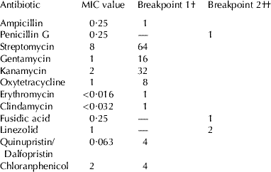
Minimum inhibitory concentrations (MICs) of 12 antimicrobial agents were determinated by microdilution using the newly developed and standardized LAB susceptibility test medium (LSM) broth formulation essentially consisting of a mixture of Iso-Sensitest (IST; Oxoid) broth (90%) and MRS broth (10%) adjusted to pH 6·7 as previously described (Klare et al. Reference Klare, Konstabel, Müller-Bertling, Reissbrodt, Huys, Vancanneyt, Swings, Goossens and Witte2005). Nine of the antimicrobials tested (ampicillin gentamicin, streptomycin, quinupristin/dalfopristin, erythromycin, clindamycin, oxytetracycline, chloramphenicol, kanamycin) were those for which the panel on additives and products or substances used in animal feed (FEEDAP) of the European Food Safety Authority (EFSA) has established microbiological breakpoints (cut-off values) that enable the distinction between Lb. fermentum strains harbouring acquired antimicrobial resistances and susceptible strains (EFSA, 2008). MICs of three additional antimicrobials agents for which tentative cut-off values for Lb. fermentum have been suggested (Klare et al. Reference Klare, Konstabel, Werner, Huys, Vankerckhoven, Kahlmeter, Hildebrandt, Müller-Bertling, Witte and Goossens2007) were also determined. The antimicrobials were tested in the concentration ranges (mg/l) given in parentheses: penicillin G (0·032-64), ampicillin (0·032-64), gentamicin (1-2048), streptomycin (2-4096), quinupristin/dalfopristin (tested as 30:70 ratio: 0·032-64), erythromycin (0·016-32), clindamycin (0·032-32), oxytetracycline (0·063-128), fusidic acid (0·063-128), linezolid (0·016-32), chloramphenicol (0·125-256), and kanamycin (1-256).
Animals
Forty male Balb/C mice aged 6–8 weeks bred at the Universidad de Granada facilities (Granada, Spain) were housed in stainless-steel cages with a 12 h light/dark cycle (8 am–8 pm) in a controlled atmosphere (temperature 22±2°C, humidity 55±2%). The animals were fed ad libitum a purified diet (Harlan, Barcelona, Spain) and had free access to water during the experimental protocol. The protocol was carried out according to the guidelines of the Helsinki declaration and was approved by the Ethic Committee of Animal Experiment of the Universidad de Granada.
Experimental Design
After 7 days of acclimation, mice were randomly assigned into two different experimental groups (n=20 per group): a control group which received orally just the vehicle and a probiotic-treated group which received orally 1010 cfu/mice/day during 28 d. Oral gavage was performed by intragastric feeding by means of a PVC tube feeding needle purchased from Vygon (Ecoue, France). Throughout the experiment, the animal's activity, behaviour and general health was observed daily. Body weight and food intake were recorded once a week. Mice were sacrificed by intraperitoneal administration of sodium pentothal (50 mg/kg), and blood was collected by cardiac puncture in EDTA containing tubes in sterile conditions. Liver and spleen, were removed in sterile conditions and weighed. Thymus, kidney, heart as well as large and small intestine were also removed and weighed.
Bacterial Translocation
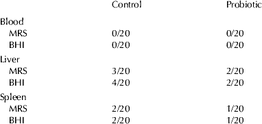
Bacterial translocation was analysed in blood, liver and spleen. 50 μl of blood were cultured in de Man, Rogosa, Sharpe (MRS) agar medium and Brain Heart Infusion (BHI) agar and incubated at 37°C during 48 h anaerobically for MRS and aerobically for BHI. Tissue samples were homogenized in buffered peptone water (1 g/ml) and 100 μl of the resulting homogenates were cultured in MRS and BHI agar as previously mentioned. After 48 h, the colonies were enumerated and the results were expressed as incidence of translocation (number of mice where cfu were detected/total number of mice). Positive growth on agar plates was defined by the presence of even a single colony of any microorganisms.
Identification of Lb. fermentum CECT5716 by PCR
For Lb. fermentum CECT5716 detection, PCR was performed directly from MRS agar colonies using the following strain-specific primers: L5716 forward 5′-GCT TGC CGC TTC TCT GGT-3′, and L5716 reverse 5′-CAA CGA CGA TGA ACA CCA CTT-3′.
Total Liver Glutathione (GSH) Concentration
A portion of 100 mg of liver from each mouse were homogenized in a 7·5% trichloroacetic acid solution by means of an Ultraturrax (Heidolph, Barcelona, Spain) and homogenates were centrifuged 3,000 g 10 min at 4°C. Total glutathione concentration was measured in the supernatants using a colorimetric commercial kit (OxisResearch, Portland, OR). Briefly, 40 μl of the homogenates or the standards were added to each well of a microtiter plate, together with 40 μl of a reducing agent (tris(2-carboxyethyl)phosphine in HCl), 40 μl of a chromogen (1-methyl-3-chloro-7-trifluoromethylquinolinium methylsulfate in HCl) and 40 μl of color developer (NaOH). After an incubation at room temperature and in the dark for 30 min., optical density was measured at 415 nm using a microplate spectrophotometer (Bio-Rad Laboratories, Hercules CA).
Malondialdehyde (MDA) Plasma Concentration
MDA plasma concentration was measured by separation with high performance liquid chromatography, using a previously described method based on thiobarbituric acid reaction and reverse phase separation with fluorescence detection (Fukunaga et al. Reference Fukunaga, Yoshida and Nakazono1998).
Evaluation of the general health status of mice
In order to evaluate possible changes in physiological functions, due to the oral treatment with bacteria, glucose concentration and glutamic-oxalacetic transaminase (GOT) activity were measured in plasma samples by means of a commercial kit (Stangest Biomedical IND, Tarragona, Spain).
Blood cell counts (white and red cells) were also analysed with an automatic haematology counter (Minos, ABX, Montpellier, France) on fresh blood, in order to evaluate possible modifications due to infection caused by the oral administration of the strain.
Statistical Analysis
All results are expressed as the mean±sd. Differences between means were tested for statistical significance using a one-way analysis of variance (ANOVA) and post hoc least significance tests. Differences between proportions (bacterial translocation incidence) were analysed with the chi-squared test. All statistical analyses were carried out with the Statgraphics 5.0 software package (STSC, Maryland), with statistical significance set at P<0·05.
Results
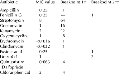
Antibiotic susceptibility profile shows that Lb. fermentum CECT5716 is sensitive to all antibiotics tested (table 1). MICs values demonstrate that the strain is sensible to β-lactam antibiotics such as penicillin G and ampicilin, to aminoglicosides such as gentamycin, to macrolids such as erythromycin, and to other broad-spectrum antibiotics such as chloramphenicol or tetracycline.
MICs and breakpoint values (μg/ml) of the antimicrobial agents tested for Lb. fermentum CECT5716
Values are means of n=3 assays
Regarding oral tolerance assay, during the experimental protocol no noticeable activity or behavioural changes were observed in the mice and no illness or death occurred. There was no difference in the animals' aspect between treatment and control groups.
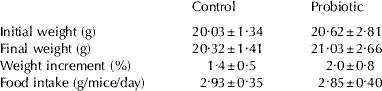
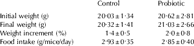
Oral administration of Lb. fermentum CECT5716 had no adverse effects on mice food intake, since there was no difference in food intake between control and treated groups throughout the experiment (Table 2). As shown in Table 2, no significant difference was observed in the body weight gain between groups.
Body weight and food intake of control and probiotic-treated mice
Values are mean±sd for n=20
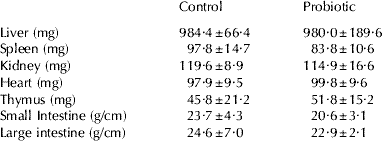
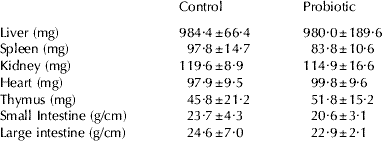
Concerning tissue weights, there were no significant differences in the weight of liver, spleen, thymus, heart and kidney between control and mice administered orally Lb. fermentum CECT5716 (Table 3). In addition, the weight/length ratio of small and large intestine did not statistically differ between the experimental groups. Apart from the weight, the coloration and aspect of the analysed tissues was normal and no difference was observed between groups.
Tissue weights and intestine weight/length ratio of control and probiotic-treated mice
Values are mean±sd for n=20
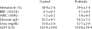
In agreement with the absence of clinical symptoms in mice, the biochemical and haematological parameters measured in blood samples did not statistically differ between control and probiotic-treated group (Table 4).
Haematology and blood biochemistry data from control and probiotic-treated mice
Values are mean±sd for n=20
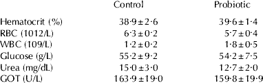
RBC, red blood cell; WBC, white blood cells; GOT, glutamic-oxalacetic transaminase
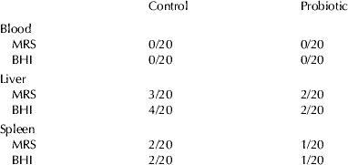
The incidence of translocation of bacteria from the gut to different tissues is shown in Table 5. No bacteraemia was observed in any of the experimental groups. Liver and spleen were aseptically removed and the presence of bacteria was analysed by culture in MRS, a lactic acid bacteria specific medium, and BHI a less specific medium. Although translocation of bacteria to liver and spleen was observed, there was no statistically significant difference in the incidence of translocation to liver or spleen between control and treated groups. In addition, colonies found on agar plates were checked by conventional PCR using specific primers and none of the colonies corresponded to the administered strain Lb. fermentum CECT5716 (data not shown).
Incidence of bacterial to blood, liver and spleen in control and probiotic-treated mice
Values represent the number of animals where translocation was observed (any bacterial growth, including one single colony, was considered as positive)/total number of mice per group
In order to detect signs of infection due to the oral probiotic treatment, the oxidative status of mice was analysed. With this aim, liver GSH concentration and plasma MDA concentration were determined. Results showed that oral administration of Lb. fermentum CECT5716 did not cause changes either in the liver content of GSH (9·5±1·0 vs 9·7±1·2 μmol/g, P>0·1) nor in the plasmatic concentration of MDA (2·4±0·2 vs 1·9±0·4 μm).
Discussion
The demonstrated health benefits of probiotic bacteria have caused an increase in both the human consumption of probiotic products and the isolation of new strains for human nutrition. Although safety aspects are included in the selection criteria of a probiotic bacteria, the high number of new isolated strains have resulted in concern about safety of probiotics. Up to now there are no general guidelines for the evaluation of this issue, but rodent studies of acute oral toxicity have been proposed as a fundamental test for the assessment of probiotic safety (Stine & Brown, Reference Stine, Brown, Stine and Brown1996) and have been applied previously in safety assessment studies (Donohue et al. Reference Donohue, Deighton, Ahokas, Salminen, Salminen and von Wright1993; Zhou et al. Reference Zhou, Shu, Rutherfurd, Prasad, Gopal and Gill2000).
A new strain of Lb. fermentum has been recently isolated from human milk. Due to its probiotics characteristics and its singular origin this strain is a good candidate for human consumption, especially for infant formulas. Lb. fermentum is included in the list of taxonomic units proposed by the European Food Safety Authority (EFSA, 2007) for the qualified presumption of safety (QPS) status. However, in this report and other by the Joint FAO/WHO Working Group (2002), it is stated that lactobacilli, although generally considered safe, may theoretically be responsible for side effects and thus safety assessments in terms of antibiotic susceptibility and virulence properties are recommended.
The pattern of antibiotic susceptibility showed that MIC values for Lb. fermentum CECT5716 are below the break points referenced by the EFSA for all the antimicrobial agents tested. Thus, Lb. fermentum CECT5716 is not able to transfer antibiotic resistance to other bacteria.
The oral toxicity assessment showed that mice treated orally with a high dose of the potential probiotic strain Lb. fermentum CECT5716 were healthy after a daily administration for 28 days. No adverse effects were observed on body weight and food intake, indicating that the potential probiotic does not exhibit gross oral toxicity and no deleterious effects were shown on the animals' health status, growth and development. In all cases clinical parameters of probiotic treated mice were similar to those of control mice, thus suggesting the absence of detrimental effects related to probiotic treatment.
Since, the infection process for most of the opportunistic indigenous lumen strains starts with translocation from gut to other tissues (Steffen & Berg, Reference Steffen and Berg1983), translocation is an important marker of probiotic toxicity. In spite of the high dose of bacteria administered to mice, there was no bacteraemia in the probiotic-treated group. We obtained bacterial cells in liver and spleen (see Table 4), but in no case Lb. fermentum CECT5716 was found and the incidence was similar in mice receiving the potential probiotic and in control mice, thus suggesting that translocation was not associated with treatment. These data suggest that a high dose of the probiotic tested, administered orally to mice, does not cause bacterial translocation to blood, spleen or liver. The presence of some bacteria in liver and spleen has been previously described in healthy mice (Ma et al. Reference Ma, Deitich, Specian, Steffen and Berg1990).
Although very scarce, some cases of Lactobacillus sepsis associated with probiotic therapy have been recently reported (Land et al. Reference Land, Rouster-Stevens, Woods, Cannon, Cnota and Shetty2005). In spite of the absence of bacteraemia and bacterial translocation to spleen or liver, we decided to evaluate possible markers of sepsis in mice consuming probiotics. In different animal models of sepsis it has been shown that 48 h after bacterial infection, total liver glutathione content is significantly increased compared with non-infected animals, probably caused by oxidative stress (Malmezat et al. Reference Malmezat, Breuillé, Pouyet, Mirand and Obled1998). It has also been shown that plasmatic concentration of MDA, a product of lipid peroxidation, is a good marker of oxidative stress (Gil et al. Reference Gil, Siems, Mazurek, Gross, Schroeder, Voss and Grune2006). In our experiment total liver glutathione concentration and plasmatic concentration of MDA were similar in probiotic-treated mice and in the control group. These data are consistent with the absence of bacteraemia and normal bacterial translocation to liver and spleen, and suggest that the probiotic strain tested do not cause either a local or systemic infection.
In agreement with the absence of any symptoms of infection, data from blood markers of physiological functions (GOT and glucose) as well as red blood cells and leukocytes counts were similar in the probiotic and control groups.
In summary feeding mice with the new LAB strain Lb. fermentum CECT5716 at a dose 10,000 times higher (per kg body weight) than that normally consumed by humans had no deleterious effects on mice. Future human clinical trials are needed to confirm safety of Lb. fermentum and to evaluate the gastrointestinal and immunological benefits that could support the inclusion of this strain in food products, such as infant formulas.
The authors are grateful to Alberto Zafra (PhD) for his help in the measurement of plasma MDA concentration. We would also like to thank Manuel Carlos Olivares for the assistance in the measurement of biochemical and haematological parameters and Julio Boza (PhD) for his help in the interpretation and discussion of results.