I. INTRODUCTION
Different considerably from their bulk properties, metal nanoparticle (M-NP) catalyst systems have aroused wide concern in many different areas including, but not limited to, environmental science, chemistry, and medical applications. Reference Aiken and Finke1–Reference Yu, Yang, Xie, Fan, Yu, Shu and Wang4 As to the synthesis of metal nanoparticle colloids, the most common methods involve wet chemical “bottom-up procedures,” particularly the chemical reduction of metal salts. Normally, it is in aqueous or organic media that the chemical reduction of transition metal salts is performed to generate zerovalent metal colloids. Reference Renato, Aline, Sébastien and Domingos5 However, metal nanoparticles (NPs) tend to aggregate during their catalytic process and can cause loss while separation after the end of reaction. These problems cause severe restrictions in using costly metal NPs as catalysts for the above-mentioned large-scale applications. Reference Jana, Dandapat and De6 Hence, stabilizers are employed to provide stability of the NPs when they are formed. Domingos et al. report an approach for effectively combining a high-throughput method using linear polyethylene imine (LPEI) with in situ screening and multivariate optimization of the synthesis conditions to produce highly catalytically stable Ag-NPs. Reference Santos, Elias, Signori, Giacomelli, Yang and Domingos7
The most commonly used stabilizer is a surfactant or a polymer matrix, which may form a layer of organic compounds present at the interface and need the further heat treatment at high temperature. Tungsten oxide and WO3−x -based materials are intensively investigated due to their interesting properties, such as tunable electronic and electrochemical properties, etc. Reference Deb8 Studies on WO3−x -supported metal particles have proved that the support can enhance the catalytic performance of the metal nanoparticles. Reference Shibuya and Miyauchi9–Reference Saha, Banis, Zhang, Li, Sun, Cai and Wagner11 Up to now, tungsten oxide with various morphologies, such as rods, wires, and mesoporous structure, has been obtained. Reference Zhang, Huang and Tao12 However, there are few reports on the preparation of high-quality tungsten oxide BUNs and metal/WO3−x BUNs heteronanocomposites. In particular, monoclinic W18O49 BUNs with the largest oxygen deficiency in the WO2.625–WO3 range have been reported as the only oxide that can be isolated in a pure form, which is not the case for other tungsten suboxides (WO3−x ).
This paper describes the formation and catalytic properties of silver nanoparticles (Ag-NPs) stabilized by tungsten oxide (WO3@W18O49) through an in situ method for the first time. As schematically illustrated in Scheme 1, as the oxidation and reduction reaction between weakly reductive W18O49 and oxidative silver nitrate proceeds, the surface of W18O49 was oxidized to WO3 and simultaneously transfers the metal nanoparticles on to the newly formed oxide support, finally producing the Ag-WO3@W18O49 BUNs heteronanocomposites. The reduction of Nip to p-aminophenol (Amp) is a model reaction, Reference Pradhan, Pal and Pal13 which has been widely used for comparison of the catalytic activity of different metal nanoparticles immobilized in a variety of carrier systems. The catalytic activity of the Ag-NPs-WO x composite is also investigated in the reduction reaction of p-nitrophenol (Nip), using NaBH4 as the reducing agent. The antibacterial activities of Ag-NPs-WO x against Escherichia coli were also studied.
II. EXPERIMENTAL
A. Materials
Tungsten(VI) hexachloride (WCl6, 99%) was from Aladdin (Shanghai, China). Silver nitrate (Cennabras, Sao Paulo, Brazil, 98%), sodium tetrahydroborate (NaBH4), absolute ethanol (A.R. grade), and para-nitrophenol were from Sinopharm Chemical Reagents Beijing Co., Ltd. (Beijing, China). Escherichia coli (DH5α), Luria–Bertani (LB) agar medium. Ultrapure water (resistivity of 18.2 mΩ cm), degassed by ultrasonic treatment, was used in all experiments. The chemicals were used as received without further purification.
B. Synthesis of bundled W18O49 nanowires
In a typical synthesis, 0.4 g of WCl6 was added into 80 mL of absolute ethanol rapidly and a clear yellow solution was formed immediately. The clear yellow solution was then loaded into a 100 mL Teflon-lined autoclave. The autoclave was sealed, heated in an oven of 180 °C for 24 h, and then naturally cooled to room temperature, and a blue flocculent product (bundled W18O49 nanowires) formed, which was then collected and washed with ethanol and distilled water several times to remove ions and possible remnants. Reference Xi, Ouyang and Li14–Reference Xi, Ye, Ma, Su, Bai and Wang16 Finally, the resultant nanowires were dried in a vacuum oven at 50 °C overnight to remove the absorbed water and ethanol for subsequent use.
C. Synthesis of Ag-WO x hybrid composite (heteronanocomposites)
A certain amount of the as-synthesized W18O49 was added into 100 mL distilled water under mild stir. In a synthesis of Ag-loaded WO x (2 wt%, 3 nm) hybrid composite, 2.5 mL of AgNO3 (0.012 M) aqueous solution was dropped into the W18O49 suspension under strong stirring. The dropping rate is about 100 μL per minute. The color of the mixture changed gradually from blue to dark gray. After 4 h, additional 4.5 mL AgNO3 solution was dropped into the suspension. After 12 h stirring, the final product was collected, washed with ethanol and distilled water several times, and dried in air at 80 °C for 12 h.
D. Catalytic reaction
The reduction of nitrophenol compound by Ag-WO x in the presence of NaBH4 was carried out to examine the catalytic activity of the Ag-WO x nanocatalysts. Amounts of 6 mL of deionized water, 120 μL of 10 mM nitrophenol, and 0.48 mL of 0.1 M NaBH4 solutions were added into followed by addition of 0.5 mg of water-soluble Ag-WO x to the mixture. The color of the solution changed gradually from yellow to transparent as the reaction proceeds. UV-vis spectrometry was used to record the change in absorbance at a certain time interval. Reference Lin and Doong17–Reference Jiang, Xie and Jiang19 Rate constant of the reaction was measured by the extinction of the peak at 400 nm as a function of time.
E. Antibacterial property
In the antibacterial test, the gram negative bacterium of Escherichia coli (DH5α) was used as the experimental strains and Luria–Bertani (LB) agar medium was used for the growth of Escherichia coli. All disks and materials were sterilized in an autoclave before experiments. The filter paper disk was dispersed in the ethanol solution with a sample concentration of 0.5 mg/mL for 5 min and then dried. Hundred microliters of the activated Escherichia coli were uniformly spread in the solid medium in the plates by a triangular spreader. The filter paper disk with samples was placed in the plates, which were finally incubated at 37 °C for 24 h to check the inhibition zone. The empty paper disk was also placed in the plates as reference. The inhibition zones were determined by measuring the areas around the filter paper disks without bacterial growth. Reference Zhao, Wang, Zhao, Li and Liu20–Reference Deng, Zhu, Peng, Chen, Sun, Gang, Jin and Wang22
F. Characterization
The crystal structure and phase purity of Ag-WO x were analyzed by x-ray diffraction (XRD) using D8 Advance XRD (Bruker AXS company, Karlsruhe, Germany) equipped with Cu Kα radiation (λ = 1.5406 Å), using a scanning rate of 7° per minute in the 2θ range from 15 to 80°. The samples for XRD analysis were obtained from dried samples. Scanning electron microscopy (SEM) and energy-dispersive x-ray spectroscopy (EDS) were performed on a scanning electron microscope (Hitachi S-4800, Tokyo, Japan) operated at an acceleration voltage of 10 kV to characterize the morphologies and the compositions of the prepared Ag-WO x . Furthermore, the morphology and size of Ag nanoparticles and tungsten oxide were also examined by transmission electron microscopy (TEM), which was recorded on a JEOL-JEM-2010 (JEOL, Tokyo, Japan) operating at 200 kV. The as-prepared Ag-WO x was washed with absolute ethyl alcohol and deionized water to remove impurities, and then dispersed in absolute ethyl alcohol. The 2–3 drops of the dispersion were dripped onto a carbon-coated TEM grid. The absolute ethyl alcohol was allowed to evaporate at room temperature for several minutes. Mutual effects between the resultant Ag nanoparticles and WO x nanowires were analyzed by Fourier transform infrared (FTIR) spectra, which were performed by the KBr wafer technique.
III. RESULTS AND DISCUSSION
A. Structural and chemical characterization
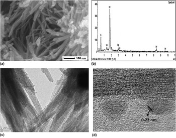
Figure 1(a) shows the representative SEM image of the sample prepared via solvothermal reaction of low concentration of WCl6 in ethanol. It is obvious that the as-synthesized W18O49 displays bundled-nanowires-like. Figure 1(b) shows the EDS of the composite, which confirms that the sample contains only the elements W, O, and Ag. It should be pointed out that nearly all the tiny nanoparticles were immobilized onto the WO x BUNs without any aggregation under experimental conditions, which was confirmed by TEM observation [Fig. 1(c)]. Furthermore, we can observe that the diameter of a single nanowire is around 1 nm and the bundle diameter is about 20 nm. From the high resolution transmission electron microscopy (HRTEM) image [Fig. 1(d)] of single Ag-WO x nanowire, we can see that the lattice spacing 0.23 nm of Ag nanoparticle is in accordance with the database.
(a) SEM and (b) EDS spectrum of the sample. (c) TEM images of the Ag-WO x sample. (d) HRTEM image of a single Ag-WO x nanowire.

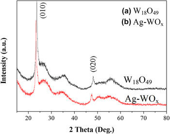
Powder XRD analysis (Fig. 2) revealed that the sample was indexed to the monoclinic phase W18O49 (JCPDS No. 36–0101). In the XRD pattern, only two obvious diffraction peaks assigned to (010) and (020) planes appeared at 23.4° and 47.5°, suggesting that the W18O49 nanowire preferentially grew along its [010] direction. Due to similarity of the XRD pattern of Ag-WO x to pure W18O49, the following analyses are given. First and the most important one is that, due to the little content and small size of metal nanoparticle, the plane of Ag is not obvious. It is therefore difficult to draw any conclusions regarding the existence of metallic Ag from the XRD measurement only. The second reason may be that the bundled nanowires are so tight that Ag+ cannot infiltrate into and react with them completely. For this reason, the bulk phase of composites retains the same pattern.
XRD patterns of the W18O49, bundled Ag-WO x nanowires.

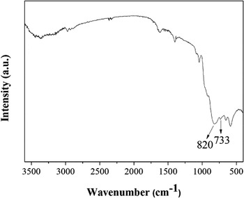
FTIR spectrum of the ultrathin Ag-WO x nanowire bundles.

FTIR was conducted, and no organics coated on the surface of WO x BUNs could be found from the spectrum (Fig. 3). The two peaks that appeared at 820 and 733 nm indicate the existence of W–O and O–W–O bands, respectively.
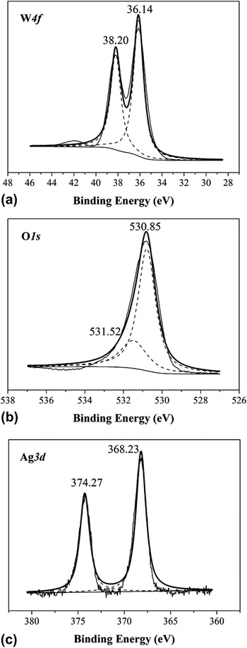
The synthesized Ag-WO x composites were further analyzed by XPS to invest the elemental composition and chemical states of elements on the surface, as shown in Fig. 4. It manifested that W species in the tungsten oxide nanowires showed only one oxidation state. The peaks at 38.20 eV and 36.14 eV were assigned to W 4f 5/2 and W 4f 7/2 of W6+ species. Combined with the characterization of XRD, this result is entirely consistent with the structure of WO3 layer wrapping W18O49, which was presented in the mechanism. The spectrum of O1s level can be decomposed into separate peaks. The peak with binding energy at 530.9 eV corresponded to O1s levels of oxygen atoms O2− in the lattice of WO x , and the other peak with weak intensity at 531.5 eV was assigned to the absorbed water molecules on the free oxide surface. As seen from the high-resolution Ag 3d XPS spectra, Ag-WO x exhibited doublet peaks located at approximately 374.3 eV and 368.2 eV corresponding to Ag 3d 3/2 and Ag 3d 5/2 levels respectively, suggesting that metal Ag0 was deposited on the WO x surface.
(a) W4f, (b) O1s, and (c) Ag3d-level XPS spectra of Ag-WO x sample.

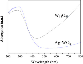
The UV-vis-NIR absorption spectrum of the as-synthesized W18O49 is shown in Fig. 5. Very interestingly, the considerably large absorption tail in the visible and NIR regions gives clear evidence that the nonstoichiometric W18O49 contains a large number of oxygen vacancies, the presence of which suggests that the W18O49 has reducing power. And then, the tail in the UV-vis absorption spectrum of the hybrid composite has disappeared after the reaction with AgNO3.
UV-vis-NIR absorption of the as-synthesized W18O49, Ag-WO x samples.

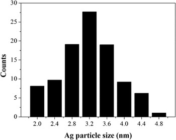
The histogram analysis shows that the Ag NPs in composites are in the range of 2.0–4.8 nm with a mean size of 3.2 nm (Fig. 6).
Size distribution of the Ag nanoparticles obtained by counting 100 particles in the TEM image.

B. Application of Ag-WO x heterostructures for catalytic reduction of p-nitrophenol to p-aminophenol
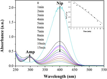
The capability to activate/catalyze some reactions that are otherwise unfeasible is the most important function of metal nanoparticles. The catalytic reduction of p-nitrophenol to their corresponding daughter derivatives, p-aminophenol, in the presence of NaBH4 was chosen as model reaction to investigate the functionality of Ag-WO x heterostructures. Such a reaction catalyzed by Ag catalysts has been reported because this reaction can be rapidly and easily characterized. The function of silver nanoparticles is virtually the electron relay, and the reaction rate is largely affected by the concentration and size of silver nanoparticles. The reaction is more efficient as the density of the silver nanoparticles is higher and/or the size of the particles decreases. Figure 7 shows the typical UV-vis spectra and concentration change of nitrophenol compounds in the presence of a certain amount of Ag-WO x composites. The original absorption peak of p-nitrophenol is centered at 400 nm after addition of freshly prepared NaBH4 solution, indicating the formation of p-nitrophenolate ions. This peak starts to decrease when the reduction proceeds in the presence of nanocatalysts. Addition of NaBH4 in the absence of Ag-WO x has little effect on the change in absorbance at 400 nm, confirming that the reduction is mainly catalyzed by Ag-WO x . In addition, the absorption peak at 400 nm decreases with the concomitant increase in peak intensity at 300 nm within 15 min after addition of Ag-WO x catalysts.
Time-dependent UV-vis spectral changes in p-nitrophenol catalyzed by Ag-WO x heterostructures. The illustration shows the linear relationship of ln(C t /C 0) versus time (t).

The pseudo-first-order kinetics can be applied to evaluate the rate constants for nitrophenol reduction because the concentration of NaBH4 is higher than those of nitrophenols and can be considered as a constant during the reaction period. According to the first order kinetic equations,
 $${{\ln C_t } \over {C_0 }} = kt\quad ,$$
where C t represents the concentration of p-nitrophenol, and C 0 signifies the initial concentration of nitrophenol. The C t /C 0 is measured from the relative intensity of absorbance (A t /A 0). The linear relationship of ln(C t /C 0) versus time (t) indicates that the reduction of nitrophenol by Ag-WO x heterostructures follows the pseudo-first-order kinetics. The rate constant for p-nitrophenol reduction namely the slope of the straight line is 0.18 min−1 by using Ag-WO x nanocatalysts (illustration in Fig. 7).
To further investigate the reduction reaction, several possible reasons of high catalytic activity for the reduction of p-nitrophenol were proposed. As is well known, the catalytic process includes two steps: adsorption and reaction. In the first step, after adding a certain amount of Ag-WO
x
nanowires with bigger surface-to-volume ratio to a mixed solution of p-nitrophenol and NaBH4, the p-nitrophenolate ions and
 ${\rm{BH}}_4^ -$
were easily adsorbed on the catalysts. In the second step, the p-nitrophenolate ions and
 ${\rm{BH}}_4^ -$
react quickly with the more electronic holes in the surface of the small Ag nanoparticles. Immediately, p-aminophenol was produced. Therefore, based on the bigger surface-to-volume ratio of Ag-WO
x
nanowires and small size of Ag-NPs with a diameter of 2.0–4.8 nm, the composites have superior catalytic activity for the reduction of p-nitrophenol.
C. Application of Ag-WO x for inhibition of the growth of bacteria
Silver and silver-based nanomaterials have been well known for their strong inhibitory and antimicrobial effects. Concerning the prospective applications of bundled Ag-WO x ultrafine nanowires as antibacterial materials, their antibacterial properties are evaluated with Escherichia coli as bacterium models.
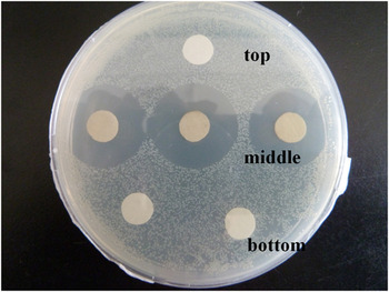
The antibacterial activities of Ag-WO x were investigated by measuring the growth inhibition of the bacteria on solid Luria–Bertani (LB) agar plates through a modified Kirba–Bauer method. Comparing with blank control (in the top of the picture), the result of inhibition zones of both the bare W18O49 nanowires (in the bottom of the picture) and the Ag-WO x (in the middle of the picture) against Escherichia coli were shown in Fig. 8. Apparently, Ag-WO x exhibits apparent antibacterial activity, and the inhibition zone is 2.60 cm, whereas the bare W18O49 exhibits no antibacterial activity.
Inhibition zones of blank filter paper, the W18O49 and the Ag-WO x against Escherichia coli.

The formation mechanism of W18O49-coated WO3 nanowires loaded with Ag nanoparticles.

Though the mode of action of bundled Ag-WO x ultrafine nanowires on the bacteria is indeterminate, its high antibacterial activity may arise from the existence of small Ag nanoparticles and the bundled ultrafine nanowires with high specific surface area, which provide better contact with microorganisms. For Escherichia coli, a short time of exposure to antibacterial concentrations of Ag nanoparticles resulted in an accumulation of envelope protein precursors. This indicates that Ag nanoparticles may target at the bacterial membrane, leading to a dissipation of the proton motive force. Reference Li, Xie, Shi, Zeng, Ouyang and Chen23–Reference Rai, Yadav and Grade25 As a consequence, the nanoparticles preferably destroy the respiratory system, cell division, and finally leading to the death of Escherichia coli.
IV. CONCLUSIONS
Using the in situ redox reaction, small Ag-NPs with a diameter of 2.0–4.8 nm and a narrow size distribution were successfully formed and stabilized by bundled tungsten oxide nanowires. The Ag-WO3@W18O49 composite was able to efficiently catalyze the p-nitrophenol reduction in water with a considerable rate. Moreover, Ag-WO3@W18O49 exhibits superior antibacterial activity against Escherichia coli to that of bare W18O49. This exciting catalytic effect and the highly efficient antibacterial property are most probably due to the moderate interaction of the Ag-NPs and WO3@W18O49, and the synergistic effect of metal-NPs and oxide semiconductor with large specific surface area. Furthermore, the heteronanostrctures of Ag-WO3@W18O49 have not been thoroughly studied, and other new applications relating to electrochemical sensors could also be developed in the near future.
ACKNOWLEDGMENTS
We acknowledge the financial supports of the National Natural Science Foundation (Grant No. 21003065).