Evaluation and performance improvement to enhance patient room cleaning is important for healthcare-associated infection prevention.Reference Han, Sullivan, Leas, Pegues, Kaczmarek and Umscheid 1 , Reference Kenters, Gottlieb and Hopman 2 The Centers for Disease Control and Prevention recommends fluorescent gel markers (FGMs) as a tool for the evaluation of environmental cleaning (EEC). 3 However, the optimal FG formulation, method of application, and evaluation of removal remains to be determined.
At the Johns Hopkins Hospital (JHH), a 1,020-bed academic hospital in Maryland, FGM has been a part of our EEC program since 2012. An FGM visible only with ultraviolet (UV) light is placed on high-touch surfaces (HTSs) in selected patient rooms and bathrooms. After a designated period, removal is assessed with UV light. Removed FGM (ie, no FGM is visible using a UV light) indicates a cleaned HTS, whereas remaining FGM indicates an uncleaned HTS. Since establishing the program, we have evaluated multiple fluorescent markers with varied compositions and application methods (eg, fluorescent gel, paint, and pen marker). Many fluorescent markers were quickly eliminated because they were clearly visible to the naked eye or because they were not removed by expected best cleaning. However, 2 FGM types did not overtly appear to have these problems. Therefore, We evaluated 2 different FGM products: a metered applicator for FGM (MA-FGM) and a generic cotton swab dipped in FGM lotion (CS-FGM). We hypothesized that use of a purpose-made MA-FGM allowing for a more standardized application would be associated with (1) decreased FGM removal because it is less visible to the naked eye and (2) less variability in size of FGM at placement and more standardized interpretation of FGM removal.
Methods
Phase 1
Over a 2-day period at JHH, we randomly selected rooms from 9 hospital units via block randomization. We placed both MA-FGM (DAZO, Ecolab, St Paul, MN) and CS-FGM (GloGerm, GloGerm, Moab, UT) simultaneously on as many of 24 HTS as possible per room. For each room, we alternated the side of the HTS assessed by each method to account for handedness that might influence the likelihood of FGM removal. The location and types of FGM for each room were recorded, and the presence of each FGM was checked 1 day later. A discharge (ie, terminal) cleaning was deemed to have occurred if there was a new patient in the room at time of FGM assessment.
Phase 2
In the Johns Hopkins biocontainment unit in an unoccupied patient room used for training and simulations, 8 trained “markers” (ie, 3 research assistants, 2 infection control epidemiologists, 1 infectious diseases physician, 1 research coordinator, and 1 infection control assistant) placed dots 3 times with each product on a bedside table, using their usual practice. The FGM dot diameters were measured, and visibility to the naked eye was assessed. The FGM dried for 2.5 hours, after which an observer wiped each FGM with disinfectant using 2 strokes to mimic real-world cleaning, and FGM removal was assessed.
The Cohen κ was used to measure agreement, and the McNemar test was used to compare the removal rates between the 2 types of FGMs. We also used logistic regression models to compare removal rates across different HTSs for each type of FGM.
Results
Phase 1
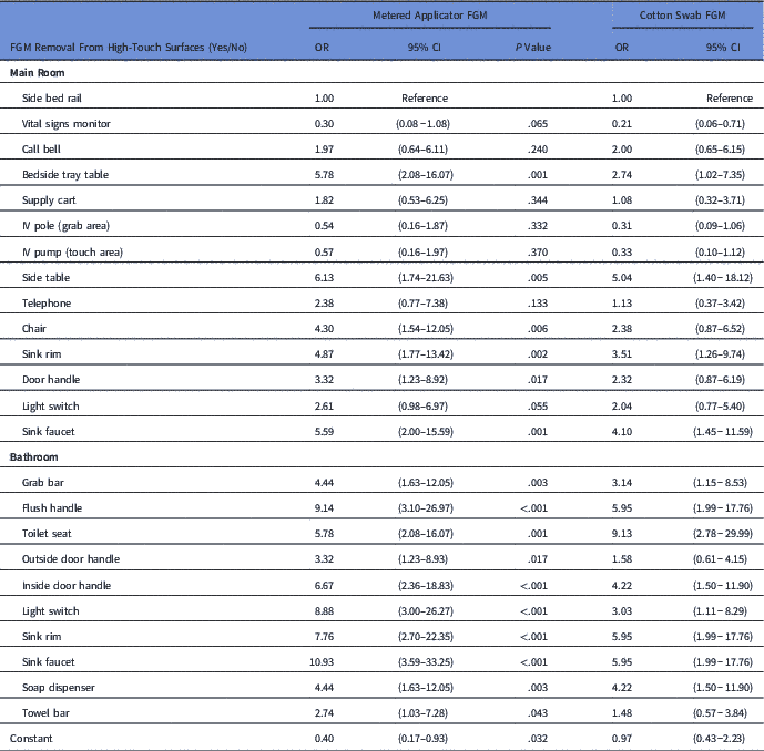
MA-FGM and CS-FGM were placed on 787 HTSs in 38 randomly selected rooms in 9 different units. The overall removal rate was higher for the CS-FGM (64.3% vs 60.5%; P<.001). Concordance between removal of the 2 products was high (κ=0.83; 95% CI, 0.79−0.87). Table 1 lists the odds of FGM removal for each type by HTS.
Odds of High-Touch Surface Fluorescent Gel Marker (FGM) Removal by TypeFootnote a

Note. OR, odds ratio; CI, confidence interval.
a From logistic regression models adjusting for daily vs. discharge cleaning, and under contact precautions (yes/no). No clustering was adjusted. Possible clustering includes the same cleaning workers, the same observer, building, unit, etc.
Phase 2
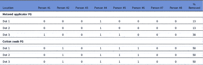
For the simulated FGM experiment, each of the 24 MA-FGM placed had a standard diameter of 0.5 cm, whereas the CS-FGM dots varied in diameter from 0.1 cm to 2 cm based on the marker. The CS-FGM appeared more visible by the naked eye than MA-FGM, as judged by the observers (see Supplemental Fig. 1a and 1d). More CS-FGMs (vs MA-FGM) were judged as having been removed by wiping: 12 of 24 (50%) versus 4 of 24 (17%) (Table 2).
Metered Applicator Fluorescent Gel (FG), Cotton Swab FG, and Removal From Bedside Table After Wiping

Note. 0=not removed; 1=removed.
Discussion
The CS-FGM product was more frequently removed from HTSs in patient rooms than the MA-FGM product (64.3% vs 60.5%). We hypothesized 2 reasons for this result. First, the CS-FGM product was more visible to the naked eye and was, therefore, preferentially removed. Second, the MA-FGM formulation was more adhesive to the HTSs and, therefore, was not as easily removed with wiping. We explored these hypotheses in phase 2 of the study. We found large variability in FGM size for the CS-FGM, demonstrating that dipping a cotton swab into the FG (vs metered applicator) is subjective and dependent on the marker’s approach, even after training in the methodology. The MA-FGM was less easily removed compared with CS-FGM (17% vs 50% removed respectively), possibly related to different gel constituents and resultant adhesiveness. The need for manual pressure to remove biofilm from dry surfaces in the patient environment is increasingly recognized and supports a less easy-to-remove FGM, which is a better surrogate for biofilm removal.Reference Vickery, Deva, Jacombs, Allan, Valente and Gosbell 4 The CS-FGM lacks standardization and reproducibility and may be a suboptimal product choice for accountability and performance improvement.
We expected a larger difference in removal rate between the 2 products. Potentially, if either FGM was visible, the full HTS was cleaned, thus removing both products. In addition, on some of the small HTSs, there was not have much distance between the 2 FGMs, and both were likely removed simultaneously. We chose this method of placing the FGM on alternating sides of the same HTS rather than each room with a different FGM method, to eliminate confounding due to cleaning practices of the individual environmental cleaning associate.
Infection control or facilities programs implementing EEC programs should carefully consider the type and method of application of FGM to standardize and optimize the measurement of FG removal.
Supplementary material
To view supplementary material for this article, please visit https://doi.org/10.1017/ice.2018.287
Acknowledgments
We thank Dr Philip Carling for providing the fluorescent gel marker DAZO. Neither Dr Carling nor EcoLab participated in the design, analysis, interpretation, or manuscript preparation of our findings. We thank Verna Scheeler for project coordination.
Financial support
This work was supported by funding from Centers for Disease Control and Prevention (CDC) Epicenters Program (C.R., A.X., Y-J.H., P.O., A.P.G., L.L.M., and S.E.C.)
Conflicts of interest
All authors report no conflicts of interest relevant to this article.