Introduction
About 90 per cent of recurrent nosebleeds originate from the anterior region of the nasal septum. However, a small but important group results from bleeding arising from the posterior branches of the sphenopalatine artery. These posterior nosebleeds are sometimes described as being more common in the older adult with hypertension or atherosclerosis.Reference Browning1, Reference Burton2
However, very little is known about the basic mechanism of idiopathic epistaxis. A single report exists in the literature describing a dissecting aneurysm at the bleeding point in the sphenopalatine artery of a patient who died from massive posterior epistaxis.Reference Shaheen3 Others have described the occurrence of blebs and haemorrhagic nodules in patients with bleeding, but the actual relationship to epistaxis is unproven.Reference Padgham and Parham4
We aimed to test the hypothesis that there are potential sites of weakness within the normal posterior distribution of the sphenopalatine artery which might, under stress, contribute to the mechanism of epistaxis. To test this hypothesis we ‘stress-tested’ the arterial system of the nasal cavity by attempting to produce a vessel rupture in unfixed cadavers, through pressure injections of the feeding arteries.
Materials and methods
This was an anatomical study using both sides of 14 cadaveric heads.
In choosing the optimal site for injection, a variety of approaches were tested and assessed for ease of approach, degree of tissue trauma and proximity to the sphenopalatine artery, as follows.
External carotid artery
This approach was the easiest by far, with minimal trauma. The cannula was inserted into the external carotid artery proximal to the origin of the facial artery, and the end of the catheter was advanced to the origin of the maxillary artery. Unfortunately, the resulting injection was unfocused, with incomplete filling of the nasal vessels in some instances.
Sphenopalatine artery
The third part of the maxillary artery was exposed transantrally via a Caldwell–Luc procedure. The bony dissection was extended laterally to increase access to the artery, and a small cannula was placed into the third part of the maxillary artery with the tip just proximal to the terminal branching. This approach enabled excellent filling of the nasal arteries, but was technically the most demanding and caused significant tissue trauma and loss of injection from injured arteries.
Maxillary artery origin
A vertical skin incision was made anterior to the tragus, and soft tissue dissection was continued to expose the masseter muscle, which was then peeled off the mandibular ramus along with the periosteum. The mandible was carefully divided at the level of the masseteric notch to allow resection of the condyle and neck. The maxillary artery was identified between the underlying periosteum and the sphenomandibular ligament, and cannulated.
Injection of the maxillary artery via the third type of access seemed to offer the best compromise, and was used in all our experimental injections. Preliminary examination of the nasal cavity was carried out prior to the injection procedures, to check for obvious signs of nasal pathology.
Nine cadaveric heads were selected for injection under high pressure. Standard embalming fluid was injected into the maxillary artery at standard pressure (375 mmHg = 7 psi = 0.5 bar). Twenty-four hours later, Indian ink with latex was injected through the same cannula at a higher pressure (620 mmHg = 12 psi = 0.8 bar) for 2–4 minutes. After the second injection, the mucosal surfaces were inspected for obvious signs of leakage, and a comprehensive, stepwise dissection of the head along the branches of the maxillary artery was carried out. Any areas showing ink leakage were excised for histological examination.
Five other cadaveric heads were used as controls. They were processed in the same way, except that the Indian ink mixture was injected at the standard embalming pressure of 375 mmHg.
Results
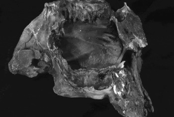
Escape of ink, ranging from mucosal staining to frank leakage (Figure 1), was found in all nine heads injected at higher pressure, and in none of the five controls injected at standard pressure.
Partly dissected specimen showing ink leakage on the posterior septum. A total of eight cases had leakage on the posterior septum; in one case, the leakage was found laterally near the sphenopalatine foramen.

Dissection of the specimens demonstrated that leakage was apparently restricted to the nasal mucosa. In eight specimens, the leakage was on the posterior septum; in the remaining case, the leakage was found on the lateral wall near the sphenopalatine foramen.
Ink leakage was not found along other branches of the maxillary artery.
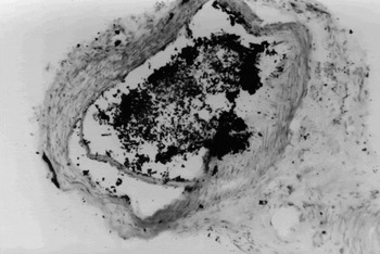
Histological examination of the leakage points demonstrated evidence of vessel disruption, with leakage of ink into the surrounding interstitium (Figure 2). Examination of sections lying just proximal to the points of vessel disruption demonstrated ink-filled cavities within the tunica media of the artery. These cavities did not have an endothelial or intimal lining.
Photomicrograph of sphenopalatine artery section just proximal to a point of ink leakage, showing a cavity within the vessel wall suggestive of a dissecting aneurysm. There are particles of ink in the surrounding interstitial tissue. (H&E; ×30)

Control specimens did not show any signs of gross ink leakage, and histological examination of comparable areas of the posterior septum showed completely intact nasal arteries with no evidence of arterial damage.
Discussion
These experimental results showed that, in cadaveric heads, high pressure injection of the maxillary artery is capable of causing consistent leakage from arteries, either in the posterior nasal septum or on the lateral wall close to the sphenopalatine foramen.
The pressures used for both embalming and for stressing the arteries were obviously much higher than normal arterial pressures present in life. Conditions in cadaveric tissue are very different from those in the living, and trial and error has shown that high pressures are needed to ensure adequate perfusion.
It is important to note that this study does not imply any role for hypertension in the occurrence of epistaxis, a topic still much discussed and far from conclusive resolution.Reference Christensen, Smith, Barnwell and Wax5 Pressure was simply used as a tool the parameters of which could be varied with reasonably good control. This is identical to the principle of ‘hydrostatic burst pressure’ used to test pipe systems in industry, whereby supra-normal stresses are used to demonstrate potential areas of weakness in a system.6 In this study, supraphysiological injection pressure was used to reveal potential points of weakness in the arterial system.
The distribution of rupture sites was consistent with the findings of many clinical investigations (Shaheen, unpublished dataReference Tucker7–Reference Chiu and McGarry10). In addition, high pressure injection produces a lesion comparable with the best current histopathological model of epistaxis, i.e. dissecting aneurysm formation.Reference Shaheen3 It is interesting that the leakage points were clustered around the tortuous early part of the sphenopalatine artery, and in one instance the dissecting aneurysm was associated with a branch point. In industrial pipe testing, ‘thrust forces’ are found in pipe systems where the direction or cross-sectional area changes, e.g. at bends, valves and branches.
This study suggests that there may be pre-existing weaknesses in the posterior nasal arterial configuration. One may speculate that factors such as degeneration of the arterial wall, endothelial trauma or a local pressure surge may expose such weaknesses, resulting in aneurysm formation with subsequent rupture.
• The mechanism of posterior epistaxis is unknown, but may involve formation of a dissecting aneursym of the nasal arteries
• Injecting the maxillary arteries of fresh cadaveric heads caused vessel rupture in the nasal cavity; other branches did not rupture
• The points of rupture were clustered in the posterior nasal cavity; in one case, the rupture was at a branch point
• Histological examination of the rupture points was consistent with formation of a dissecting aneurysm
This experimental recognition of the existence of sites of particular weakness underpins the clinical impression that there are specific vascular sites at which bleeding is likely to occur.Reference McGarry8 This re-emphasises the need to direct attention to these sites, in the investigation and treatment of posterior epistaxis.