INTRODUCTION
Pearls are calcareous deposits (nacreous or non-nacreous) that can be found naturally in the soft tissues of some shell-bearing molluscs (Landman et al., Reference Landman, Mikkelsen, Bieler and Bronson2001). Nacre is composed mainly of aragonite, one of several polymorphs of calcium carbonate, and biological macromolecules such as the polysaccharide chitin and proteins (Suzuki et al., Reference Suzuki, Saruwatari, Kogure, Yamamoto, Nishimura, Kato and Nagasawa2009). Pearl formation is presumed to occur due to lesions caused by an intruder, a parasite or any other physical process that causes the displacement of the shell secreting outer epithelial cells of the mantle into surrounding connective tissue (Nagai, Reference Nagai2013). These epithelial cells will then form a pearl sac that will produce a pearl. Other putative causes of pearl formation such as abnormal growth or physiological disorders of mantle epithelium cells due to abiotic factors have not been addressed. Commercial pearl producing species, such as pearl oysters of the Pinctada genus and freshwater mussels of the Hyriopsis genus have generally been used to study the cellular mechanisms of pearl formation. In pearl oysters, pearl formation can be artificially induced by inserting a piece of mantle tissue (graft) of a donor and an inorganic bead (nucleus) into the gonad or mantle of a recipient pearl oyster (Awaji & Machii, Reference Awaji and Machii2011). As soon as the mantle graft and nuclei are introduced in the host, hemocytes form a sheet that encapsulates the foreign body. The outer epithelial cells of the mantle graft proliferate around the nuclei forming the pearl sac (Awaji & Machii, Reference Awaji and Machii2011). When the pearl sac is formed, layers of calcium carbonate crystals are produced by the epithelial cells to cover the nuclei. A recent transcriptome analysis confirmed that the implanted mantle tissue is primarily responsible for the expression of biomineralization genes of the pearl sac (McGinty et al., Reference McGinty, Zenger, Jones and Jerry2012). In freshwater mussels, insertion of the donor mantle tissue into the mantle of the recipient host is sufficient to induce pearl formation in a similar way as described above but in the absence of a nucleus (Taylor & Strack, Reference Taylor, Strack, Southgate and Lucas2008).
Most natural pearls that have been studied are composed mainly of aragonite, although some freshwater forms contain vaterite, a highly metastable polymorph (Soldati et al., Reference Soldati, Jacob, Wehrmeister and Hofmeister2008; Ma et al., Reference Ma, Berland, Andrieu, Feng and Bedouet2013) while others have at least patches or layers of calcite (Pérez-Huerta et al., Reference Pérez-Huerta, Cuif, Dauphin and Cusack2014). Although there have been a range of microstructures reported, both natural and cultured (artificially induced) pearls are typically nacreous, i.e. the outer layer is made up of nacre (Cuif et al., Reference Cuif, Dauphin, Howard, Nouet, Rouzière and Salomé2011), and it is this structure which gives pearls their characteristic optical properties. Natural pearls have been reported and illustrated in a broad range of bivalve (Arcidae, Malleidae, Margaritiferidae, Mytilidae, Ostreidae, Pectinidae, Pinnidae, Placunidae, Pteriidae, Spondylidae, Tridacnidae, Unionidae and Veneridae) and gastropod (Cassidae, Fasciolariidae, Haliotidae, Muricidae, Strombidae, Trochidae, Turbinidae and Volutidae) families and in one cephalopod (Nautilidae) family (Landman et al., Reference Landman, Mikkelsen, Bieler and Bronson2001; Strack, Reference Strack2006). However, the occurrence of pearls in the ‘true’ oysters, the Ostreioidea, is poorly documented despite the intense interest in oysters because of their enormous commercial value. Studies on pearl formation in a wide selection of taxa can contribute to shed more light on both common and distinctive aspects of pearl composition and formation across them. This article aims to contribute to this matter by investigating natural pearls in oysters of the Crassostrea genus. Previous reports about Crassostrea pearls appear limited to those that were presumably found in the Pacific oyster Crassostrea gigas (Thunberg, 1793) (Zwaan & Groenenboom, Reference Zwaan and Groenenboom2014). True oysters are closely but not directly related to the pteriids (which contains the pearl forming Pinctada genus) and their shell mineralogy and microstructure are distinctly different (Bieler et al., Reference Bieler, Mikkelsen, Collins, Glover, González, Graf, Harper, Healy, Kawauchi, Sharma, Staubach, Strong, Taylor, Tëmkin, Zardus, Clark, Guzmán, McIntyre, Sharp and Giribet2014). The present study reports the unusual occurrence of natural pearls in oysters of the Crassostrea genus collected in southern Portugal and describes the tissue where they were found and their microstructure.
MATERIALS AND METHODS
Biological material and parasitological analysis
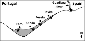
Oysters of the Crassostrea genus were collected in southern Portugal in the Guadiana estuary in the breakwater on the west side of the river (N = 798) and in several different sites (near Faro, Olhão, Fuseta and Tavira) in the Ria Formosa lagoon (N = 446), between November 2010 and July 2011 (Figure 1). All oysters were collected in the intertidal zone. The live weight and height (distance between the umbo and the ventral valve margin) of all oysters were recorded and they were opened and examined macroscopically for the presence of pearls, ectoparasites, shell abnormalities, signs of disease and abnormal physiological conditions. For oysters containing pearls, cross-sections of 4–5 mm that included the mantle and several organs (digestive gland, gill, gonad, intestine and stomach) were collected, fixed in Davidson's solution for 48 h and then preserved in 70% ethanol. Fixed tissues were then embedded in paraffin and sections (4 µm) cut on a rotary microtome, placed on glass slides and stained with haematoxylin and eosin (H&E). All pearls were photographed and length and width determined using ImageJ (straight-line tool). The weight of the pearls was established using a Mettler Toledo MX5 microbalance.
Sites (stars) where the oysters were collected in Ria Formosa lagoon (grey area) and Guadiana estuary (Gulf of Cadiz, Southern Portugal).

Pearl mineralogy and microstructure
The mineralogy of one pearl was determined by grinding it into a fine powder and subjecting the sample to X-ray diffraction (XRD) analysis using a Bruker D8 Advance diffractometer with a Sol-X scintillation counter detector (scan from 20° to 90° with a 0.02° step and a 4 s dwell time). Details of the microstructure were determined by setting selected pearls in epoxy resin and sectioning them across their diameter. The cut surface was then polished, etched in 1% HCl for ~ 25 s, gold coated and then viewed using a Jeol 820 scanning electron microscope.
RESULTS
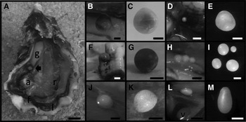
The oysters analysed from the Guadiana estuary had a mean (±SD) height of 73 (±23) mm and a mean live weight of 76 (±60) g. The oysters analysed from the Ria Formosa lagoon had a mean (±SD) height of 80 (±18) mm and a mean live weight of 68 (±39) g. The oysters in which pearls were found had a mean (±SD) height of 112 (±18) mm and a mean live weight of 168 (±42) g. Pearls were identified in 12 out of the 798 oysters analysed from the Guadiana estuary (online Table 1, Appendix). No pearls were observed in the 446 oysters analysed from the Ria Formosa lagoon. The majority (N = 9) of the oysters in which pearls were found only had one pearl. However, four pearls were found in one oyster and two other oysters had two pearls each. Hence, a total of 17 pearls were retrieved. Fifteen of the pearls were located on the edge of the right mantle lobe (Figure 2D, F and H), 13 on the anterior side while two were on the posterior side. Of the remaining two pearls one was present in the adductor muscle (Figure 2E) and the other in the interior part of the right mantle close to the pericardial cavity (Figure 2L and M). The biggest pearl was found on the edge of the right mantle lobe close to the gonad (Figure 2A) and was 190 mg in weight and had a maximum length of 5.5 mm (Figure 2B, C). The smallest pearl weighed 0.50 mg and had a maximum length of ~ 0.9 mm (Figures 2J, K). The majority of the pearls were spherical (e.g. Figure 2C, G, I), although two were shaped like a drop (Figure 2M, L). One of the drop-like pearls was found near the pericardial cavity (Figure 2L) and the other one was found at the edge of the mantle very close to the fusion of the mantle lobes. Almost all pearls had a white-cream colouration with the exception of two pearls that had a black-brown colour (e.g. Figure 2G). All pearls had little or no lustre and a smooth surface with the exception of one that had a rough and granular surface (Figure 2J, K).
Pearls found in Crassostrea sp.: (A) pearl no. 1 found in the mantle close to the gonad measured 5.5 × 5.1 mm (arrow); Abbreviations: a, adductor muscle; f, fusion of mantle lobes; g, gonad; l, left mantle lobe; r, right mantle lobe; (B) Higher magnification of pearl no. 1 in the pearl sac; (C) Pearl no. 1 after removal from the pearl sac; (D) Pearl no. 12 in the pearl sac in the anterior side of the right mantle lobe in the inner side (the mantle was folded back); (E) Pearl no. 7 found in the adductor muscle; (F) Pearl no. 8 in the pearl sac in the anterior side of the right mantle lobe; (G) Pearl no. 8 has a black-brown colouration; (H) Pearls no. 2, 3 and 4 in the pearl sac in the anterior side of the right mantle lobe, a fourth pearl (no. 5) was close but it is not visible in the picture; (I) pearls no. 2, 3, 4 and 5 after removal from their respective pearl sacs; (J) Pearl no. 9 in the pearl sac in the anterior side of the right mantle lobe; (K) Pearl no. 9 has a rough and granular surface; (L) pearl no. 10 in the pearl sac close to pericardial cavity (v, ventricle); (M) pearl no. 10 has a drop-like shape after being removed from the pearl sac. Scale bars: A, 10.0 mm; B, C, D, F, G, H and L, 2.0 mm; E, J and K, 0.5 mm; I and M, 1.0 mm.

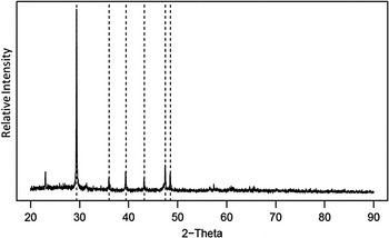
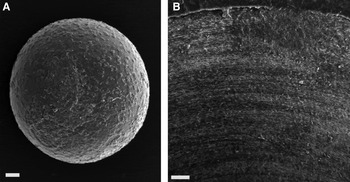
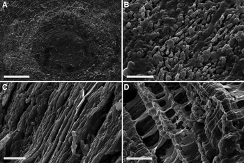
The results of the XRD confirmed that the mineralogy of the pearl analysed was wholly calcitic (Figure 3) with no indication of either aragonite or vaterite present. Scanning electron microscopy (SEM) of the surface of the pearls showed them to be uneven rather than smooth with small, raised features (Figure 4A). SEM of sections through the pearls revealed concentric growth rings (Figure 4B). The pattern of etching shows the fabric of crystals interspersed with an organic matrix and reveals that the latter is unevenly distributed, with prominent layers of organics associated with growth lines. Pearls had distinctive microstructures with blocky crystals in the central zone with more laminar fabrics in the outer shell layers (Figure 5). The latter are composed of thin sheets. There was no sign of stacked hexagonal units associated with nacreous fabrics (Cuif et al., Reference Cuif, Dauphin, Howard, Nouet, Rouzière and Salomé2011).
X-ray diffractometer scan of pearl no. 4. The dot lines mark the expected peak positions for calcite.

Scanning electron microscopy (SEM) of pearls: (A) SEM of pearl no. 5; (B) SEM of a section of pearl no. 8 revealing concentric growth lines. Scale bars: A, 50 µm; B, 20 µm.

Scanning electron micrographs showing the diversity of pearl microstructures. Polished and etched cut surface of pearl no. 8: (A) General shot showing the central nucleus region, an inner ‘blocky’ layer and outer foliated layer; (B) Details of blocky layer; (C) Details of foliated layer; (D) Over-etched surface of pearl no. 6 reveals prismatic units marked here by thick organic sheaths around the spaces formerly occupied by calcite. Note the prism ‘spaces’ are covered by thin organic films that presumably mark growth lines within them. Scale bars: A, 40 µm; B, 4 µm; C, 3 µm; D, 10 µm.

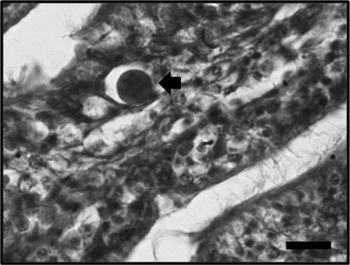
Shell warts characteristic of foot disease (also known as shell disease or ‘maladie du pied’) caused by the fungus Ostracoblabe implexa was observed in 6.3 and 0.3% of the oysters analysed from the Guadiana estuary and the Ria Formosa lagoon, respectively. The ectoparasitic copepod Myicola ostreae was observed in the gill of oysters collected both from the Guadiana estuary and the Ria Formosa lagoon. The prevalence of M. ostreae did not differ in oysters from the Guadiana estuary and the Ria Formosa lagoon and was 56%. Mud worms of the genus Polydora were also observed in oysters collected in both locations but the prevalence was not recorded. Histological examination did not reveal the presence of parasites in the 12 oysters that contained pearls with the exception of Haplosporidium-like plasmodia (Figure 6) several of which were detected in one of the oysters in which a pearl was found (in oyster F). The Haplosporidium-like plasmodia were observed mainly in the labial palps (52% of the plasmodia), but also in the gills (14%), epithelium of the stomach (27%) and intestine (7%), but not in the mantle.
Haplosporidium-like plasmodium observed in the labial palps of oyster F. Scale bar 20 µm.

DISCUSSION
Despite the fact that oysters of the Crassostrea genus are the bivalve species most produced in the world there have been very few reports about the occurrence of natural pearls in these species. One of the few reports about the occurrence of pearls in true oysters was presented recently by Zwaan & Groenenboom (Reference Zwaan and Groenenboom2014). They reported the occurrence of pearls that were presumably found in C. gigas in the Netherlands, although the origin of the pearls identified was uncertain. In other bivalve and gastropod species the occurrence of pearls is also usually rare. However, there are some reports about the occurrence of natural pearls. This is the case of the blue mussel Mytilus edulis in which 94% of the mussels analysed in some sites contain pearls, sometimes as high as 264 pearls per individual (Fernandes & Seed, Reference Fernandes and Seed1983; Ambariyanto & Seed, Reference Ambariyanto and Seed1991). Similarly in the windowpane oyster Placuna placenta natural pearls were reported in 26% of the individuals analysed (Murty, Reference Murty1976). Despite the fact that we have analysed several thousand oysters of the Crassostrea genus over the last 30 years in Portugal this is the first time we have found pearls. It is worth noting that we previously rarely analysed oysters from the Guadiana estuary. Nevertheless, the number of oysters in which pearls were found in the present study in one of the studied sites (Guadiana estuary) was relatively low (in 12 out of the 798 oysters analysed). The majority of the pearls that we found were located in the anterior side of the right mantle lobe close to the gills, i.e. in the inhalant chamber of the mantle cavity. However, pearls were also found in the posterior side of the right mantle lobe, although at much lower frequency. Commercial pearls are mostly composed of aragonite, specifically in the form of nacre. Indeed it is the stacking of thin nacre tablets which scatter incident light that gives pearls their characteristic lustre (Landman et al., Reference Landman, Mikkelsen, Bieler and Bronson2001). Other pearls, such as those described herein, are generally composed of calcite or vaterite and lack the characteristic lustre of pearls and are less sought after. It is often noted that pearls tend to show the same mineralogy and microstructure of the host bivalve shell (Cox in Moore [Reference Moore1969]), but laid down in the ‘reverse order’. So, for example, in Pinctada where the host shell has an outer calcite prismatic layer and a thick inner nacreous layer, the pearls show central prismatic layers with nacreous layers on the outside (Taylor & Strack, Reference Taylor, Strack, Southgate and Lucas2008). Ostreid shell layers are solely calcitic, with aragonite restricted to the myostracum and ligament only. The arrangement of the calcite within the shells is a very thin foliated prismatic outer layer, with the thick inner layers being foliated (Esteban-Delgado et al., Reference Esteban-Delgado, Harper, Checa and Rodrigez-Navarro2008). This is essentially reflected in the pearl mineral and microstructure described here although in the report by Zwaan & Groenenboom (Reference Zwaan and Groenenboom2014) one of the pearls they examined was aragonitic. The occurrence of pearls in mollusc bivalves has often been associated with parasites such as trematode larvae, copepods, cestode larvae and nematopsis oocysts (Sindermann & Rosenfield, Reference Sindermann and Rosenfield1967). In the present study, none of these parasites were observed. Although the parasitic copepod M. ostreae was identified in 56% of the oysters collected from the Guadiana estuary and the Ria Formosa lagoon, the identification of oysters containing pearls only in the former site suggests this parasite is unlikely to be the causal factor. In fact M. ostreae is generally found in the gills of C. gigas and C. angulata (Batista et al., Reference Batista, Boudry, dos Santos, Renault and Ruano2009), although very rarely it can also be found attached to the mantle. Significant differences were observed in the prevalence of foot disease between oysters collected in the Ria Formosa lagoon and the Guadiana estuary (chi-square value with Yates’ correction of 25.0, P < 0.0001). Since shell warts characteristic of foot disease were not observed in the oysters in which pearls were found it seems unlikely that this disease is involved in pearl formation. The only parasite observed by histology in the oysters with pearls was Haplosporidium-like plasmodia, several of which were observed in one individual and not in the mantle. It is also possible that pearls may have been formed due to abnormal growth or a physiological disorder of mantle epithelium cells, which can be linked to the environmental conditions. Guadiana estuary is a narrow and relatively deep estuary with strong currents and a high amount of suspended sediment (Garel et al., Reference Garel, Pinto, Santos and Ferreira2009). The salinity in Guadiana estuary varies considerably, namely between 2 and 37 ppt (Garel et al., Reference Garel, Pinto, Santos and Ferreira2009), whereas the Ria Formosa lagoon has salinity close to that of open-sea water with small fluctuations (Newton & Mudge, Reference Newton and Mudge2003). The amount of suspended sediment and currents in Ria Formosa lagoon is generally low (Neumeier & Ciavola, Reference Neumeier and Ciavola2004), which contrasts with Guadiana estuary. It is therefore possible that the occurrence of pearls in Guadiana estuary could be linked to its specific environmental conditions. Hence, further studies are needed to determine the causes of pearl formation in members of the Ostreidae family and why they are apparently much less common than in other bivalve species.
SUPPLEMENTARY MATERIAL
The supplementary material for this article can be found at http://dx.doi.org/10.1017/S0025315416000382.
ACKNOWLEDGEMENTS
Oscar Branson assisted with the XRD determination. We would like to thank the two anonymous reviewers for their comments that contributed to improve this manuscript.
FINANCIAL SUPPORT
This work was partially funded by the PROSANI (31-03-01-FEP-002, PROMAR) project and by the European Union Atlantic Area Transnational Programme (2007–2013), project SEAFARE, Grant agreement No. 2009–1/123.