Introduction
The egg-laying monotremes are the most basal extant mammals and today comprise only five species: the duck-billed platypus (Ornithorhychus anatinus; Shaw, Reference Shaw1799), the short-beaked echidna (Tachyglossus aculeatus; Shaw, Reference Shaw1792), and three species of long-beaked echidnas (Zaglossus attenboroughi; Flannery & Groves, Reference Flannery and Groves1998, Z. bartoni; Thomas, Reference Thomas1907, and Z. brujini; Peters & Doria, Reference Peters and Doria1876). All living monotremes are nocturnal, homeothermic, and possess a low rate of reproduction (Dawson et al., Reference Dawson, Grant and Fanning1979; Rismiller, Reference Rismiller1999; Werneburg & Sánchez-Villagra, Reference Werneburg and Sánchez-Villagra2010). However, some diurnal activity has also been occasionally observed for echidnas (T. aculeatus multiaculeatus) (Rismiller & McKelvey, Reference Rismiller and McKelvey2009). Overall, monotremes exhibit an intriguing mosaic of reptilian and mammalian characters in terms of anatomy, physiology, and reproduction (Griffiths, Reference Griffiths, Walton and Richardson1989; Campbell & Reece, Reference Campbell and Reece2009). They are endemic to the Australia–New Guinea shelf (Dawson et al., Reference Dawson, Grant and Fanning1979; Rismiller, Reference Rismiller1999), with the short-beaked echidna being the most widely distributed extant monotreme and found both in Australia and in New Guinea (Griffiths, Reference Griffiths, Walton and Richardson1989; Nicol & Andersen, Reference Nicol and Andersen2006). The origin of Monotremata presumably occurred sometime in the Late Triassic/Early Jurassic, a date supported by fossil as well as molecular data (Luo et al., Reference Luo, Kielan-Jaworowska and Cifelli2002; Woodburne et al., Reference Woodburne, Rich and Springer2003; Phillips et al., Reference Phillips, Bennett and Lee2009). Based on fossil evidence, the divergence of modern platypus and echidna lineages occurred in the Early Cretaceous (Rowe et al., Reference Rowe, Rich, Vickers-Rich, Springer and Woodburne2008).
Rhodopsin is the visual pigment responsible for dim light vision in the rod photoreceptors of vertebrates (Menon et al., Reference Menon, Han and Sakmar2001). While visual pigment (opsin) gene sequences have been isolated in a variety of vertebrates, including fish (Chinen et al., Reference Chinen, Hamaoka, Yamada and Kawamura2003), amphibians (Starace & Knox, Reference Starace and Knox1998), reptiles including birds (Kawamura & Yokoyama, Reference Kawamura and Yokoyama1998), and mammals (Nathans & Hogness, Reference Nathans and Hogness1983; Carvalho et al., Reference Carvalho, Cowing, Wilkie, Bowmaker and Hunt2006), functional studies of mammalian rhodopsins have been largely limited to bovine (Sakmar et al., Reference Sakmar, Menon, Marin and Awad2002; Yan et al., Reference Yan, Kazmi, De, Chang, Selbert, Marin, Mathies and Sakmar2002; Natochin et al., Reference Natochin, Gasimov, Moussalf and Artemyev2003) and human (Tam & Moritz, Reference Tam and Moritz2009; Pulagam & Palczewski, Reference Pulagam and Palczewski2010), both diurnal therians. Recent evidence, however, suggests that female bovine may not follow a strictly diurnal activity pattern (Betteridge et al., Reference Betteridge, Costall, Balladur, Upsdell and Umemura2010). Although gene sequences of echidna cone opsins and platypus visual pigments have been isolated (Davies et al., Reference Davies, Carvalho, Cowing, Beazley, Hunt and Arrese2007; Wakefield et al., Reference Wakefield, Anderson, Chang, Wei, Kaul, Graves, Grützner and Deeb2008), neither study included a thorough functional characterization of the expressed photopigments. Furthermore, there has been a notable lack of functional studies of nocturnal mammalian rhodopsins, which is surprising given that the molecular evolution of rhodopsin is thought to be linked to its photic environment (Zhao et al., Reference Zhao, Ru, Teeling, Faulkes, Zhang and Rossiter2009; Shen et al., Reference Shen, Liu, Irwin and Zhang2010).
In this study, we isolate and functionally characterize the rhodopsin from the short-beaked echidna (T. aculeatus), a monotreme, which are the most basal living mammals, in order to investigate a number of functions known to differ among visual pigments, most notably between rod and cone opsins, including hydroxylamine stability (Kawamura & Yokoyama, Reference Kawamura and Yokoyama1998; Starace & Knox, Reference Starace and Knox1998) and the rate of retinal release upon photoactivation (Farrens & Khorana, Reference Farrens and Khorana1995; Yan et al., Reference Yan, Kazmi, De, Chang, Selbert, Marin, Mathies and Sakmar2002). We perform site-directed mutagenesis in order to identify residues underlying functional differences found in the echidna rhodopsin in comparison with bovine rhodopsin, a largely diurnal therian mammal. Characterizing the rhodopsin of the echidna, one of the most basal of the living mammals, is crucial for understanding the evolution of mammalian rhodopsins.
Materials and methods
Rhodopsin cloning and site-directed mutagenesis
Blood samples from a female short-beaked echidna (T. aculeatus) were obtained from the Toronto Zoo (Toronto, Canada) and stored in Queen’s Lysis buffer (Shaw et al., Reference Shaw, Wilson and White2003). Genomic DNA was extracted from these blood samples using the DNeasy Blood and Tissue Kit (Qiagen, The Netherlands), and a Genome Walker library was synthesized according to the manufacturer’s protocol (Universal GenomeWalker Kit; Clontech, Madison, WI). The echidna rhodopsin sequence was isolated from the Genome Walker library by polymerase chain reaction (PCR) using Genome Walker adapter primers (AP1 and AP2) combined with degenerate primers previously designed to target the platypus rhodopsin (Davies et al., Reference Davies, Carvalho, Cowing, Beazley, Hunt and Arrese2007), as well as degenerate primers designed from other tetrapod rhodopsins (). The following cycling conditions were used: an initial 1-min denaturation at 95°C followed by 7 cycles of denaturation at 94°C for 25 s and primer annealing at 72°C for 3 min; another 32 cycles of denaturation at 94°C for 25 s and primer annealing at 67°C for 3 min; product extension was at 67°C for 7 min.
Site-directed mutagenesis primers were designed to generate the mutants N83D, T158A, and F169A () using the following primers (). N83D was selected because it was previously shown to be involved in both spectral tuning (Fasick & Robsinson, Reference Fasick and Robsinson1998) and meta intermediate kinetics (Sugawara et al., Reference Sugawara, Imai, Nikaido, Imamoto and Okada2010). Both T158 and F169 are amino acids unique to the echidna rhodopsin, with F169 being of particular interest, as it potentially interacts with the β-ionine ring of all-trans-retinal (Borhan et al., Reference Borhan, Souto, Imai, Shichida and Nakanishi2000; Palczewski et al., Reference Palczewski, Kumasaka, Hori, Behnke, Motoshima, Fox, Le Trong, Teller, Okada, Stenkamp, Yamamoto and Miyano2000). Amino acid numbering used in this study corresponds to bovine rhodopsin (Sakmar et al., Reference Sakmar, Menon, Marin and Awad2002).
Phylogenetic analyses
The echidna rhodopsin gene sequence (GenBank accession: JX103830) was aligned to 12 other tetrapod rhodopsins downloaded from the NCBI database () and subjected to Bayesian phylogenetic analyses in MrBayes 3.2 (Ronquist & Huelsenbeck, Reference Ronquist and Huelsenbeck2003). The analysis was conducted using the GTR + I + G model, the best fitting model as determined by JModelTest (Posada, Reference Posada2008), and was run for 1,000,000 generations sampling every 100 generations. Trees that were generated before stationary was reached were discarded (burn in = 1000), and the remaining trees were used to construct a consensus tree. Nodal support was assessed by posterior probability values (≥95% = statistical support).
Protein expression and purification
Once the gene sequence had been determined, the full coding sequence (minus the introns) of echidna rhodopsin was synthesized by GeneArt AG (Regensburg, Germany). The artificially synthesized sequence was then inserted into the p1D4-hrGFP II expression vector and thereby tagged with a nine amino acid sequence (TETSQVAPA) at the carboxy terminus to allow for immunoaffinity purification of expressed proteins from HEK293T cells (Morrow & Chang, Reference Morrow and Chang2010).
Expression vectors containing echidna rhodopsin and the mutants as well as bovine rhodopsin (8 μg per 10-cm plate) were transiently transfected into cultured HEK293T cells using Lipofectamine 2000 (Invitrogen, Carlsbad, CA) and harvested 48 h after transfection. Visual pigments were regenerated with 11-cis-retinal, generously provided by Dr. Rosalie Crouch (Medical University of South Carolina), then solubilized in 1% dodecylmaltoside, and purified with the 1D4 monoclonal antibody as previously described (Morrow & Chang, Reference Morrow and Chang2010; Morrow et al., Reference Morrow, Lazic and Chang2011).
Spectroscopic assays
The ultraviolet-visible absorption spectra of purified visual pigments were recorded using a Cary 4000 double beam spectrophotometer (Aglient, Santa Clara, CA). Dark-light difference spectra were calculated by subtracting light-bleached absorbance spectra from respective dark spectra. Pigments were photoexcited with light from a fiber optic lamp for 60 s at 25°C. Reactivity to hydroxylamine was determined by incubating visual pigments in 50 mM hydroxylamine (Sigma-Aldrich, St. Louis, MO) at 25°C. Absorption spectra were taken every 3–5 min for 30 min and every 15 min for another 90 min. The rate of retinal release from light-activated rhodopsin was monitored using a Cary Eclipse fluorescence spectrophotometer (Aglient). This rate was measured by monitoring the increase in fluorescence due to the exit of retinal from the chromophore-binding pocket after photoactivation; a process that has previously been referred to as meta II decay (Farrens & Khorana, Reference Farrens and Khorana1995). Excitation and emission wavelengths were set to 295 and 330 nm, respectively, with excitation and emission slit widths set to 1.5 and 10 nm. Fluorescence measurements were taken every 30 s for 45 min at 20°C. Data for retinal release assays was fit to a first-order exponential curve ( $\,y = y_0 + a(1 - e^{ - bx})$), with half-life values being calculated based on rate constant “b” (
 $t_{1/2} = \ln 2/b$).
Results and discussion
Echidna rhodopsin sequence analysis
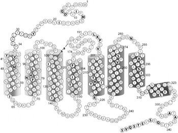
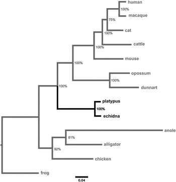
Using a combination of degenerate primers designed from other mammalian rhodopsins, and Genome Walker adapter primers, we were able to isolate a full-length genomic sequence of echidna rhodopsin. This sequence contained four introns (). The intron–exon boundaries are highly similar to the ones in another monotreme, the platypus (Davies et al., Reference Davies, Carvalho, Cowing, Beazley, Hunt and Arrese2007). The translated amino acid echidna rhodopsin sequence contained many of the conserved motifs known to be important for visual pigment function (Sakmar et al., Reference Sakmar, Menon, Marin and Awad2002), including a lysine residue (Lys 296) in the seventh transmembrane domain serving as the site of covalent attachment of the retinal chromophore, its counterion (Glu 113), a pair of cysteine residues known to form a structurally important disulphide bridge between helices 3 and 4 and another pair at the end of helix 8 known to be palmitoylated in bovine rhodopsin (Fig. 1). Phylogenetic analyses of the echidna rhodopsin coding sequence aligned with other tetrapod rhodopsin gene sequences placed the echidna as basal to all other mammalian sequences, as expected from its phylogenetic position within mammals (Fig. 2).
Two-dimensional structure of the echidna rhodpsin (modified from Sakmar et al., Reference Sakmar, Menon, Marin and Awad2002). Residues shared with bovine rhodopsin are in light gray, whereas residues differing from bovine rhodopsin are highlighted in black. Note the insert at the C-terminus.

Phylogenetic analysis of 13 tetrapod rhodopsin sequences, conducted in MrBayes 3.2. using the GTR + I + G model. Percentages indicate posterior probabilities. As expected, monotremes are situated basal to all other mammals.

At highly conserved sites, the echidna rhodopsin bears the same residues shared with all other taxa (). Interestingly, at more variable sites, it shares a surprising number of residues in common with reptiles and amphibians, as opposed to therian mammals (). Shared residues between monotreme and reptile and amphibian rhodopsins include 7, 8, 13, 225, 346, and 348 (). Furthermore, serine and threonine residues at the C-terminus (349–353) are shared in common with amphibians and reptiles but were lost in placentals and marsupials (). These additional residues could be targets for phosphorylation, which in turn could affect the binding affinity of arrestin, a regulator of rhodopsin photochemistry (Sommer & Farrens, Reference Sommer and Farrens2006). In contrast, the echidna sequence only shares four residues with therian mammals at more variable sites, where amino acids differ between Theria and nonmammals: 99, 100, 228, and 308 (). Substitutions unique to monotremes are found at residues 39, 169, and 344 (). Although site 169 is not very conserved among tetrapods, both echidna and platypus bear unique residues at this site (). At site 158, which is not highly conserved either, the echidna rhodopsin bears a unique substitution ().
A mosaic of derived and plesiomorphic characters in monotremes, as exemplified by our analysis of the echidna rhodopsin sequence, has also been reported from anatomic, genomic, physiological, and developmental studies (Bolk et al., Reference Bolk, Göppert, Kallius and Lubosch1934; Gresser & Noback, Reference Gresser and Noback1935; Griffiths, Reference Griffiths, Walton and Richardson1989; Young & Pettigrew, Reference Young and Pettigrew1991; Warren et al., Reference Warren, Hillier, Graves, Birney, Ponting, Grützner, Belov, Miller, Clarke, Chinwalla, Yang, Heger, Locke, Miethke, Waters, Veyrunes, Fulton, Fulton, Graves, Wallis, Puente, López-Otín, Ordóñez, Eichler, Chen, Cheng, Deakin, Alsop, Thompson, Kirby, Papenfuss, Wakefield, Olender, Lancet, Huttley, Smit, Pask, Temple-Smith, Batzer, Walker, Konkel, Harris, Whittington, Wong, Gemmell, Buschiazzo, Vargas Jentzsch, Merkel, Schmitz, Zemann, Churakov, Kriegs, Brosius, Murchison, Sachidanandam, Smith, Hannon, Tsend-Ayush, McMillan, Attenborough, Rens, Ferguson-Smith, Lefèvre, Sharp, Nicholas, Ray, Kube, Reinhardt, Pringle, Taylor, Jones, Nixon, Dacheux, Niwa, Sekita, Huang, Stark, Kheradpour, Kellis, Flicek, Chen, Webber, Hardison, Nelson, Hallsworth-Pepin, Delehaunty, Markovic, Minx, Feng, Kremitzki, Mitreva, Glasscock, Wylie, Wohldmann, Thiru, Nhan, Pohl, Smith, Hou, Renfree, Mardis and Wilson2008; Werneburg & Sánchez-Villagra, Reference Werneburg and Sánchez-Villagra2010). This aspect of the echidna rhodopsin sequence would support the yet controversial Theria hypothesis that monotremes are sister to marsupials and placentals (Janke et al., Reference Janke, Magnell, Wieczorek, Westerman and Arnason2002; Rowe et al., Reference Rowe, Rich, Vickers-Rich, Springer and Woodburne2008). Moreover, the odd mosaic pattern in the echidna amino acid sequence might suggest unexpected structural and functional similarities with amphibian and reptilian rhodopsins, a hypothesis which would require further investigation.
Protein expression and spectroscopic assays
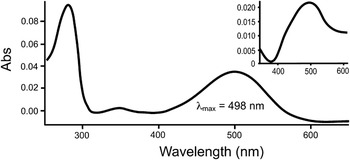
Echidna rhodopsin was successfully expressed in vitro, immunoaffinity purified, and regenerated with the 11-cis-retinal chromophore to produce a stable photopigment with an absorption maximum (λ max) at 497.9 ± 1.1 nm (Fig. 3). The λ max value of the platypus, the other extant monotreme, has been found to be 498 nm (Davies et al., Reference Davies, Carvalho, Cowing, Beazley, Hunt and Arrese2007). Both are well within the characteristic range of λ max values for mammalian rhodopsins, which tend to be at around 500 nm (Menon et al., Reference Menon, Han and Sakmar2001; Bowmaker, Reference Bowmaker2008). Bovine rhodopsin, which was expressed alongside, showed a λ max of 500 nm, which falls within the published range (Oprian et al., Reference Oprian, Molday, Kaufmann and Khorana1987; Stavenga et al., Reference Stavenga, Smits and Hoenders1993). We created three mutants to see if sites of interest cause any shift in properties known to differ between rod and cone opsins. Mutations of echidna-specific residues, T158A and F169A, were found to have λ max values similar to wild type of 498.0 ± 1.3 nm and 499.4 ± 0.1 nm, respectively. However, the N83D mutant caused a significant redshift in the absorbance spectrum, with a λ max of 503.8 ± 1.5 nm, compared to D83 wild-type echidna rhodopsin, which has its λ max at 497.9 ± 1.1 nm. This is consistent with previous studies that identified N83 as the cause of a blueshifted λ max in the rhodopsins of some marine mammals (Fasick & Robinson, Reference Fasick and Robinson2000). Both wild-type and mutant pigments as well as bovine rhodopsin converted to their biologically active meta II intermediates when activated with light, noted by a shift in λ max to approximately 380 nm, the characteristic absorbance maximum of metarhodopsin II (Fig. 3, Inset). This confirms that all synthesized visual pigments are able to properly activate in response to light.
Dark absorption spectra of the rhodopsin of the echidna (T. aculeatus). Note the λ max at 498 nm. Inset, difference spectra generated by taking an absorbance spectrum after bleaching with a fiber optic lamp for 30 s and subtracting it from the dark spectrum.

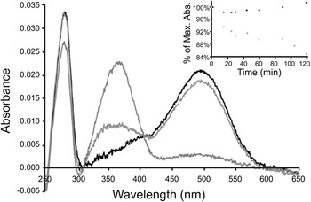
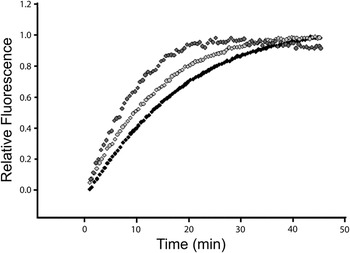
The wild-type echidna rhodopsin and its mutants, together with bovine rhodopsin, were subjected to two spectroscopic assays shown in previous studies to differ between rod and cone visual pigments, hydroxylamine reactivity, and retinal release following photoactivation (Kawamura & Yokoyama, Reference Kawamura and Yokoyama1998; Starace & Knox, Reference Starace and Knox1998; Chen et al., Reference Chen, Kuemmel, Birge and Knox2012). Both assays revealed significant differences between echidna and bovine rhodopsins and unexpected functional properties reminiscent of cone visual pigments. Monitoring hydroxylamine reactivity by absorption spectroscopy revealed that, unlike bovine and other mammalian rhodopsins, wild-type echidna rhodopsin, along with T158A and F169A mutants, are in fact sensitive to hydroxylamine (Fig. 4). Moreover, fluorescence assays monitoring the decay of light-activated meta intermediates found a substantially faster rate of retinal release in echidna rhodopsin (t 1/2 = 9.5 ± 2.6 min−1) in comparison to bovine rhodopsin (t 1/2 = 13.5 ± 1.3 min−1; Fig. 5). The echidna N83D mutation resulted in even faster decay rates, decreasing the half-life to 5.1 ± 0.1 min−1 (Fig. 5), whereas mutations of echidna-specific substitutions (T158A, F169A) had little effect (data not shown).
Absorption spectra of echidna rhodopsin before and after 1 m hydroxylamine treatment. One mole of freshly prepared hydroxylamine diluted in phosphate buffered saline (PBS) was added at 25°C. Spectra were recorded with a scan rate of 400 nm/min, average and integration time of 0.1 and 0.12 s, respectively, data interval of 0.667 nm, and a slit width of 2 nm. Black curve shows dark absorption spectrum before hydroxylamine treatment. Light gray shows rhodopsin absorption after treatment for 2 h. Dark gray curve shows absorption after photoexcitement for 30 s with a fiber optic lamp. Inset, percentage of maximum absorbance over time for echidna wild type (light gray dots) and bovine rhodopsin (black dots) after hydroxylamine treatment. Note the decrease seen in echidna rhodopsin compared to the bovine.

Fluorescence increase of retinal release following Schiff base hydrolysis due to photobleaching of rhodopsin. Excitation and emission wavelengths were 295 and 330 nm respectively, monitoring the decrease in fluorescent quenching of Trp265 by all-trans-retinal as the latter is released from the chromophore-binding pocket of rhodopsin. The t 1/2 values for bovine rhodopsin (black), echidna rhodopsin (light gray), and echidna rhodopsin N83D (dark gray) were 13.5, 9.5, and 5.1 min−1, respectively. Data were collected for 45 min after photoactivation, at 30-s interval, with an integration time of 2 s.

Previous studies have shown that nonmammalian rhodopsins sometimes display more cone-like functional characteristics relative to bovine rhodopsin. For example, rhodopsin of the anole (Anolis carolinensis) is sensitive to hydroxylamine, yielding a complete reaction in around 8.5 h, whereas bovine rhodopsin is virtually unaffected for over 12 h (Kawamura & Yokoyama, Reference Kawamura and Yokoyama1998). Meanwhile, the decay rate of chicken metarhodopsin II is about twice as fast compared to both bovine and human rhodopsin (Imai et al., Reference Imai, Imamoto, Yoshizawa and Shichida1995; Lewis et al., Reference Lewis, van Kuijk, Carruthers and Kliger1997; Okada et al., Reference Okada, Matsuda, Kandori, Fukada, Yoshizawa and Shichida1994; Janz & Farrens, Reference Janz and Farrens2004).
Our study is the first to reveal such functional characteristics in a mammalian rhodopsin, with the echidna rhodopsin both reacting to hydroxylamine and having a faster rate of retinal release, a property connected to metarhodopsin decay rates, than bovine rhodopsin. It should also be noted that these differences among vertebrate rhodopsins are not as large as the more striking contrasts between rhodopsins and cone opsins, which are largely thought to be mediated by substitutions at two residues, Q122E and P189I (Imai et al., Reference Imai, Kojima, Oura, Tachibanaki, Terakita and Shichida1997, Reference Imai, Imamoto, Yoshizawa and Shichida2007; Kuwayama et al., Reference Kuwayama, Imai, Hirano, Terakita and Shichida2002, Reference Kuwayama, Imai, Morizumi and Shichida2005), which are invariant among vertebrate rhodopsins. However, our results contribute to the idea that intriguing functional differences exist within rhodopsins, not just between rhodopsins and cone opsins, and are worth investigating, especially in enigmatic and phylogenetically relevant species such as the echidna.
A recent study has suggested that N83 may in fact be an adaptation to dim light environments, by increasing the rate of formation of the biologically active meta II rhodopsin intermediate, and thereby increasing photosensitivity in dim light in cichlid fishes and nocturnal bats (Sugawara et al., Reference Sugawara, Imai, Nikaido, Imamoto and Okada2010). Echidna rhodopsin also has N83. When we mutated this residue to aspartic acid (D), the most common identity of site 83 in mammals, our results indicate a faster rate of retinal release compared to wild-type echidna rhodopsin. Although the precise mechanisms by which N83 affect meta intermediate kinetics remain unclear, previous studies have suggested that greater stability of one or more meta intermediates of light-activated echidna rhodopsin may allow for further transducin activation, sustaining signaling activity, and possibly even increasing the sensitivity of the photoreceptor cells (Heck et al., Reference Heck, Schadel, Maretzki, Bartl, Ritter, Palczewski and Hofmann2003; Lamb & Pugh, Reference Lamb and Pugh2004; Imai et al., Reference Imai, Kefalov, Sakurai, Chisaka, Ueda, Onishi, Morizumi, Fu, Ichikawa, Nakatani, Honda, Chen, Yau and Shichida2007). Increased photosensitivity could be advantageous, particularly for vision at low light levels. Therefore, our investigations of echidna rhodopsin function suggest that although its rhodopsin is sensitive to hydroxylamine and has a faster rate of retinal release upon light activation relative to other mammalian rhodopsins, it does possess a substitution, N83, which might be an adaptation to dim light environments.
In this study, we present the first detailed functional characterization of a rhodopsin from a most basal mammal, the echidna (T. aculeatus). Surprisingly, our results indicate that the rhodopsin of the echidna displays functional characteristics such as sensitivity to hydroxylamine and a faster metarhodopsin decay rate (as compared to bovine rhodopsin), which are more similar to nonmammalian rhodopsins, and, to a lesser extent, cone opsins. These results are intriguing and show that significant variation can occur even within mammalian rhodopsins.
Finally, although the rhodopsin of the echidna did not display overall functional characteristics in comparison with the more diurnal bovine rhodopsin that might be expected of a nocturnal pigment, our data do suggest nonetheless that N83 might be an adaptation to dim-light vision. In conclusion, the rhodopsin of the echidna appears as enigmatic as the animal itself displaying unusual biochemical and functional characteristics, which differ from all other mammalian rhodopsins investigated, and may be due to the mosaic pattern of derived and plesiomorphic characters within its amino acid sequence.
Acknowledgments
We thank G. Crawshaw (Toronto) for the echidna blood samples. We are thankful to all members of the Chang Lab, especially to I. van Hazel (Toronto, Canada), as well as to M. Meixner, R. Schreiber, and A. Sonntag (all Berlin, Germany) for help in the lab. We thank E.C. Kirk (Austin, TX) for discussion on bovine activity patterns, as well as L.A. Tsuji, R.K. Schott, and the two anonymous reviewers for their constructive comments, which helped to improve an earlier version of the manuscript. This work was funded by the Deutsche Forschungsgemeinschaft (DFG Mu 1760/2-3, to C.B. and J.M.), a University Health Network (University of Toronto) Vision Science Research Program fellowship (J.M.M.), a National Sciences and Engineering Research Council Discovery grant (to B.S.W.C.), and a Government of Ontario Early Researcher Award (to B.S.W.C.).