Introduction
Fallot-type double outlet right ventricle and tetralogy of Fallot are among the most common forms of cyanotic CHD in children. tetralogy of Fallot is characterised by four cardinal anatomical features: right ventricular hypertrophy, an overriding aorta, a large nonrestrictive ventricular septal defect, and obstruction of the right ventricular outflow tract. Fallot-type double outlet right ventricle represents a spectrum in which both great arteries arise predominantly from the right ventricle, with the lesion being classified as such when the aortic override exceeds 50%. This anatomical configuration leads to compromised pulmonary blood flow, thereby exacerbating cyanosis. Reference Gibbs, Uzun and Blackburn1
Figure 1.

The natural history of uncorrected tetralogy of Fallot or Fallot-type double outlet right ventricle includes chronic hypoxaemia, progressive right ventricular hypertrophy, polycythaemia, failure to thrive, and a significant risk of sudden death during hypoxic spells. Reference Stumper, Ramchandani and Noonan2,Reference Bentham, Zava and Harrison3 The best course of action for these lesions is still the final surgical repair, but when to do so can still be clinically challenging, especially for newborns and infants with concomitant conditions like low birth weight or preterm, sepsis, extracardiac anomalies, or unfavourable coronary anatomy. Reference Morray, McElhinney and Jones4,Reference Prakoso, Citra Dewi and Mendel5 These conditions make early corrective surgery a high-risk endeavour, prompting the need for palliative strategies to stabilise the patient, promote pulmonary artery growth, and defer surgical correction until the child is older and physiologically more stable. Reference Krasemann, Kehl and Vogt6
Traditionally, the modified Blalock–Taussig shunt, which connects the subclavian artery to the pulmonary artery using a prosthetic graft, has been employed as the palliative procedure of choice. Reference Sandica, Reineker and Wiegand7 Although the modified Blalock–Taussig shunt is effective in enhancing systemic oxygenation, but it carries several limitations. These include thoracotomy-related morbidity, unbalanced pulmonary blood flow, distortion of the pulmonary arteries, and potential complications such as infection, thrombosis, and shunt occlusion. Reference Celebi, Karpuz and Yilmaz8,Reference Bentham, Shivaprakasha and Rao9 Additionally, the procedure often necessitates a sternotomy at the time of corrective repair, increasing cumulative surgical trauma.
Over the past two decades, percutaneous catheter-based interventions have emerged as promising alternatives to surgical palliation. These include ductal stenting, balloon valvuloplasty of the pulmonary valve, and, more recently, right ventricular outflow tract stenting. Reference Cools, Boshoff, Heying, Rega, Meyns and Gewillig10,Reference Berdat and Carrel11 In patients with native right ventricular outflow tract obstruction, right ventricular outflow tract stenting is becoming more and more popular as a practical method of increasing pulmonary blood flow while preserving central pulmonary artery anatomy and facilitating uniform pulmonary artery growth. Reference Michel-Behnke, Ewert and Nagdyman12
The principle behind right ventricular outflow tract stenting involves the placement of a bare-metal stent across the obstructed infundibular region or pulmonary valve annulus to create a low-resistance channel from the right ventricle to the pulmonary artery. This reestablishes pulsatile antegrade flow and effectively bypasses the obstruction, leading to increased pulmonary perfusion, the reduction of right ventricular hypertrophy, and an increase in systemic oxygen saturation. Reference Abqari, Kidwai and Kamran13 Furthermore, right ventricular outflow tract stenting allows time for the infant to grow, gain weight, and develop more favourable surgical anatomy before undergoing definitive intracardiac repair. Reference Sandoval, Chaturvedi, Benson, Morgan, Van Arsdell and Honjo14
Several observational studies have established the safety and efficacy of right ventricular outflow tract stenting in symptomatic neonates with tetralogy of Fallot and Fallot-type double outlet right ventricle. In a landmark study by Gibbs et al., four patients who underwent right ventricular outflow tract stenting showed a significant reduction in right ventricular systolic pressure and a marked improvement in systemic oxygen saturation, without encountering major procedural complications. Reference Castleberry, Gudausky, Berger, Tweddell and Pelech15 In a similar vein, Dohlen et al. found that nine infants who received right ventricular outflow tract stenting showed notable improvements in arterial oxygen saturation and pulmonary artery Z-scores, with no early mortality. Reference Dohlen, Chaturvedi, Benson, Ozawa, Van Arsdell and Fruitman16 Barron et al., in a larger group of 32 patients, confirmed improved pulmonary artery development and a favourable substrate for later surgical repair after right ventricular outflow tract stenting. Reference Barron, Ramchandani, Murala, Stumper, De Giovanni and Jones17
In 2013, Stumper et al. published one of the largest single-centre experiences, involving 52 patients who underwent right ventricular outflow tract stenting due to high surgical risk. They reported a procedural success rate of 94%, with significant improvements in pulmonary artery diameters and oxygenation. Importantly, the study highlighted the importance of stent selection and deployment technique to minimise the risks of stent migration, tricuspid valve injury, and coronary artery compression. Reference Abumehdi, Sasikumar, Chaudhari, Bhole, Botha and Mehta18
Despite increasing global evidence supporting right ventricular outflow tract stenting, data from the Indian subcontinent are limited. Indian children often present late with low body weight, complex anatomy, and restricted access to early surgery, highlighting the need to assess the safety and effectiveness of right ventricular outflow tract stenting in this context.
Furthermore, most existing studies are retrospective in nature, often limited to small cohorts, and focus primarily on procedural feasibility and short-term oxygenation parameters. There is limited prospective data examining the spectrum of anatomical substrates, procedural nuances, and immediate complications in a real-world clinical setting. Reference Laurentius, Wiyono, Subali and Siagian19
The present study was therefore undertaken to fill this knowledge gap by systematically analysing both retrospective and prospective cohorts of symptomatic children (<5 years) with Fallot-type physiology undergoing right ventricular outflow tract stenting at a high-volume tertiary-care centre in India. The primary objective was to assess the immediate clinical and procedural outcomes of right ventricular outflow tract stenting in terms of oxygenation, pulmonary artery growth, and technical success. Secondary objectives included identifying common procedural complications and analysing the anatomical and clinical predictors of success and failure.
By comprehensively examining patient selection, procedural technique, stent characteristics, and short-term outcomes, this study aims to contribute meaningful clinical insights to the growing body of literature on right ventricular outflow tract stenting and to explore its potential as a standard palliative approach in resource-limited, high-burden paediatric cardiac settings.
Materials and methods
Study design and setting
The Department of Paediatric Cardiology at the Narayana Institute of Cardiac Sciences in Bengaluru, India, was the site of this single-centre observational study. Both prospective and retrospective designs were used in the investigation. For instances from August 2017 to July 2020, retrospective data were gathered; for cases from August 2020 to November 2021, prospective data were gathered. The Institutional Ethics Committee previously approved the study (Ref: NHH/AEC-CL-2020-505), and all potential enrolled children’s guardians provided written informed permission.
Study population and inclusion criteria
Children under five years of age with tetralogy of Fallot, Fallot-type double outlet right ventricle, or similar lesions featuring normally related great arteries and restricted antegrade pulmonary blood flow due to a narrowed right ventricular outflow tract were included in the study. All of the kids exhibited noticeable symptoms and risk factors for corrective surgery. Both clinical (such as low body weight, preterm, young age, or neonates) and morphological (such as tiny branch pulmonary arteries, large coronary crossing the right ventricular outflow tract, and the presence of substantial comorbidities such as cyanotic episodes) risk factors were present.
Exclusion criteria
Children were excluded if they had:
-
1. Great arteries not normally related,
-
2. Membranous pulmonary atresia with ventricular septal defect requiring radiofrequency perforation before stenting,
-
3. Associated lesions mandating open-heart surgery, or (patients with significant branch pulmonary artery origin stenosis were excluded from this study)
-
4. Guardians who declined consent for right ventricular outflow tract stenting.
Data collection and variables
For the retrospective cohort, data were collected from hospital records. For the prospective cohort, data were recorded at baseline and at discharge. Demographic information such as age, sex, and weight was documented. Pre-procedural echocardiography and CT angiography were evaluated for anatomical and physiological assessment. Outcome parameters included oxygen saturation, Z-scores of branch pulmonary arteries, procedural duration, ICU and hospital stay, need for blood transfusion, complications, and duration of ventilation.
Device characteristics and stent selection
Stents used included Herculink Elite (Abbott Vascular, USA), Visi-Pro (Boston Scientific, Ireland), and Cook Formula (Cook Medical, USA). Right ventricular outflow tract infundibular length and pulmonary valve annulus size were used to select stents. It was decided to use a premounted balloon-expandable bare-metal stent that was about 2 mm wider than the narrowest right ventricular outflow tract segment. The length of the stent ensured coverage from the pulmonary annulus proximally into the infundibulum (across the pulmonary valve). Stent availability and deliverability through a 5F or 6F sheath guided the final choice.
Procedural technique
All procedures were performed under general anaesthesia with arterial line monitoring, central venous access, and endotracheal intubation. The preferred vascular access was the right internal jugular vein, though femoral access was used when necessary. Intra-procedural echocardiography guided stent placement and assessed for pericardial effusion or ventricular dysfunction.
A right ventricular angiogram was obtained using a multipurpose catheter in multiple views (30° RAO, 30° LAO, 20° cranial, and AP) to assess right ventricular outflow tract dimensions, which were compared with echocardiographic measurements. A 0.014“ coronary wire was advanced into the distal pulmonary artery, and the stent was delivered via a Judkins Right guiding catheter. After confirming positioning with contrast, the stent was deployed using manual balloon inflation.
Post-deployment angiography and echocardiography confirmed stent position and ruled out complications. Patients were monitored in the ICU with strategies to prevent pulmonary overcirculation, including permissive hypercapnia. Once stable, they were extubated and monitored using chest X-ray, ECG, and echocardiography. Our protocol included heparin infusion for 72 hours post-procedure, followed by aspirin and clopidogrel until corrective surgery.
Post-procedure assessment
Post-procedural monitoring included assessment of oxygen saturation, ECG for conduction abnormalities, echocardiography for stent position and function, and chest X-ray for stent integrity and position. Complications such as heart block, local haematoma, pericardial effusion, ventricular dysfunction, worsening tricuspid regurgitation, and stent migration or embolisation were actively monitored. A successful procedure was defined as accurate preoperative anatomical selection and technically successful right ventricular outflow tract stent placement with improvement in clinical parameters.
Statistical assessment
Data were analysed using SPSS v23.0. Continuous variables were compared using unpaired t-tests or Mann–Whitney U tests based on distribution. Paired t-tests or Wilcoxon signed-rank tests assessed within-group changes. Results were reported as mean ± SD or median (IQR). Spearman correlation analysed associations between stent size, body surface area, and other variables. A p-value <0.05 was considered statistically significant.
Results
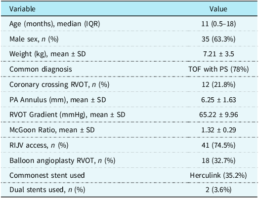
During the study period, 55 patients aged 2 to 60 months underwent right ventricular outflow tract stenting. The median age was 11 months (IQR: 0.5–18 months), with 63.3% being male. Tetralogy of Fallot with pulmonary stenosis was the most common diagnosis (78%), followed by double outlet right ventricle with pulmonary stenosis and functional pulmonary atresia. Over half of the cohort (56.3%) belonged to the 0–12 months age group (Group A). Baseline demographic and clinical characteristics are detailed in Table 1.
Baseline and procedural characteristics

Cyanosis was the most common presenting symptom, present in all cases. Difficulty in feeding (52.7%), failure to thrive (49.1%), and a history of cyanotic spells (80%) were also frequent. A sizeable percentage had a history of congenital cardiac disease in their families (21.8%) or sibling death (18.2%). Clubbing was present in 96.4% of cases. Neurological manifestations were observed in 9 patients, and two patients had VACTERL anomalies.
Pre-procedural echocardiography showed a mean pulmonary artery annulus of 6.25 ± 1.63 mm with a Z-score of –3.18 and a mean right ventricular outflow tract gradient of 65.22 ± 9.96 mmHg. The McGoon ratio was 1.32 ± 0.29. Abnormal coronary anatomy crossing the right ventricular outflow tract was present in 12 patients (21.8%). Associated anomalies such as right aortic arch, hemitruncus, and hypertrophic cardiomyopathy were also noted.
Right ventricular outflow tract stenting was performed via the right internal jugular vein in 74.5% of patients, with femoral access in 20%. Balloon angioplasty of the right ventricular outflow tract was performed in 32.7%, and of branch PAs in 12.7%. The most commonly used stent was Herculink (35.2%), followed by VisioPro (25.9%). The median stent diameter and length were 7 mm and 25 mm, respectively.
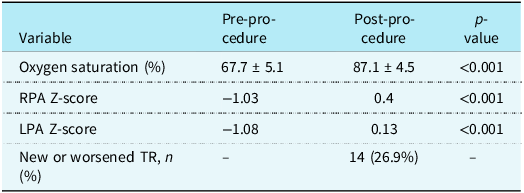
The mean procedural duration was 151.2 ± 73.7 minutes, with a median fluoroscopy time of 24 minutes and a radiation dose of 120 cGy/cm2. A significant increase in systemic oxygen saturation was observed, rising from a pre-procedural mean of 67.7% ± 5.1 to 87.1% ± 4.5 post-procedure (p < 0.001). A significant increase was observed in branch pulmonary artery dimensions post-stenting: the left pulmonary artery Z-score improved from −1.08 to 0.13, and the right pulmonary artery Z-score from −0.33 to 0.4 (both p < 0.001). Table 2 summarises key peri-procedural outcomes, including procedure duration, fluoroscopy exposure, and changes in haemodynamic and anatomical parameters pre- and post-stenting.
Postprocedural outcomes

The median hospital stay was 7 days (IQR: 5–10), with a median ICU stay of 4 days (IQR: 3–6). Pulmonary oedema occurred in 47.3% of patients, and furosemide was administered in 54.5% for its management. The median duration of post-procedural mechanical ventilation was 12.5 hours (IQR: 6–36).
Procedure-related complications included mild pericardial effusion in 12 patients (21.8%), stent embolisation in 4 patients (7.2%), and persistent desaturation in 4 patients (7.3%). Three patients (5.5%) died—one due to cardiac arrest within 24 hours, one following stent embolisation and atrial septectomy, and one due to aspiration in the ward. The overall procedural success rate was 83.6% (46 out of 55 patients). Outcome failures (n = 9) were primarily attributed to stent displacement (n = 3), persistent desaturation (n = 2), dynamic infundibulum (n = 1), and inability to cross a subinfundibular spasm (n = 1).
Right ventricular outflow tract stenting led to a significant increase in branch pulmonary artery dimensions. The median right pulmonary artery Z-score improved from −1.03 (IQR: −2.52 to 0.28) to 0.4 (IQR: −0.61 to 1.7) (p < 0.001), and the left pulmonary artery Z-score increased from −1.08 (IQR: −2.18 to −0.15) to 0.13 (IQR: −0.74 to 0.95) (p < 0.001).
Analysis of prolonged ICU stay (defined as >3 days) showed no significant association with age, weight, pulmonary oedema, or stent/BSA ratio. However, the use of furosemide was significantly associated with longer ICU stays (p = 0.009). The stent/BSA ratio did not significantly correlate with pulmonary oedema (p = 0.586), Lasix use (p = 0.942), or ICU stay (p = 0.756). Although the use of femoral venous access was associated with a higher incidence of new-onset tricuspid regurgitation, this difference did not reach statistical significance (p = 0.119). Similarly, procedures performed via the femoral vein route demonstrated a longer duration compared to those using the right internal jugular vein approach, but this difference was also not statistically significant (p = 0.227).
Out of the four cases of stent embolisation, one required surgical retrieval via sternotomy, and another had intraoperative tricuspid valve repair. In all cases, follow-up interventions included central shunts or corrective surgeries. Follow-up was done at 1 month, 6 months, and 1 year, continuing up to 2 years or until corrective surgery, with many visits conducted online due to travel constraints.
Discussion
The present study assessed the effectiveness and outcomes of right ventricular outflow tract stenting as a primary palliative intervention in symptomatic children with Fallot-type physiology. The findings revealed a significant improvement in systemic oxygen saturation and branch pulmonary artery growth following the procedure, with a complication profile deemed acceptable. These findings support the growing role of right ventricular outflow tract stenting as a feasible alternative to conventional systemic-to-pulmonary shunt surgery, especially in carefully selected high-risk paediatric patients.
In our cohort of 55 patients, the procedural success rate was 83.6%, which is slightly lower than some published series but still clinically meaningful. For example, according to Quandt et al., 92% of procedures were successful in their comparative study of right ventricular outflow tract stenting versus Blalock–Taussig shunt. Reference Quandt, Ramchandani and Penford20 Haas et al. similarly observed success in 90% of their cohort. Reference Haas, Happel and Moysich21 The slight difference in outcomes in our study is likely reflective of institutional learning curve effects and case complexity.
A key finding of our study was a significant increase in oxygen saturation, from a mean of 67.7% pre-procedure to 87.1% post-procedure (p < 0.001). This aligns with the data reported by Quandt et al., where a similar degree of saturation improvement was observed following stenting in cyanotic infants. Reference Quandt, Penford, Ramchandani, Bhole, Mehta and Stumper22 The Z-scores of branch pulmonary arteries also improved significantly, indicating measurable anatomical benefit beyond symptomatic relief. This is important, as adequate pulmonary artery growth is essential for safe future surgical repair.
Anatomically, most patients had tetralogy of Fallot with pulmonary stenosis (tetralogy of Fallot-PS), which was followed by double outlet right ventricle variations. Notably, 11 patients (20%) had coronary artery anomalies with right ventricular outflow tract crossing, which would have made a traditional Blalock–Taussig shunt or surgical repair high-risk or contraindicated. Reference Morray, McElhinney and Jones4,Reference Krasemann, Kehl and Vogt6,Reference Bugeja, Grech and DeGiovanni23 In such patients, right ventricular outflow tract stenting offers a physiologically superior and anatomically safer approach by maintaining antegrade flow and promoting balanced pulmonary artery growth.
The most frequent complications were pulmonary oedema (47%) and new-onset or worsened tricuspid regurgitation (26.9%), consistent with published experiences by Tanıdır İC et al. and Luxford JC et al. Reference Tanıdır, Bulut, Kamal, Öztürk, Yücel and Güzeltaş24,Reference Luxford, Adams, Roberts and Mervis25 Stent embolisation, although infrequent (7.2%), was a serious event with two cases requiring surgical retrieval and one resulting in mortality. These complications underscore the need for careful stent sizing and deployment techniques. Our data showed that higher stent/BSA ratios (≥30 mm/m2) were associated with prolonged ICU stay, though this did not reach statistical significance. Similar observations have been made by Aurigemma D et al., suggesting that aggressive oversizing may contribute to post-procedural volume overload. Reference Aurigemma, Moore, Vaughn, Moiduddin and El-Said26
The access route also influenced procedural dynamics. The right internal jugular vein was the most commonly used access (74.5%) and was associated with shorter procedural time compared to the femoral approach. Other research has confirmed that the right internal jugular vein is the best access because it aligns coaxially with the right ventricular outflow tract, making stent deployment and catheter manipulation easier. Reference Bigdelian, Sedighi and Sabri27
The success of right ventricular outflow tract stenting is strongly influenced by appropriate patient selection and thorough anatomical assessment. In our study, echocardiography and CT angiography played a pivotal role in determining suitability, guiding both the approach and stent sizing. Cases with hypoplastic PAs, multiple ventricular septal defects, or severe infundibular narrowing posed specific technical challenges. A notable learning point was the importance of pre-procedure imaging to identify patients with unrecognised dynamic right ventricular outflow tract obstruction or significant valve regurgitation, as these were associated with suboptimal outcomes.
Statistical analysis did not reveal significant correlations between age, weight, stent/BSA ratio, or pulmonary oedema with prolonged ICU stay or procedural failure. However, furosemide use emerged as an indirect indicator of fluid overload and ICU duration, highlighting the role of postoperative haemodynamics over purely anatomical predictors. While prior studies, such as those by Haas and Aurigemma D et al., have noted similar associations, Reference Haas, Happel and Moysich21,Reference Aurigemma, Moore, Vaughn, Moiduddin and El-Said26 our findings suggest that individualised postoperative care is equally critical to outcomes.
Though the stent/BSA ratio was not significantly associated with adverse events, it remains a valuable intraoperative guide. McGovern E et al. have suggested higher ratios may predict haemodynamic compromise, but our results did not replicate this trend. Reference McGovern, Morgan, Oslizlok, Kenny, Walsh and McMahon28
This study provides real-world insights from an Indian tertiary-care context, where late presentations and resource constraints often complicate management. In this setting, right ventricular outflow tract stenting remains a cost-effective and less invasive bridge to definitive repair, particularly in neonates with sepsis, prematurity, or low weight. Reference McGovern, Morgan, Oslizlok, Kenny, Walsh and McMahon28
Key limitations include a modest sample size, lack of a comparator group, and limited long-term follow-up. These factors constrain broader generalisation and necessitate further prospective, multicentric studies. Hardware advancements and experience will likely reduce complications and optimise outcomes. Reference Bang, Ko, Yu, Han, Kim, Ko and Park29,Reference Bertram, Emmel, Ewert, Grohmann, Haas and Jux30
Our study contributes real-world data from an Indian tertiary-care setting, where socioeconomic factors and delayed presentations often complicate management. In this context, right ventricular outflow tract stenting offers a cost-effective and less invasive alternative to surgical palliation. Particularly in neonates with sepsis, prematurity, or low weight—where the mortality of Blalock–Taussig shunt surgery is substantial—right ventricular outflow tract stenting can bridge patients to a more stable physiological state before definitive repair. Reference McGovern, Morgan, Oslizlok, Kenny, Walsh and McMahon28
Conclusion
Right ventricular outflow tract stenting is an effective palliative treatment option for children with Fallot-type physiology who are not candidates for surgical repair. The technique improves systemic oxygenation and promotes branch pulmonary artery growth, hence improving the anatomical and physiological circumstances required for decisive surgical intervention.
This study highlights the importance of careful patient selection and thorough pre-procedural evaluation with echocardiography and cardiac CT. The slightly lower success rate compared to published data likely reflects the initial institutional learning curve, which is expected to improve with experience and standardised protocols.
The right internal jugular vein was the preferred access route, offering better alignment and shorter procedural time than femoral access. Right ventricular outflow tract stenting proved especially valuable in high-risk patients with complex anatomy or comorbidities, offering a stable bridge to later repair.
In conclusion, in certain high-risk infants, right ventricular outflow tract stenting need to be regarded as a first-line palliative measure. Larger multicentric studies with extended follow-up are needed to refine indications and develop regional practice guidelines.