Introduction
Marinaite Cu2Fe3+O2(BO3), a new mineral, has been found in a hot fracture on a cooling lava field of the 2012–2013 Tolbachik fissure eruption, 10 months after the eruption ceased. It was the second fissure eruption in this area during the last 50 years (Belousov et al., Reference Belousov, Belousova, Edwards, Volynets and Melnikov2015). The previous 1975–1976 Great Tolbachik Fissure Eruption (GTFE) lasted 1.5 years, produced ∼2,2 km3 of lava and pyroclastics and left behind several scoria cones, which are famous for abundant exhalative mineralization (Fedotov and Markhinin, Reference Fedotov and Markhinin1983; Fedotov, Reference Fedotov1984) This mineralization at the cones of the Northern Breakthrough of the Great Tolbachik Fissure Eruption results from a long-lasting post-eruptive degassing and has been studied for many years (Vergasova and Filatov, Reference Vergasova and Filatov2016; Pekov et al., Reference Pekov, Koshlyakova, Zubkova, Lykova, Britvin, Yapaskurt, Agakhanov, Shchipalkina, Turchkova and Sidorov2018, Reference Pekov, Agakhanov, Zubkova, Koshlyakova, Shchipalkina, Sandalov, Yapaskurt, Turchkova and Sidorov2020a). Mineralogy of the exhalative crusts has been proven to be so diverse that, despite its relatively small size, the Second scoria cone of the Northern Breakthrough of the Great Tolbachik Fissure Eruption currently holds the world record for the number of new mineral species discovered per unit area. Over 350 mineral species have been identified in the Great Tolbachik Fissure Eruption fumaroles, of which more than 150 new minerals have been discovered there. The scoria cones of the Northern Breakthrough of the Great Tolbachik Fissure Eruption are considered as a ‘reference object’ for oxidizing-type fumarolic systems (Pekov et al., Reference Pekov, Agakhanov, Zubkova, Koshlyakova, Shchipalkina, Sandalov, Yapaskurt, Turchkova and Sidorov2020a). The Northern Breakthrough of the Great Tolbachik Fissure Eruption mineralization is mainly represented by oxysalts with only a few boron minerals among them. These are chubarovite KZn2(BO3)Cl2 (Pekov et al., Reference Pekov, Zubkova, Pautov, Yapaskurt, Chukanov, Lykova, Britvin, Sidorov and Pushcharovsky2015), rhabdoborite-(V), rhabdoborite-(Mo) and rhabdoborite-(W) forming a new group of borate minerals with the general formula Mg12M 1⅓O6[(BO3)6–x(PO4)xF2–x] (M = V5+, Mo6+ or W6+ and x < 1) (Pekov et al., Reference Pekov, Zubkova, Koshlyakova, Belakovskiy, Agakhanov, Vigasina, Britvin, Sidorov and Pushcharovsky2020b), ludwigite and yuanfuliite (Bulakh et al., Reference Bulakh, Pekov, Koshlyakova and Sidorov2022b), fredrikssonite (Bulakh et al. Reference Bulakh, Britvin, Zubkova, Yapaskurt, Agakhanov, Anikin and Pekov2025), and OH-free fluoborite Mg3(BO3)F3 (Bulakh et al., Reference Bulakh, Pekov, Chukanov, Yapaskurt, Koshlyakova, Britvin and Sidorov2022a). Despite the fact that copper minerals make up a significant part of the Tolbachik fumarolic mineralization, no copper borate was found in fumaroles of the Northern Breakthrough of the Great Tolbachik Fissure Eruption.
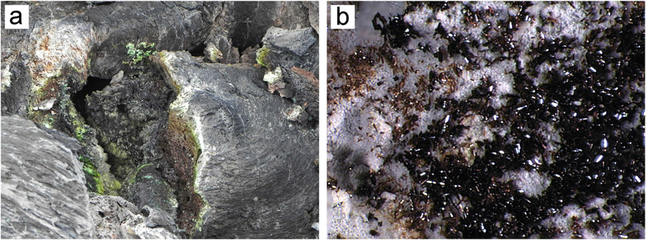
Occurrence of marinaite. (a) The hot crack ∼30–40 cm wide on the surface of the lava field – the place of marinaite sampling (photo A. Belousov); (b) marinaite crystals on sulfate material covering lava fragments. Specimen number 2714/2, Field of view ∼1.5×2 mm.

Copper borates are in general rare in nature. Only five borate minerals containing copper as a species-defining element have been reported. Copper metaborate santarosaite CuB2O4 was found only in the oxidation zone of the Santa Rosa hydrothermal base-metal vein deposit in Iquique Province, Chile (Schlüter et al., Reference Schlüter, Pohl and Golla-Schindler2008). Four other minerals contain an OH group, namely bandylite Cu[B(OH)4]Cl, which occurs as a secondary mineral in a leached zone above massive iron sulfates near the surface at Mina Quetena, Calama, El Loa Province, Chile (Palache and Foshag, Reference Palache and Foshag1938); henmilite Ca2Cu(OH)4[B(OH)4]2 (Nakai et al., Reference Nakai, Okada, Masutomi, Koyama and Nagashima1986) and numanoite Ca4Cu(B4O6(OH)6)(CO3)2 (Ohnishi et al., Reference Ohnishi, Kusachi, Kobayashi, Yamakawa, Tanabe, Kishi and Yasuda2007) from a low-temperature hydrothermal assemblage related to calcic marble near the gehlenite–spurrite skarns at the Fuka mine, Fuka, Okayama Prefecture, Japan; and jacquesdietrichite Cu2[BO(OH)2](OH)3, which was discovered in secondary products of manganese ores at the Tachgagalt Mine, Zagora Province, Morocco and is also known from the Caledonia copper mine in Michigan, USA (Kampf and Favreau Reference Kampf and Favreau2004).
Numerous natural and synthetic borates (oxoborates) with ludwigite-like structures have the general formula M2+2M3+O2(BO3) in which M2+ = Mg, Fe, Cu, Co, Ni; and M3+ = Mn, Cr, Fe, Al, Ga, Ti4+. They are characterized by wide isomorphism, including heterovalent substitutions, different degrees of ordering of metal cations and, as a consequence, specific physical, especially magnetic properties. Synthetic Cu2FeBO5 crystals were first obtained and studied using single-crystal X-ray diffraction (XRD) by Schäfer and Bluhm (Reference Schäfer and Bluhm1995). The complex magnetic behaviour of this compound was characterized on a powder sample (Continentino et al., Reference Continentino, Fernandes, Guimaraes, Borges, Sulpice, Tholence, Siqueira and Dos Santos1999) and later on single crystals (Petrakovsky et al., Reference Petrakovsky, Bezmaternykh, Velikanov, Vorotynov, Bayukov and Schneider2009). A dozen Cu-containing synthetic ludwigites Cu2MBO5 with homometallic and heterometallic trivalent subsystems, where M = Al3+, Ga3+, Fe3+, Mn3+, Cr3+, (Sn4+0.5Cu2+0.5)3+, (Fe1−xVx)3+, (Mn1−xFex)3+, and (Mn1−xGax)3+ are known. They demonstrate lowering of symmetry from orthorhombic, typical for Cu-free ludwigites, to monoclinic: Pnma → P21/c. In recent years, new properties of Cu-ludwigites have been found, making them promising functional materials for future use (Moshkina et al., Reference Moshkina, Belskaya, Molokeev, Bovina, Shabanova, Kokh, Seretkin, Velikanov, Eremin, Krylov and Bezmaternykh2023 and references therein). Thermal and magnetic properties of compounds with the ludwigite-type structures were studied in connection with their potential use as materials for non-volatile computer memory cells (Biryukov et al., Reference Biryukov, Zinnatullin, Cherosov, Shablinskii, Yusupov, Bubnova, Vagizov, Filatov, Avdontceva and Pekov2021).
The new mineral marinaite (Cyrillic: маринаит) is named in honour of Marina Vsevolodovna Kuznetsova (Cyrillic: Марина Всеволодовна Кзнецова) (1948–2015), a Russian mineralogist who worked in the Institute of Geology of Ore Deposits, Petrography, Mineralogy and Geochemistry of the Russian Academy of Sciences (IGEM RAS), Moscow. Being a unique specialist in mineralogical studies, which required the separation of micro-quantities of individual minerals from complex polymineralic samples, she was involved in many scientific projects. Marina Kuznetsova was first to extract this new mineral from a heavy fraction of a bulk sample of fumarolic sublimates.
Both the new mineral and its name (symbol Mr) have been approved by the Commission on New Minerals, Nomenclature and Classification of the International Mineralogical Association (IMA-CNMNC), IMA2016–021 (Chaplygin et al., Reference Chaplygin, Yudovskaya, Pekov, Zubkova, Britvin, Vigasina, Pushcharovsky, Belakovskiy, Griboedova, Kononkova and Rassulov2016b). Type material is deposited in the systematic collection of the Fersman Mineralogical Museum of the Russian Academy of Sciences, Moscow, Russia with catalogue number 95615. No mineral close to marinaite is present in the lists of both valid and invalid unnamed species. This paper provides the description of marinaite and its crystal structure.
Occurrence and association
The samples containing marinaite were collected on June 22, 2014, ten months after the 2012–2013 Tolbachik Fissure Eruption had terminated. A couple of porous basaltic fragments covered with coloured incrustations were taken from the vent of a crack glowing red on the surface of cooling lava flow above the main east-orientated lava tube feeding the Toludskoe lava field (55°45ʹ22.58ʹʹN, 160°19ʹ35.94ʹʹE) (Fig. 1a). The temperature that we measured inside the mineralized crack with a type K thermocouple ranged from 400 to 600°C.
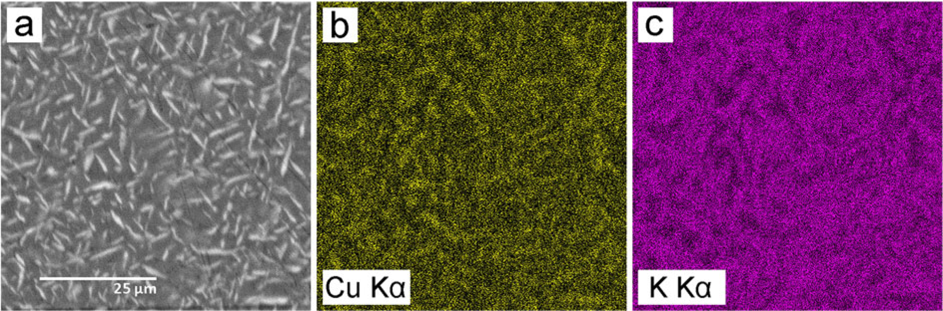
Element-distribution maps of the sulfate material covering lava fragments with marinaite. (a) SEM image in BSE; (b and c) copper and potassium distribution.

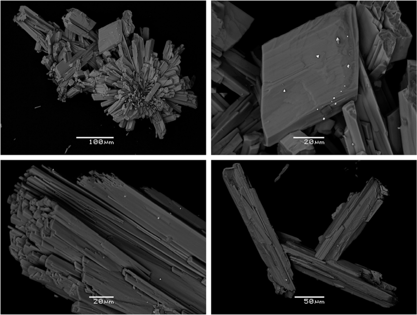
Crystals and aggregates of marinaite. White triangular spots are minute crystals of native gold. SEM BSE images.

The associated minerals are tenorite and minor hematite, anglesite and native gold. The walls of the fractures and vesicles in lava are in some places completely covered by crusts (to several mm thick) or films of whitish or pale-green sulfate material. Marinaite crystals form clusters on sulfate crusts (Fig. 1b). Marinaite most probably crystallized after the deposition of sulfates, although individual marinaite crystals are also found within the sulfate material. The composition of the sulfate matrix was studied in a polished sample on scanning electron microscope (SEM) equipped with energy dispersive X-ray spectroscopy (EDS). The average composition of the sulfate matrix is close to the aphthitalite-group mineral belomarinaite, ideally KNaSO4 (Filatov et al., Reference Filatov, Shablinskii, Vergasova, Saprikina, Bubnova, Moskaleva and Belousov2019; Shchipalkina et al., Reference Shchipalkina, Pekov, Chukanov, Belakovskiy, Zubkova, Koshlyakova, Britvin and Sidorov2020), with a CuO content ranging from 3 to 9 wt.% and averaging ∼5 wt.%. However, this sulfate substance turned out to be inhomogeneous. Under the scanning electron microscope at higher magnifications, exsolution structures are observed (Fig. 2a). Elemental mapping showed that the lamellae of a mineral which showed brighter in back-scattered electrons (BSE) are enriched in copper and depleted in potassium (Fig. 2b, c). A possible explanation involves decomposition of a complex sulfate upon cooling that led to the formation of pure belomarinaite KNaSO4 and Na-K-Cu sulfate. The exsolution pattern is very similar to ones described for aphthitalite-group sulfates from fumaroles of the Northern Breakthrough of the Great Tolbachik Fissure Eruption by Shchipalkina et al. (Reference Shchipalkina, Pekov, Britvin, Koshlyakova and Sidorov2021). The sample contains tenorite, which forms clusters of well-faceted black plate-like crystals up to 0.5 mm in size.
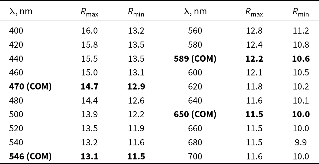
Reflectance values (R, %) of marinaite

Table 1 Long description
Reflectance values are listed as Rmax and Rmin in percent at wavelengths labeled lambda in nanometers, shown in two side by side sets of columns. Wavelengths run from 400 to 700 nm, including entries marked 470 COM, 546 COM, 589 COM, and 650 COM. At 420 nm, Rmax is 15.8 and Rmin is 13.5; at 440 nm, Rmax is 15.5 and Rmin is 13.5; at 460 nm, Rmax is 15.0 and Rmin is 13.1; at 480 nm, Rmax is 14.4 and Rmin is 12.6; at 500 nm, Rmax is 13.9 and Rmin is 12.2; at 520 nm, Rmax is 13.5 and Rmin is 11.9; at 540 nm, Rmax is 13.2 and Rmin is 11.6; at 560 nm, Rmax is 12.8 and Rmin is 11.2; at 580 nm, Rmax is 12.4 and Rmin is 10.8; at 600 nm, Rmax is 12.1 and Rmin is 10.5; at 620 nm, Rmax is 11.8 and Rmin is 10.2; at 640 nm, Rmax is 11.6 and Rmin is 10.1; at 660 nm, Rmax is 11.5 and Rmin is 10.0; at 680 nm, Rmax is 11.5 and Rmin is 9.9. At 700 nm, Rmax is 11.6 and Rmin is 10.0.
Note: The values required by the Commission in Ore Mineralogy ere given in bold.
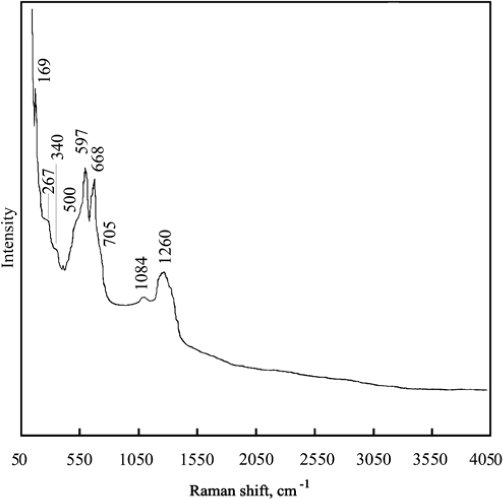
The Raman spectrum of marinaite. Labelled peaks are at 169, 267, 340, 500, 597, 668, 705, 1084 and 1260.

General appearance, physical properties and optical data
Marinaite forms prismatic, elongated parallel to [001], lath-like to tabular or lamellar, and acicular crystals up to 0.06 x 0.06 x 0.3 mm. Crystals are separate or combined in parallel, sheaf-like, radiating or chaotic aggregates up to 3 mm across on the surface of fresh basaltic lava covered by a sulfate encrustation of 0.5–1 mm thick. Some marinaite crystals are skeletal, box-like, or slightly divergent (Fig. 3). Twinning is not observed. Colour is dark-brown to black, the thinnest needles are brown to golden-brown. Streak is yellow to light-brown and the lustre is submetallic. Marinaite is opaque in the thickest crystals and translucent in thin lamellae or needles. It is non-fluorescent.
The micro-indentation hardness range is 670–965, with a mean of 839 kg mm–2 (VHN load 20 g). Marinaite is brittle. Cleavage is not observed. Fracture is splintery. Density could not be measured because of the small grain size. The calculated density based on the single-crystal XRD data and the empirical formula Cu2.07Fe0.56Al0.27Ti0.10Si0.01(B0.98O3)O2 is 5.026 g·cm–3.
Under the microscope in reflected light marinaite is grey with distinct reddish-brown internal reflections. Bireflectance is distinct with ∆R = 1.6% (589 nm) (Table 1). Anisotropism is moderate. The reflectance values measured in air using the Si standard are given in Table 1.
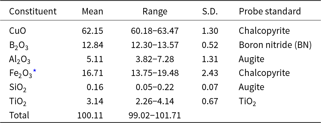
Chemical composition of marinaite (wt.%)

Table 2 Long description
Chemical constituents are reported in weight percent with columns for Mean, Range, S. D., and Probe standard. CuO has mean 62.15, range 60.18 to 63.47, S. D. 1.30, probe standard Chalcopyrite. B2O3 has mean 12.84, range 12.30 to 13.57, S. D. 0.52, probe standard Boron nitride, BN. Al2O3 has mean 5.11, range 3.82 to 7.28, S. D. 1.31, probe standard Augite. Fe2O3* has mean 16.71, range 13.75 to 19.48, S. D. 2.43, probe standard Chalcopyrite. SiO2 has mean 0.16, range 0.05 to 0.22, S. D. 0.07, probe standard Augite. TiO2 has mean 3.14, range 2.26 to 4.14, S. D. 0.67, probe standard TiO2. Total has mean 100.11, range 99.02 to 101.71, with S. D. and probe standard not shown.
* Fe is calculated as Fe3+ in accordance with the structure data. S.D. – standard deviation.
Raman spectroscopy
The Raman spectrum of marinaite (Fig. 4) was obtained at the Dept. of Mineralogy, Lomonosov Moscow State University, using an EnSpectr R532 spectrophotometer with a green laser (532 nm) at room temperature. The output power of the laser beam was ∼10 MW. The spectrum was processed using the EnSpectr expert mode program in the range from 100 to 4000 cm–1 with the use of a holographic diffraction grating with 1800 mm–1 and a resolution equal to 6 cm–1. The diameter of the laser spot on the sample was ∼10 μm. The spectrum was obtained from a randomly orientated crystal.
The bands within the region 1000–1400 cm–1 correspond to stretching vibrations of triangular BO33– anions (Nakamoto, Reference Nakamoto1986). The mode at 1260 cm–1 represents a doubly degenerate in-plane stretching vibration of B–O bonds. For the planar triangular group BO3 with symmetry D 3h this mode is designated as E′(ν3). The band with maximum at 1084 cm–1 corresponds to the non-degenerate symmetric stretching vibration of B–O bonds – mode A 1′(ν1) of the triangle. Two bands at 597 and 668 cm–1 belong to the split of the doubly degenerate in-plane B–O–B bending vibrations of BO3 groups – mode E′(ν4) of the triangle. Taking into account the chemical composition of the mineral, the shoulders at 500 and 705 cm–1 most likely correspond to Fe3+–O and Al–O stretching vibrations in octahedra. Bands with frequencies lower than 400 cm–1 correspond to Cu–O stretching vibrations and lattice modes. The absence of bands with frequencies higher than 1400 cm–1 indicates the absence of groups with O–H, C–H, C–O, N–H and N–O bonds in marinaite.
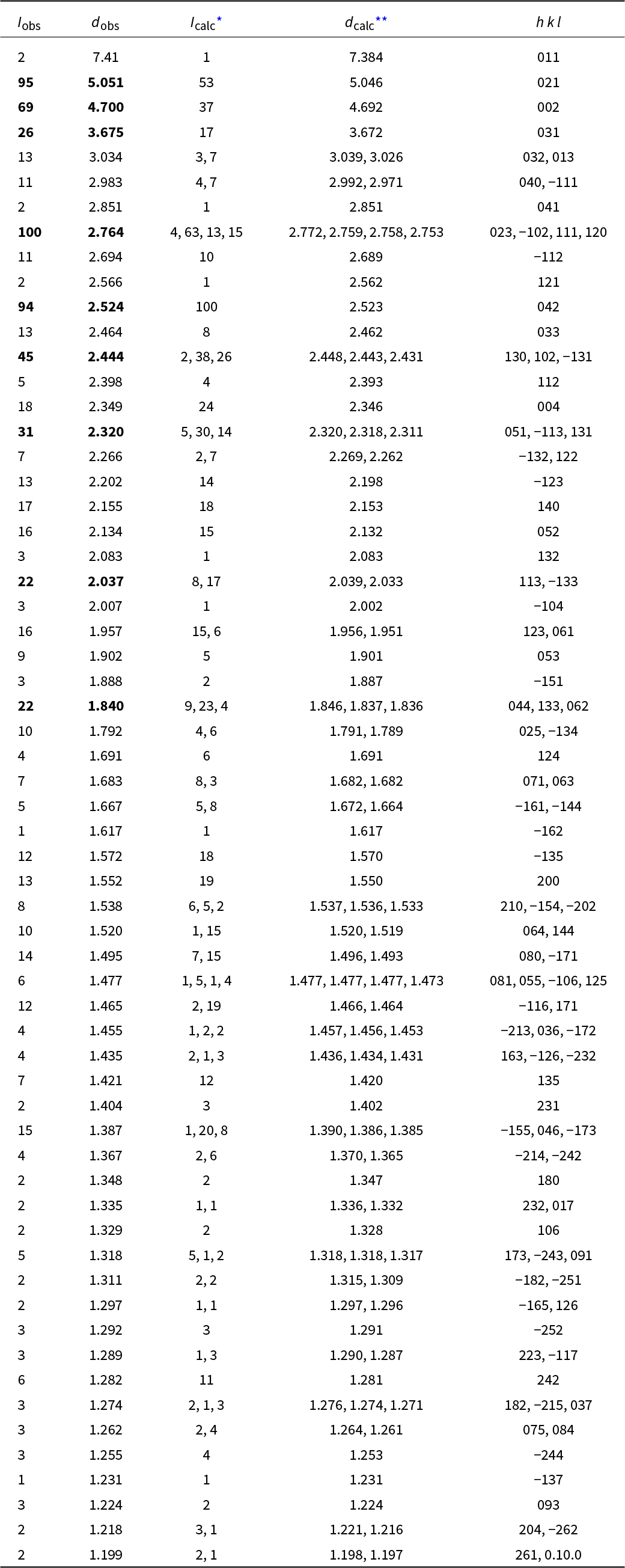
Powder X-ray diffraction data (d in Å) of marinaite

Table 3 Long description
Powder X ray diffraction reflections are listed with observed intensity, observed d spacing in angstroms, calculated intensity, calculated d spacing, and h k l indices. Observed intensities range from 1 to 100, with dobs values from 7.41 down to 1.199 angstroms. The highest Iobs is 100 at dobs 2.764 angstroms, paired with Icalc values 4, 63, 13, and 15 at dcalc 2.772, 2.759, 2.758, and 2.753 angstroms for h k l 023, minus 102, 111, and 120. Other prominent observed peaks include Iobs 95 at dobs 5.051 angstroms with Icalc 53 at dcalc 5.046 for h k l 021, Iobs 94 at dobs 2.524 angstroms with Icalc 100 at dcalc 2.523 for h k l 042, and Iobs 69 at dobs 4.700 angstroms with Icalc 37 at dcalc 4.692 for h k l 002. Several rows list multiple calculated reflections for one observed line, for example dobs 3.034 angstroms with Icalc 3 and 7 at dcalc 3.039 and 3.026 for h k l 032 and 013, and dobs 2.444 angstroms with Icalc 2, 38, and 26 at dcalc 2.448, 2.443, and 2.431 for h k l 130, 102, and minus 131. Many remaining entries have low observed intensities between 2 and 22 across dobs values from about 2.7 to 1.2 angstroms, each paired with one or more calculated intensities and corresponding h k l indices.
* for the calculated pattern, only reflections with intensities ≥1 are given;
** for the unit-cell parameters calculated from single-crystal data.
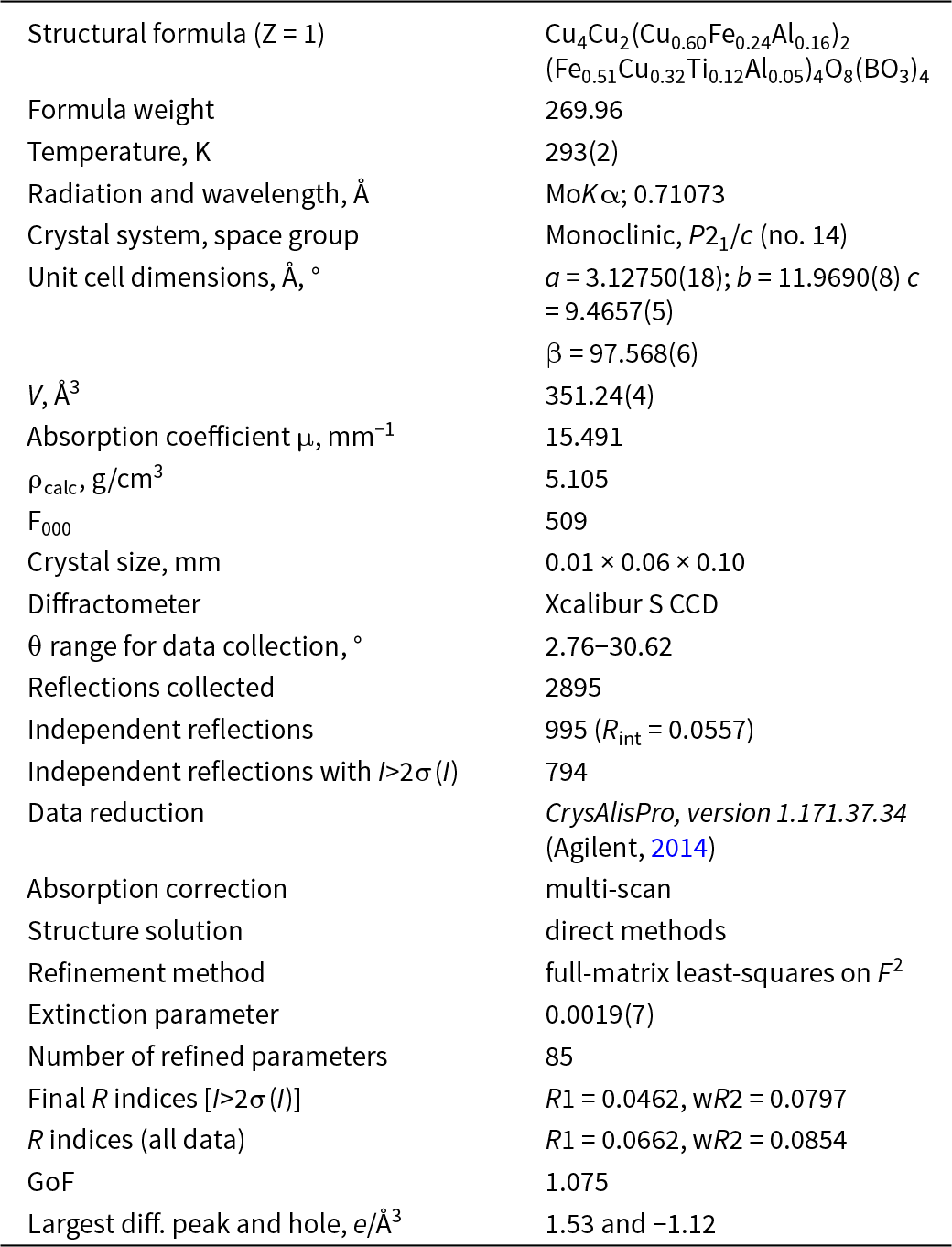
Crystal data, data collection information and structure refinement details

Table 4 Long description
Crystal data and refinement details are listed for a copper, iron, aluminum, titanium borate oxide with a structural formula given for Z 1 and a formula weight of 269.96. Measurement temperature is 293 with an uncertainty of 2 K, using MoK alpha radiation with wavelength 0.71073 angstrom. Crystal system and space group are monoclinic, P21/c, number 14, with unit cell parameters a 3.12750 with uncertainty 0.00018 angstrom, b 11.9690 with uncertainty 0.0008 angstrom, c 9.4657 with uncertainty 0.0005 angstrom, and beta 97.568 with uncertainty 0.006 degrees; cell volume is 351.24 with uncertainty 0.04 cubic angstrom. Absorption coefficient mu is 15.491 per millimeter, calculated density is 5.105 grams per cubic centimeter, and F000 is 509; crystal size is 0.01 by 0.06 by 0.10 millimeters. Data were collected on an Xcalibur S CCD diffractometer over a sigma range from 2.76 to 30.62 degrees, with 2895 reflections collected and 995 independent reflections with Rint 0.0557; independent reflections with intensity greater than two sigma of intensity are 794. Data reduction used CrysAlisPro version 1.171.37.34, absorption correction is multi scan, structure solution used direct methods, and refinement used full matrix least squares on F squared with an extinction parameter of 0.0019 with uncertainty 0.0007 and 85 refined parameters. Final R indices for data above two sigma are R1 0.0462 and wR2 0.0797, and for all data are R1 0.0662 and wR2 0.0854; goodness of fit is 1.075, and the largest difference peak and hole are 1.53 and minus 1.12 electrons per cubic angstrom.
Chemical data
The preliminary study of marinaite was performed at IGEM RAS on a JEOL JXA-8200 electron probe microanalyser equipped with one energy dispersive (ED) and five wavelength dispersive (WD) X-ray spectrometers. Totals for the preliminary analyses were significantly less than 100 wt.%, which indicated the possible presence of light elements. Electron microprobe analyses including boron determination (5) were carried out with a Camebax SX100 microanalyser with four vertical spectrometers (WDS mode, 15 kV, 40 nA, 2 μm beam diameter) at Vernadsky Institute of Geochemistry and Analytical Chemistry RAS (analyst N.N. Kononkova). Boron was analysed using a PC2 crystal. Accumulation of intensity for boron was 40 sec. Chemical composition of marinaite is given in Table 2. The empirical formula calculated based on 5 O atoms per formula unit (apfu) is Cu2.07Fe0.56Al0.27Ti0.10Si0.01B0.98O5. The simplified formula is Cu2(Fe3+,Al,Ti,Cu)O2(BO3). The idealized, end-member formula is Cu2Fe3+O2(BO3), which requires (wt.%) 58.11 CuO, 29.17 Fe2O3, 12.72 B2O3 at a total 100.
X-ray crystallography and crystal structure
Powder X-ray diffraction (XRD) data for marinaite (Table 3) were collected with a Rigaku R-AXIS Rapid II diffractometer equipped with cylindrical image plate detector (radius 127.4 mm) using Debye-Scherrer geometry, CoKα radiation (rotating anode with VariMAX microfocus optics), λ = 1.7892 Å, 40 kV, 15 mA, and exposure 12 min. The angular resolution of the detector is 0.045°2θ (pixel size 0.1 mm). The data were integrated using the software package Osc2Tab (Britvin et al., Reference Britvin, Dolivo-Dobrovolsky and Krzhizhanovskaya2017). The monoclinic unit cell parameters calculated from the powder XRD data are a = 3.131(1), b = 11.972(2), c = 9.466(2) Å, β = 97.71(2)°, V = 351.6(2) Å3.
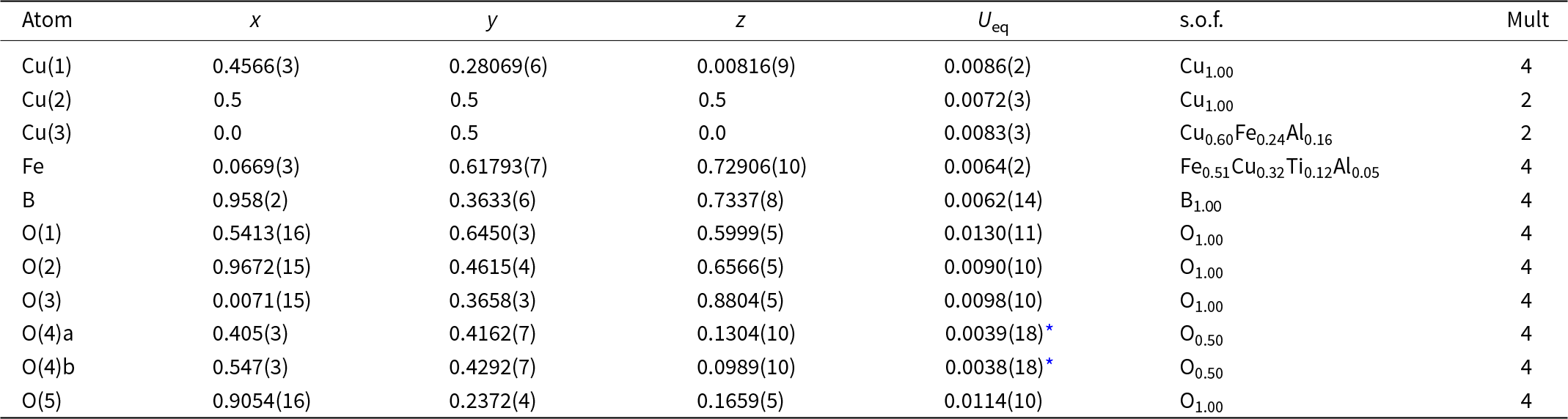
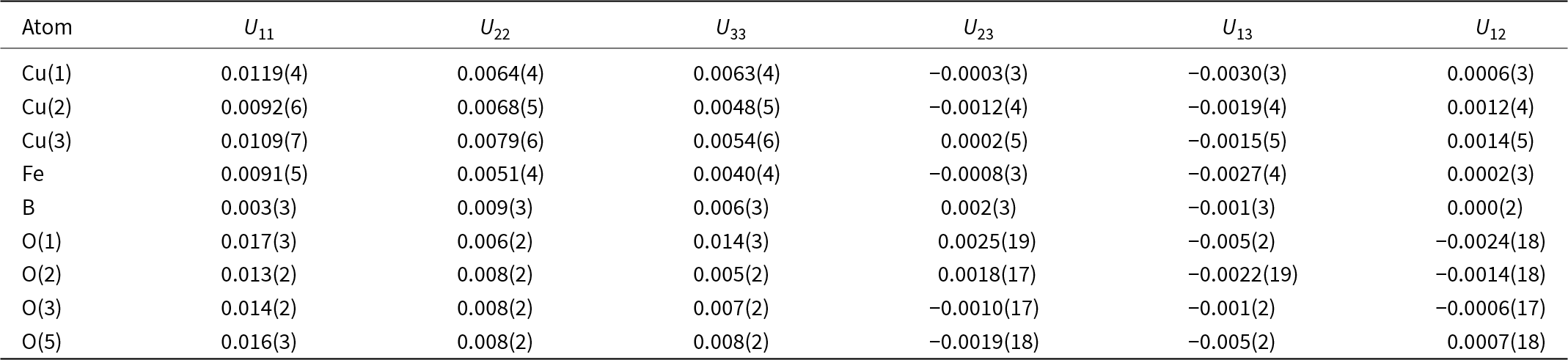
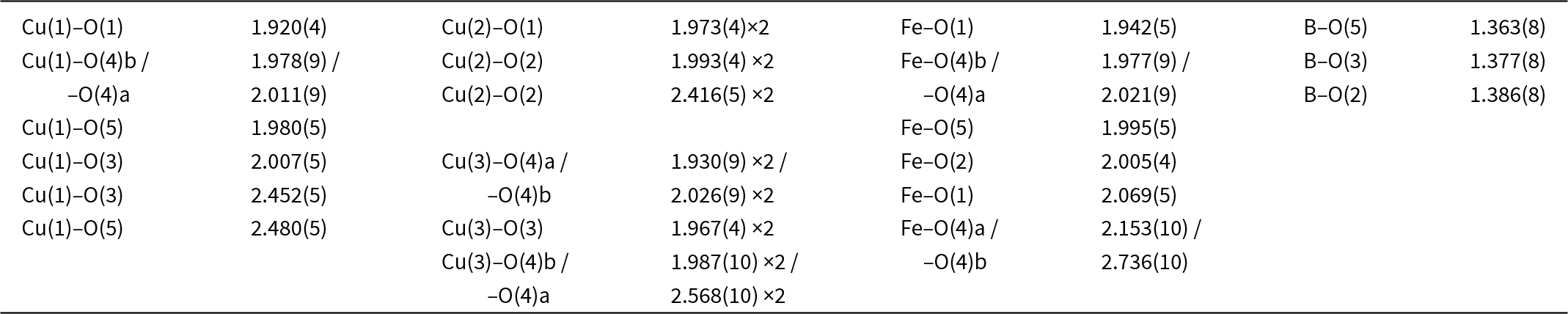
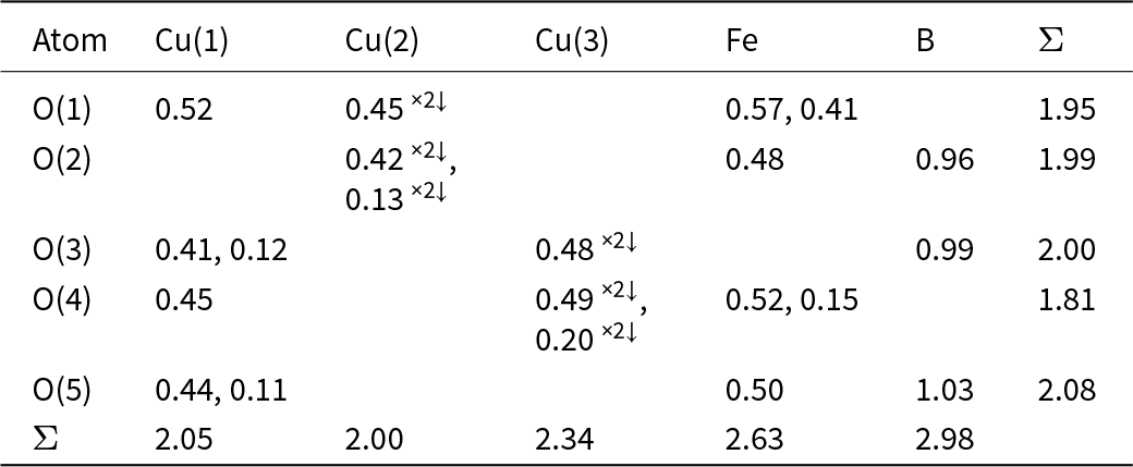
Single-crystal XRD studies of marinaite were carried out at room temperature using an Xcalibur S diffractometer equipped with a CCD detector (MoKα-radiation, λ = 0.71073 Å). More than a hemisphere of three-dimensional data was collected. Data reduction was performed using CrysAlisPro Version 1.171.37.34 (Agilent, 2014). The data were corrected for Lorentz factor and polarization effects. The crystal structure was solved by direct methods and refined using the SHELX software package (Sheldrick, Reference Sheldrick2015) to R = 0.0462 for 794 independent reflections with I > 2σ(I). The crystal data and the experimental details are presented in Table 4, atom coordinates and thermal displacement parameters in Table 5, anisotropic displacement parameters (Å2) of atoms in Table 6, selected interatomic distances in Table 7 and bond valence calculations in Table 8. The crystallographic information file has been deposited and is available as Supplementary material (see below).
Atom coordinates (x,y,z), equivalent thermal displacement parameters (U eq, Å2), site occupancies (s.o.f.) and site multiplicities (Mult) for marinaite

Table 5 Long description
Columns list Atom, x, y, z, Ueq, s.o.f., and Mult for Cu, Fe, B, and O sites. Cu(1) has x 0.4566(3), y 0.28069(6), z 0.00816(9), Ueq 0.0086(2), s.o.f. Cu1.00, Mult 4. Cu(2) is at x 0.5, y 0.5, z 0.5, Ueq 0.0072(3), s.o.f. Cu1.00, Mult 2. Cu(3) is at x 0.0, y 0.5, z 0.0, Ueq 0.0083(3), s.o.f. Cu0.60Fe0.24Al0.16, Mult 2. Fe is at x 0.0669(3), y 0.61793(7), z 0.72906(10), Ueq 0.0064(2), s.o.f. Fe0.51Cu0.32Ti0.12Al0.05, Mult 4. B is at x 0.958(2), y 0.3633(6), z 0.7337(8), Ueq 0.0062(14), s.o.f. B1.00, Mult 4. Oxygen sites O(1), O(2), O(3), and O(5) each have s.o.f. O1.00 and Mult 4, with Ueq values 0.0130(11), 0.0090(10), 0.0098(10), and 0.0114(10), respectively. O(4)a has x 0.405(3), y 0.4162(7), z 0.1304(10), Ueq 0.0039(18) with an asterisk, s.o.f. O0.50, Mult 4, and O(4)b has x 0.547(3), y 0.4292(7), z 0.0989(10), Ueq 0.0038(18) with an asterisk, s.o.f. O0.50, Mult 4.
* U iso
Anisotropic displacement parameters (Å2) of atoms in marinaite

Table 6 Long description
Atoms Cu(1), Cu(2), Cu(3), Fe, B, O(1), O(2), O(3), and O(5) are listed with anisotropic displacement parameters in angstrom squared for columns, U11, U22, U33, U23, U13, and U12. For Cu(1), values are U11 0.0119(4), U22 0.0064(4), U33 0.0063(4), U23 minus 0.0003(3), U13 minus 0.0030(3), and U12 0.0006(3). For Cu(2), values are U11 0.0092(6), U22 0.0068(5), U33 0.0048(5), U23 minus 0.0012(4), U13 minus 0.0019(4), and U12 0.0012(4). For Cu(3), values are U11 0.0109(7), U22 0.0079(6), U33 0.0054(6), U23 0.0002(5), U13 minus 0.0015(5), and U12 0.0014(5). For Fe, values are U11 0.0091(5), U22 0.0051(4), U33 0.0040(4), U23 minus 0.0008(3), U13 minus 0.0027(4), and U12 0.0002(3). For B, values are U11 0.003(3), U22 0.009(3), U33 0.006(3), U23 0.002(3), U13 minus 0.001(3), and U12 0.000(2). For O(1), values are U11 0.017(3), U22 0.006(2), U33 0.014(3), U23 0.0025(19), U13 minus 0.005(2), and U12 minus 0.0024(18). For O(2), values are U11 0.013(2), U22 0.008(2), U33 0.005(2), U23 0.0018(17), U13 minus 0.0022(19), and U12 minus 0.0014(18). For O(3), values are U11 0.014(2), U22 0.008(2), U33 0.007(2), U23 minus 0.0010(17), U13 minus 0.001(2), and U12 minus 0.0006(17). For O(5), values are U11 0.016(3), U22 0.008(2), U33 0.008(2), U23 minus 0.0019(18), U13 minus 0.005(2), and U12 0.0007(18).
Selected interatomic distances (Å) in the crystal structure of marinaite

Table 7 Long description
Interatomic distances in angstroms are listed for Cu(1), Cu(2), Cu(3), Fe, and B bonded to oxygen atoms labeled O(1) through O(5), with some oxygen labels marked a or b. Cu(1) distances include O(1) 1.920(4), O(4)b 1.978(9), O(4)a 2.011(9), O(5) 1.980(5) and 2.480(5), and O(3) 2.007(5) and 2.452(5). Cu(2) distances include O(1) 1.973(4) with a multiplicity of two, O(2) 1.993(4) with a multiplicity of two, and O(2) 2.416(5) with a multiplicity of two. Cu(3) distances include O(4)a 1.930(9) with a multiplicity of two, O(4)b 2.026(9) with a multiplicity of two, O(3) 1.967(4) with a multiplicity of two, O(4)b 1.987(10) with a multiplicity of two, and O(4)a 2.568(10) with a multiplicity of two. Fe distances include O(1) 1.942(5) and 2.069(5), O(4)b 1.977(9), O(4)a 2.021(9) and 2.153(10), O(5) 1.995(5), O(2) 2.005(4), and O(4)b 2.736(10). B distances include O(5) 1.363(8), O(3) 1.377(8), and O(2) 1.386(8).
Bond valence calculations for marinaite

Table 8 Long description
Bond valence values are listed by oxygen row, O(1) through O(5), across atom columns Cu(1), Cu(2), Cu(3), Fe, B, plus a rightmost column labeled with the sigma symbol for each oxygen total. For O(1), Cu(1) is 0.52, Cu(2) is 0.45 with a times two down arrow marker, Fe has 0.57 and 0.41, and the oxygen total is 1.95. For O(2), Cu(2) has 0.42 with a times two down arrow marker and 0.13 with a times two down arrow marker, Fe is 0.48, B is 0.96, and the oxygen total is 1.99. For O(3), Cu(1) has 0.41 and 0.12, Cu(3) is 0.48 with a times two down arrow marker, B is 0.99, and the oxygen total is 2.00. For O(4), Cu(1) is 0.45, Cu(3) has 0.49 with a times two down arrow marker and 0.20 with a times two down arrow marker, Fe has 0.52 and 0.15, and the oxygen total is 1.81. For O(5), Cu(1) has 0.44 and 0.11, Fe is 0.50, B is 1.03, and the oxygen total is 2.08. The bottom row labeled with the sigma symbol gives column totals of 2.05 for Cu(1), 2.00 for Cu(2), 2.34 for Cu(3), 2.63 for Fe, and 2.98 for B.
Notes: For Cu(3) and Fe positions bond valences were calculated taking into account site occupancy factors, i.e. Cu(3) = Cu0.60Fe0.24Al0.16; Fe = Fe0.51Cu0.32Ti0.12Al0.05. An averaged O(4) site was considered for the calculations. Bond-valence parameters were taken from Gagné and Hawthorne (Reference Gagné and Hawthorne2015).
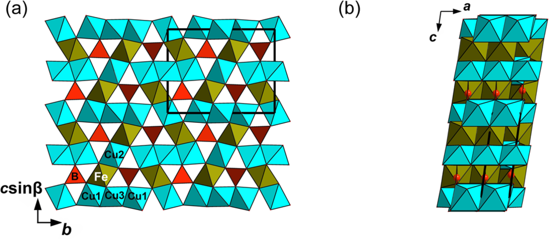
The crystal structure of marinaite projected along the a (a) and b (b) axes. The unit cell is outlined. Drawn using Diamond, Version 3.2k.

Figure 5 Long description
Panel (a) illustrates the crystal structure of marinaite projected along the a axis, featuring a network of polyhedra. The unit cell is outlined and cations are labeled as Cu1, Cu2, Cu3, Cu4 and Fe, positioned within the polyhedra. Arrows indicate the directions of the b axis and c sin beta. Panel (b) shows the structure projected along the b axis, emphasizing stacked layers with arrows marking the a and c axes. The arrangement highlights atomic connectivity and cation sites in two crystallographic views. No scale bar or numeric measurements are present. The diagram′s purpose is to illustrate the spatial arrangement and connectivity of cations within the marinaite structure.
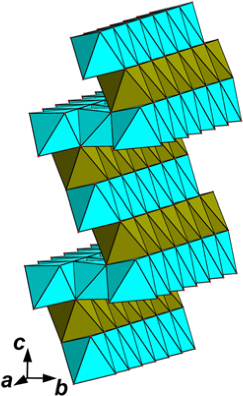
Octahedral sheets in the structure of marinaite. For legend see Fig. 5.

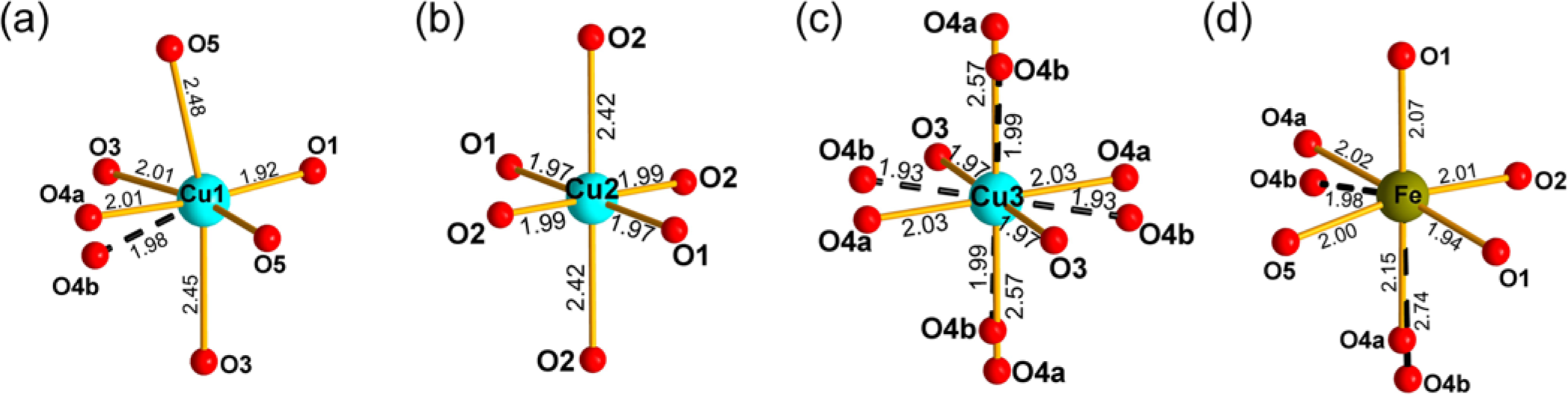
Octahedrally coordinated Cu(1–3) (a, b and c, correspondingly) and Fe (d) sites. Bond lengths are given in Å. Bonds to the O4b subsite are given as black dashed lines.

Figure 7 Long description
An octahedrally coordinated Cu site labeled Cu1 at the center, surrounded by six oxygen atoms labeled O1 to O5 and O4b. Bond lengths are indicated, with Cu1 to O1 measuring 2.01 angstroms, Cu1 to O2 measuring 2.42 angstroms, Cu1 to O3 measuring 2.01 angstroms, Cu1 to O4b measuring 2.45 angstroms, Cu1 to O5 measuring 1.92 angstroms and Cu1 to O4 measuring 2.01 angstroms. The image B showing another octahedrally coordinated Cu site labeled Cu2 at the center, surrounded by six oxygen atoms labeled O1 to O5 and O4b. Bond lengths are indicated, with Cu2 to O1 measuring 1.99 angstroms, Cu2 to O2 measuring 2.42 angstroms, Cu2 to O3 measuring 1.97 angstroms, Cu2 to O4b measuring 2.42 angstroms, Cu2 to O5 measuring 1.97 angstroms and Cu2 to O4 measuring 1.99 angstroms. The image C showing an octahedrally coordinated Cu site labeled Cu3 at the center, surrounded by six oxygen atoms labeled O1 to O5 and O4b. Bond lengths are indicated, with Cu3 to O1 measuring 2.03 angstroms, Cu3 to O2 measuring 2.57 angstroms, Cu3 to O3 measuring 1.98 angstroms, Cu3 to O4b measuring 2.03 angstroms, Cu3 to O5 measuring 1.97 angstroms and Cu3 to O4a measuring 1.98 angstroms. The image D showing an octahedrally coordinated Fe site labeled Fe at the center, surrounded by six oxygen atoms labeled O1 to O5 and O4b. Bond lengths are indicated, with Fe to O1 measuring 2.07 angstroms, Fe to O2 measuring 2.15 angstroms, Fe to O3 measuring 1.94 angstroms, Fe to O4b measuring 2.74 angstroms, Fe to O5 measuring 2.07 angstroms and Fe to O4a measuring 2.15 angstroms.
Marinaite is isostructural with synthetic Cu2Fe3+O2(BO3), Cu2GaO2(BO3) (Schäfer and Bluhm, Reference Schäfer and Bluhm1995) and Cu2AlO2(BO3) (Hriljac et al., Reference Hriljac, Brown, Cheetham and Satek1990), which crystallize as a monoclinically distorted ludwigite-like structure. The lowering of symmetry from orthorhombic, which is characteristic of ludwigite, Mg2FeO2(BO3), to monoclinic is caused by the Jahn-Teller distortion of Cu2+-centred octahedra. The oxygen atom O(4) was characterized by high values of U 11 (0.071(5) Å2) and U 33 (0.034(4) Å2) that indicated possible splitting of the O(4) site. The same situation was described in the structures of all known Cu-bearing synthetic ludwigite-type compounds (Hriljac et al., Reference Hriljac, Brown, Cheetham and Satek1990; Schäfer and Bluhm, Reference Schäfer and Bluhm1995). An attempt to model this splitting was successful, and the O(4) site was split into two subsites 0.59 Å apart. For better clarity for Figs 5 and 6 and bond-valence calculations we report the data for the averaged O(4) site. The crystal structure of marinaite is shown in Fig. 5. Three of four crystallographically independent metal cation sites are Cu-dominant. Cu(1) and Cu(2) cations occupy [4+2] octahedra significantly distorted by the Jahn-Teller effect: four short Cu–O distances that range from 1.920(4) to 2.011(9) Å [Cu(1)] and from 1.973(4) to 1.993(4) Å [Cu(2)] while elongated Cu–O distances are 2.452(5) and 2.480(5) Å for Cu(1) and two equal to 2.416(5) Å distances for Cu(2). Taking into account the splitting of O(4) into two subsites, the Cu(3) site is at the centre of the octahedron with Jahn-Teller distortion in one case, with four short Cu(3)–O bonds and two elongated bonds with the distance 2.568(10) Å and close to a regular octahedron in the other (Table 7). This fact together with electron microprobe data, refined numbers of electrons, and bond valence sums (BVS), and taking into account the analogy with isostructural synthetic Cu2Fe3+O2(BO3), characterized by partial cation ordering [the structural formula (Z = 1) is Cu4Cu2(Cu0.6Fe0.4)2(Fe3+0.8Cu0.2)4O8(BO3)4: Schäfer and Bluhm, Reference Schäfer and Bluhm1995), demonstrate that the Cu(3) site is Cu-dominant with significant admixtures of Fe3+ and Al3+ (Table 5). An Fe-centred polyhedron can be considered as square pyramid with five Fe–O distances varying from 1.942(5) to 2.069(5) Å or as a distorted octahedron with one strongly elongated bond with the distance 2.736(10) Å with O(4)b or as an octahedron with a slightly elongated distance of 2.153(10) with O(4)a. On the basis of the same reasons as for the Cu(3) site, the Fe site was considered as Fe3+-dominant with the admixtures of Cu, Ti and Al. The Fe–O distances (Table 7), as well as BVS (Table 8) undoubtedly demonstrate the trivalent state of iron that is consistent with strongly oxidizing conditions necessary for crystallization of marinaite. We should note that very close cation distribution and the split character of the O(4) site were recently reported for the synthetic monoclinic ludwigite-type oxoborate Cu2FeO2(BO3) (Gokhfeld et al., Reference Gokhfeld, Kazak, Bel’skaya, Molokeev, Gudim, Kondratiev, Eremin, Knyazev, Velikanov and Ovchinnikov2023; Biryukov et al., Reference Biryukov, Bubnova, Shablinskii, Gokhfeld, Kazak, Avdontceva, Krzhizhanovskaya and Vereshchagin2025). Figure 7 demonstrates four octahedrally coordinated sites in the structure of marinaite. The structural formula of marinaite is (Z = 1): Cu(1)Cu4Cu(2)Cu2Cu(3)(Cu0.60Fe3+0.24Al0.16)2Fe(Fe3+0.51Cu0.32Ti0.12Al0.05)4O8(BO3)4.
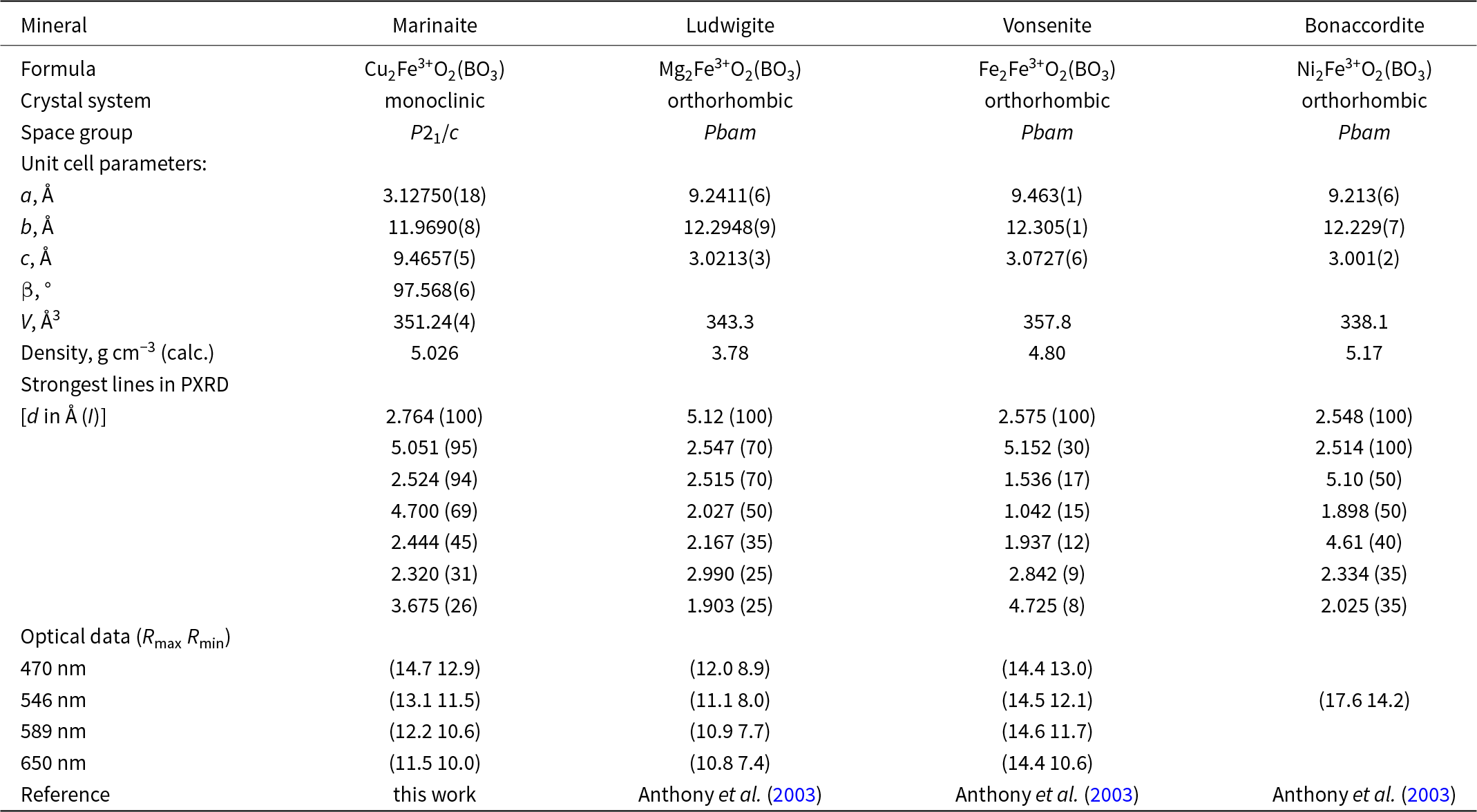
Comparative data for marinaite and the related minerals ludwigite, vonsenite and bonaccordite – ludwigite-group members with species-defining Fe3+

Table 9 Long description
Four minerals are compared across formula, crystal system, space group, unit cell parameters, calculated density, strongest lines in PXRD, and optical data at selected wavelengths. Formulas are Cu2Fe3+O2(BO3) for marinaite, Mg2Fe3+O2(BO3) for ludwigite, Fe2Fe3+O2(BO3) for vonsenite, and Ni2Fe3+O2(BO3) for bonaccordite. Unit cell parameters list a, b, c, beta, and V: marinaite has a 3.12750(18) angstrom, b 11.9690(8) angstrom, c 9.4657(5) angstrom, beta 97.568(6) degrees, and V 351.24(4) angstrom cubed; ludwigite has a 9.2411(6), b 12.2948(9), c 3.0213(3), and V 343.3; vonsenite has a 9.463(1), b 12.305(1), c 3.0727(6), and V 357.8; bonaccordite has a 9.213(6), b 12.229(7), c 3.001(2), and V 338.1. Calculated densities in grams per cubic centimeter are 5.026 for marinaite, 3.78 for ludwigite, 4.80 for vonsenite, and 5.17 for bonaccordite. Strongest lines in PXRD are given as d in angstrom with intensity in parentheses: marinaite lists 2.764 (100), 5.051 (95), 2.524 (94), 4.700 (69), 2.444 (45), 2.320 (31), and 3.675 (26); ludwigite lists 5.12 (100), 2.547 (70), 2.515 (70), 2.027 (50), 2.167 (35), 2.990 (25), and 1.903 (25). Vonsenite lists 2.575 (100), 5.152 (30), 1.536 (17), 1.042 (15), 1.937 (12), 2.842 (9), and 4.725 (8); bonaccordite lists 2.548 (100), 2.514 (100), 5.10 (50), 1.898 (50), 4.61 (40), 2.334 (35), and 2.025 (35). Optical data labeled Rmax Rmin are provided at 470, 546, 589, and 650 nanometers for marinaite, ludwigite, and vonsenite, while bonaccordite has values only at 546 nanometers, 17.6 and 14.2.
Like ludwigite (Takeuchi et al., Reference Takeuchi, Watanabe and Ito1950), the crystal structure of marinaite is built by dense zig-zag sheets, or ‘walls’ of edge-sharing octahedra (Fig. 6). Neighbouring sheets are connected via common vertices of the octahedra to form triangular tunnels containing BO33– triangle anionic groups.
Relation to other species
Marinaite is structurally related to orthorhombic oxoborates belonging to the ludwigite and the orthopinakiolite groups that have the same type of formula M(1)2M(2)O2(BO3) and similar unit cell dimensions. Pinakiolite-group oxoborates with the same general formula are characterized by different configuration of monoclinic unit cell (Table 9). Unlike all members of these three groups, marinaite contains species-defining Cu2+ that causes its structural difference from related minerals. We propose including marinaite into the ludwigite group as its first monoclinic member, a mineral with monoclinically distorted ludwigite-type structure. This mineral group also includes, besides the members with species-defining M 3+ = Fe3+ listed in Table 9, azoproite, ideally Mg2(Mg0.5Ti0.5)O2(BO3) (Biryukov et al., Reference Biryukov, Zinnatullin, Levashova, Shablinskii, Cherosov, Bubnova, Vagizov, Krzhizhanovskaya, Filatov, Shilovskikh and Pekov2022), fredrikssonite, ideally Mg2Mn3+O2(BO3) (Dunn et al., Reference Dunn, Peacor, Simmons and Newbury1983), and savelievaite, ideally Mg2CrO2(BO3) (Pekov et al., Reference Pekov, Vakhrusheva, Zubkova, Yapaskurt, Shelukhina, Erokhin, Bulakh, Britvin, Kasatkin, Turchkova and Pushcharovsky2024).
The Cu-rich end-member of marinaite is a natural analogue of synthetic Cu2Fe3+O2(BO3) (Schäfer and Bluhm, Reference Schäfer and Bluhm1995).
Genesis of marinaite
Marinaite was discovered inside a hot degassing crack on the cooling lava field several months after the eruption terminated. The surface of the lava field was cold, but some cracks were still hot and degassing in many places, indicating elevated temperatures of the lava flow interior. Temperature, being still high due to the heat capacity of lavas, was probably further increased and maintained by secondary oxidative processes. The oxidation was caused by air entraining into the lava flows, as evidenced by the noticeable air suction at the entrances of some lava tubes.
All major elements included in marinaite show strong enrichment in high-temperature condensates, which were sampled during the 2012–2013 Tolbachik eruption from a window into a tube with flowing lava (1030°C) and a fissure on the frozen lava field (690°C). The condensates contain, 9.7 and 1180 ppm Cu, 18.7 and 1.2 ppm Fe, 109 and 25 ppm B respectively (Chaplygin et al., Reference Chaplygin, Lavrushin, Dubinina, Bychkova, Inguaggiato and Yudovskaya2016a). Given the similar temperature of up to 600°C in the fissure at the marinaite sampling point we argue that the gaseous fluid from degassing lava was the source of copper, iron and boron in the volcanic exhalations. We suggest that marinaite formed as a result of a gas transport reaction, involving boric acid, Cu and Fe chlorides, and water vapour:
 \begin{align*}& {\mathrm {H_3BO_{3{(g)}}}} + {\mathrm {2CuCl_{2{(g)}}}} + {\mathrm {FeCl_{3{(g)}}}} + {\mathrm {2H_2O_{(g)}}}\\ & \quad\qquad\quad\ = {\mathrm {Cu_2FeO_2(BO_3)_{(s)}}} + {\mathrm {7HCl_{(g)}}}\end{align*}
where g – gas, s – solid.
As marinaite is considered to crystallize from a gas phase originated from cooling and degassing lavas, it can be classified, along with fluoborite, ludwigite, yuanfuliite, fredrikssonite (Bulakh et al., Reference Bulakh, Pekov, Chukanov, Yapaskurt, Koshlyakova, Britvin and Sidorov2022a, Reference Bulakh, Pekov, Koshlyakova and Sidorov2022b, Reference Bulakh, Britvin, Zubkova, Yapaskurt, Agakhanov, Anikin and Pekov2025), chubarovite and rhabdoborites (Pekov et al., Reference Pekov, Zubkova, Pautov, Yapaskurt, Chukanov, Lykova, Britvin, Sidorov and Pushcharovsky2015, Reference Pekov, Zubkova, Koshlyakova, Belakovskiy, Agakhanov, Vigasina, Britvin, Sidorov and Pushcharovsky2020b), as a borate of fumarolic sensu stricto, i.e. sublimate genesis. Ludwigite, fredrikssonite, yuanfuliite and rhabdoborites were found at the Second scoria cone of the Northern Breakthrough of the Great Tolbachik Fissure Eruption in the lower part of the section at a depth of ∼2 metres in the anhydrite zone lacking copper minerals. Above this zone the arsenate and sulfate zones rich in copper mineralization are located (Pekov et al., Reference Pekov, Koshlyakova, Zubkova, Lykova, Britvin, Yapaskurt, Agakhanov, Shchipalkina, Turchkova and Sidorov2018). The gas flow and temperature gradient at the Second scoria cone are mild to barely noticeable, thus leading to the formation of the broad mineral zoning and separate deposition of B and Cu. In the degassing fissures on the cooling lava field, the gas temperature is comparable to that observed in the fumarole fields of the Northern Breakthrough of the Great Tolbachik Fissue Eruption (up to 500°C; Pekov et al., Reference Pekov, Agakhanov, Zubkova, Koshlyakova, Shchipalkina, Sandalov, Yapaskurt, Turchkova and Sidorov2020a), but the gas flow is concentrated (clearly noticeable) and the thermal gradient is obviously higher. This generally leads to the formation of a narrower mineral zonation, hindering separation of copper and boron, and facilitating the crystallization of marinaite. Fumarolic mineralization contains many water-soluble minerals, which could be dissolved as the conditions change. Fumarolic mineral associations of the Second scoria cone have been formed and preserved over more than 50 years due to the steady heat flow that dries the overlying cinder crust. At the same time active lava fields cool more rapidly with their ephemeral mineralization having a much shorter life span.
Crystals of ludwigite-like compounds are commonly grown for laboratory use by flux synthesis (e.g. Moshkina et al., Reference Moshkina, Belskaya, Molokeev, Bovina, Shabanova, Kokh, Seretkin, Velikanov, Eremin, Krylov and Bezmaternykh2023). The discovery of fumarolic borates suggests an alternative way of producing borate crystals by chemical vapour deposition (CVD) reactions.
Supplementary material
The supplementary material for this article can be found at https://doi.org/10.1180/mgm.2025.10179.
Acknowledgements
We are grateful to our colleagues from the Institute of Volcanology and Seismology FEB RAS Alexander Belousov and Marina Belousova for the invitation to join the fieldwork at the new Tolbachik lava fields in 2013 and 2014. We thank Edward Grew and the anonymous reviewers for their suggestions that greatly improved the paper. This study was partly performed at IGEM RAS under the state assignment. Lidiya Levitskaya of IGEM RAS performed SEM images and EDS analyses. The crystal chemical and Raman spectroscopy studies were conducted under the state assignment of Lomonosov Moscow State University (state task No. 121061600049-4). The powder XRD investigation was carried out at the Centre for X-Ray Diffraction Research of the Science Park of St. Petersburg State University within the framework of project 125021702335-5.
Competing interests
The authors declare none.