Introduction
Emotion dysregulation, which is characterized by abnormalities in emotional sensitivity and abnormalities in emotion regulation (Crowell et al. Reference Crowell, Beauchaine and Linehan2009), has been suggested to be a core feature of borderline personality disorder (BPD), accounting for most if not all, symptoms of BPD, such as self-injurious behaviour, suicide attempts, aggressive outbursts and turbulent relationships (Crowell et al. Reference Crowell, Beauchaine and Linehan2009). Functional imaging studies indicate that a dysfunctional interplay between (para-)limbic and prefrontal brain regions may lead to emotion dysregulation in BPD (Mauchnik & Schmahl, Reference Mauchnik and Schmahl2010; Krause-Utz et al. Reference Krause-Utz, Winter, Niedtfeld and Schmahl2014). During the processing of emotional stimuli, BPD patients frequently show hyperactivity in (para-)limbic brain regions (e.g. Herpertz et al. Reference Herpertz, Dietrich, Wenning, Krings, Erberich, Willmes, Thron and Sass2001; Niedtfeld et al. Reference Niedtfeld, Schulze, Kirsch, Herpertz, Bohus and Schmahl2010; Schulze et al. Reference Schulze, Domes, Kruger, Berger, Fleischer, Prehn, Schmahl, Grossmann, Hauenstein and Herpertz2011; Hazlett et al. Reference Hazlett, Zhang, New, Zelmanova, Goldstein, Haznedar, Meyerson, Goodman, Siever and Chu2012), such as the amygdala and hypoactivity in prefrontal brain regions (e.g. Koenigsberg et al. Reference Koenigsberg, Fan, Ochsner, Liu, Guise, Pizzarello, Dorantes, Guerreri, Tecuta, Goodman, New and Siever2009a , Reference Koenigsberg, Siever, Lee, Pizzarello, New, Goodman, Cheng, Flory and Prohovnik b ; Niedtfeld et al. Reference Niedtfeld, Schulze, Kirsch, Herpertz, Bohus and Schmahl2010; Schulze et al. Reference Schulze, Domes, Kruger, Berger, Fleischer, Prehn, Schmahl, Grossmann, Hauenstein and Herpertz2011), such as the anterior cingulate cortex (ACC), the orbitofrontal and dorsolateral prefrontal cortex (OFC, PFC). Moreover, the functional connectivity between these brain regions is also often impaired in BPD patients (e.g. New et al. Reference New, Hazlett, Buchsbaum, Goodman, Mitelman, Newmark, Trisdorfer, Haznedar, Koenigsberg, Flory and Siever2007; Silbersweig et al. Reference Silbersweig, Clarkin, Goldstein, Kernberg, Tuescher, Levy, Brendel, Pan, Beutel, Pavony, Epstein, Lenzenweger, Thomas, Posner and Stern2007; Niedtfeld et al. Reference Niedtfeld, Kirsch, Schulze, Herpertz, Bohus and Schmahl2012), indicating that the interplay between prefrontal and (para-)limbic brain region is, indeed, disturbed in BPD. Structural imaging studies suggest that this dysfunctional interplay may be due to macrostructural grey-matter alterations in prefrontal and (para-)limbic brain regions (e.g. Schmahl et al. Reference Schmahl, Vermetten, Elzinga and Bremner2003; Tebartz van Elst et al. Reference Tebartz van Elst, Hesslinger, Thiel, Geiger, Haegele, Lemieux, Lieb, Bohus, Hennig and Ebert2003; Hazlett et al. Reference Hazlett, New, Newmark, Haznedar, Lo, Speiser, Chen, Mitropoulou, Minzenberg, Siever and Buchsbaum2005; Weniger et al. Reference Weniger, Lange, Sachsse and Irle2009; Bruehl et al. Reference Bruehl, Preissler, Heuser, Heekeren, Roepke and Dziobek2013; Niedtfeld et al. Reference Niedtfeld, Schulze, Krause-Utz, Demirakca, Bohus and Schmahl2013), in particular in the ACC, OFC and amygdala. Microstructural white-matter alterations may also account for the dysfunctional interplay between these brain regions, implying that impairments in structural connectivity may account for impairments in functional connectivity. An analysis of structural alterations in white-matter tracts connecting (para-)limbic and prefrontal brain regions may help to elucidate whether impairments in structural and functional connectivity are, in fact, related to one another. The uncinate fasciculus (UF), the major white-matter tract connecting the amygdala to the ACC and PFC (Wakana et al. Reference Wakana, Jiang, Nagae-Poetscher, van Zijl and Mori2004), may be the ideal candidate to test this hypothesis; in particular because structural alterations in the UF have been linked to emotion dysregulation in mental disorders that share common features with BPD, such as social anxiety disorder (SAD; e.g. Phan et al. Reference Phan, Orlichenko, Boyd, Angstadt, Coccaro, Liberzon and Arfanakis2009), bipolar disorder (BD; e.g. Versace et al. Reference Versace, Almeida, Hassel, Walsh, Novelli, Klein, Kupfer and Phillips2008) or post-traumatic stress disorder (PTSD; e.g. Fani et al. Reference Fani, King, Jovanovic, Glover, Bradley, Choi, Ely, Gutman and Ressler2012). Of note, previous studies investigating white-matter alterations in BPD have rarely focused on the UF (e.g. Grant et al. Reference Grant, Correia, Brennan-Krohn, Malloy, Laidlaw and Schulz2007; Rusch et al. Reference Rusch, Luders, Lieb, Zahn, Ebert, Thompson, Toga and van Elst2007a , Reference Rusch, Weber, Il'yasov, Lieb, Ebert, Hennig and van Elst b ; Carrasco et al. Reference Carrasco, Tajima-Pozo, Diaz-Marsa, Casado, Lopez-Ibor, Arrazola and Yus2012; New et al. Reference New, Carpenter, Perez-Rodriguez, Ripoll, Avedon, Patil, Hazlett and Goodman2013), leaving open whether the UF is similarly implicated in BPD as in SAD, BD or PTSD.
The aim of the present study was to investigate the structural integrity of the UF in BPD by means of diffusion tensor imaging (DTI). DTI allows the measurement of the magnitude and direction of water diffusion in white-matter tracts like the UF (Mori & Zhang, Reference Mori and Zhang2006). Fractional anisotropy (FA), an index provided by DTI, represents the coherence of water diffusion in these tracts. Although FA is the primary index for structural integrity of white-matter tracts (Mori & Zhang, Reference Mori and Zhang2006), it does not differentiate between different causes for structural alterations in these tracts. Secondary and tertiary integrity indices, like axial diffusivity (AD; water diffusion along these tracts) and radial diffusivity (RD; water diffusion perpendicular to these tracts), on the contrary, are related to distinct structural alterations (e.g. Song et al. Reference Song, Sun, Ramsbottom, Chang, Russell and Cross2002, Reference Song, Sun, Ju, Lin, Cross and Neufeld2003; Sun et al. Reference Sun, Liang, Trinkaus, Cross, Armstrong and Song2006), allowing a detailed depiction and quantification of white-matter alterations (e.g. axonal injuries, myelin degeneration). Such a detailed depiction and quantification of white-matter alterations, in particular in the UF, is still lacking in BPD, encouraging us to assess several diffusion measures (FA, AD, RD) in our study. Previous studies using these diffusion measures revealed increased FA and decreased RD of the UF in mental disorders associated with emotion dysregulation (e.g. Versace et al. Reference Versace, Almeida, Hassel, Walsh, Novelli, Klein, Kupfer and Phillips2008; Phan et al. Reference Phan, Orlichenko, Boyd, Angstadt, Coccaro, Liberzon and Arfanakis2009; Fani et al. Reference Fani, King, Jovanovic, Glover, Bradley, Choi, Ely, Gutman and Ressler2012). Considering that emotion dysregulation appears to be the core feature of BPD, we expected to find similar structural alterations in BPD. In addition, we expected to find these structural alterations to be related to emotion dysregulation in BPD.
Method
Participants
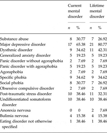
Twenty-six healthy females (healthy controls; HC) and 26 females with a DSM-IV diagnosis of BPD participated in the study. BPD participants were included in the study if they met at least five of nine DSM-IV criteria for a diagnosis of BPD, including criteria associated with emotional instability. BPD participants who met DSM-IV criteria for bipolar disorder, schizoaffective disorder or schizophrenia were excluded from the study. BPD participants who were included in the study met DSM-IV criteria for several other mental disorders, including substance-related disorders, mood disorders, anxiety disorders, somatoform disorders and eating disorders (see Table 1). HC participants were excluded from the study if they met DSM-IV criteria for any mental disorder. Additional exclusion criteria for all participants were severe cognitive impairment and mental retardation. None of the HC participants but nine of the BPD participants were using psychotropic medication at the time of the study (neuroleptics: n = 2; antidepressives: n = 8; mood stabilizers: n = 1). All participants provided written informed consent and were paid for their participation.
Co-morbid mental disorders among participants with borderline personality disorder

The study was carried out in accordance with the Declaration of Helsinki and was approved by the ethics committee of the University of Greifswald.
Measures
Structured clinical interviews were administered for the assessment of Axis I (Composite International Diagnostic Interview, CIDI; Wittchen & Pflister, Reference Wittchen and Pflister1997) and Axis II (Structured Clinical Interview for DSM-IV Axis II Disorders, SCID-II; Fydrich et al. Reference Fydrich, Renneberg, Schmitz and Wittchen1997) disorders in HC and BPD participants. In addition, self-report questionnaires were applied to assess the severity of participants’ anxious (State Trait Anxiety Inventory – Trait Version, STAI; Laux et al. Reference Laux, Glanzmann, Schaffner and Spielberger1981), depressive (Beck Depression Inventory, BDI; Hautzinger et al. Reference Hautzinger, Bailer, Worall and Keller1995) and borderline (Borderline Symptom List 23, BSL-23; Bohus et al. Reference Bohus, Kleindienst, Limberger, Stieglitz, Domsalla, Chapman, Steil, Philipsen and Wolf2009) symptoms. Besides this, a multiple choice vocabulary test was used as a proxy for participants’ level of general intelligence (Multiple Choice Vocabulary Test, MWT; Lehrl et al. Reference Lehrl, Triebig and Fischer1995)
Magnetic resonance imaging
Data acquisition
Magnetic resonance imaging was performed on a 3-T whole-body MR scanner (Verio, Siemens, Germany) equipped with a 32-channel head coil. Diffusion-weighted images were recorded with a multi-directional diffusion weighting sequence [repetition time (TR): 15 300 ms; echo time (TE): 107 ms; flip angle (FA): 90°; field of view (FOV): 230 × 230 mm2; matrix: 128 × 128]. In all, 80 axial slices (slice thickness: 2 mm, no gap; voxel size: 1.8 × 1.8 × 2 mm3) were acquired along 64 gradient directions with two b values (0 and 1000 mm/s2) and one repetition. Additionally, high-resolution structural images were recorded with a T1-weighted coronal oriented magnetization-prepared rapid gradient echo (MPRAGE) sequence (TR: 2900 ms; TE: 2.52 ms; FA: 25°; FOV: 256 × 256 mm2; matrix: 256 × 256), leading to the acquisition of 176 sagittal slices (voxel size: 1 × 1 × 1 mm3).
Data processing
Following established procedures (Domin et al. Reference Domin, Langner, Hosten and Lotze2014), the diffusion-weighted images were preprocessed with tools from the Functional Magnetic Resonance Imaging of the Brain Software Library (http://www.fmrib.ox.ac.uk/fsl). After correction of eddy currents and head motion, skull stripping was performed with bet. Next, flirt was used for a linear co-registration of the diffusion-weight image and the structural image as well as for a linear co-registration of the structural image and the MNI template. Then, nflirt was used for a nonlinear co-registration of the structural image and the MNI template. The transformation information obtained during the linear and nonlinear co-registration was combined using convertwarp and subsequently inversed using invwarp. The inversed transformation information was used for the de-normalization of the ICBM-DTI-81 white-matter labels atlas (Mori et al. Reference Mori, Oishi, Jiang, Jiang, Li, Akhter, Hua, Faria, Mahmood, Woods, Toga, Pike, Neto, Evans, Zhang, Huang, Miller, van Zijl and Mazziotta2008, Oishi et al. Reference Oishi, Zilles, Amunts, Faria, Jiang, Li, Akhter, Hua, Woods, Toga, Pike, Rosa-Neto, Evans, Zhang, Huang, Miller, van Zijl, Mazziotta and Mori2008), which contained anatomical masks in MNI space. De-normalization of this atlas was necessary because the diffusion-weighted images were deliberately left in native space to avoid unnecessary image transformations during normalization to MNI space. Finally, the gradient vectors for the diffusion weighting gradients were corrected by the rotation matrix calculated during motion correction and linear co-registration.
Data analysis
JavaDTI (Domin et al. Reference Domin, Langner, Hosten and Lotze2014) was used for diffusion tensor calculation and diffusion tensor tractography. For diffusion tensor calculation, a diffusion tensor model was fitted at each voxel to provide a voxel-wise calculation of FA, AD and RD. For diffusion tensor tractography, tracts were determined by a continuous path originating from a seed voxel in a region of interest (ROI) and following the tensor direction. Parameter thresholds for tract determination were FA values greater than 0.20 and turning angles smaller than 45°. Tracts were determined in pre-defined ROIs using de-normalized masks of the ICBM-DTI-81 white-matter labels atlas (Mori et al. Reference Mori, Oishi, Jiang, Jiang, Li, Akhter, Hua, Faria, Mahmood, Woods, Toga, Pike, Neto, Evans, Zhang, Huang, Miller, van Zijl and Mazziotta2008; Oishi et al. Reference Oishi, Zilles, Amunts, Faria, Jiang, Li, Akhter, Hua, Woods, Toga, Pike, Rosa-Neto, Evans, Zhang, Huang, Miller, van Zijl, Mazziotta and Mori2008). In line with previous procedures (e.g. Versace et al. Reference Versace, Almeida, Hassel, Walsh, Novelli, Klein, Kupfer and Phillips2008; Zhang et al. Reference Zhang, Leow, Ajilore, Lamar, Yang, Joseph, Medina, Zhan and Kumar2012), the bilateral UF comprised the primary ROI, whereas the bilateral cingulum (CG) served as a secondary ROI to clarify the specificity of findings. The mean FA, AD and RD were extracted from each of these ROIs and used for the statistical analyses.
Data visualization
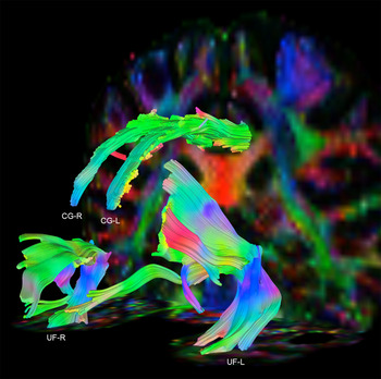
For visualization purposes (see Fig. 1), tracts of the bilateral UF and bilateral CG were projected on the diffusion-weighted image of one HC participant using DSI Studio (http://dsi-studio.labsolver.org/).
Tracts of the bilateral uncinate fasciculus (UF) and the bilateral cingulum (CG) projected on the diffusion-weighted image of one healthy control (HC) participant. Red, green, and blue represent left to right, anterior to posterior and superior to inferior directions, respectively.

Statistical analyses
All statistical analyses were conducted using SPSS v. 22 (SPSS Inc., USA). Differences in HC and BPD participants’ demographic (age, intelligence) and clinical (anxious, depressive, borderline symptoms) characteristics were analysed with χ2 tests and analyses of variance (ANOVAs). Differences in diffusion measures (FA, AD, RD) indicating structural alterations in the bilateral UF or CG of HC and BPD participants were analysed by means of mixed-design ANOVAs with group as between-subjects factor and hemisphere as within-subjects factor. Diffusion measures (FA, AD, RD) that differed between HC and BPD participants were considered as covariates in the above-mentioned ANOVAs. Associations between diffusion measures (FA, AD, RD) indicating structural alterations in the bilateral UF or CG and clinical characteristics were analysed with Pearson's product-moment correlations, separately for HC and BPD participants. The significance level for these analyses was set at p < 0.05 (two-tailed) and if necessary corrected for multiple comparisons by applying the Bonferroni method. In addition to the significance level p, the effect size measures η p 2 and d are reported to facilitate interpretation of (marginally) significant effects.
Results
Group differences in demographic and clinical characteristics
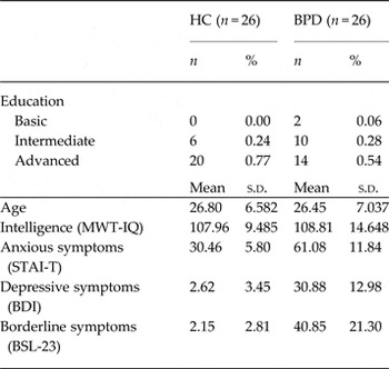
Several ANOVAs and χ2 tests were run to investigate whether HC and BPD participants differed in their demographic and clinical characteristics (see Table 2). HC and BPD participants did not differ in terms of age (F 1,50 = 0.03, p = 0.85, η p 2 = 0.001), education [χ2(1, N = 52) = 4.06, p = 0.13] or intelligence (F 1,50 = 0.06, p = 0.81, η p 2 = 0.001). BPD participants, however, reported more anxious (F 1,50 = 140.18, p < 0.001, η p 2 = 0.74), depressive (F 1,50 = 115.16, p < 0.001, η p 2 = 0.70) and borderline (F 1,50 = 84.31, p < 0.001, η p 2 = 0.63) symptoms than HC participants.
Group differences in demographical and clinical characteristics

HC, Healthy control group; BPD, borderline personality disorder group; MWT-IQ, Multiple Choice Vocabulary Test – Intelligence quotient (Lehrl et al. Reference Lehrl, Triebig and Fischer1995); STAI-T, State Trait Anxiety Inventory – Trait Version (Laux et al. Reference Laux, Glanzmann, Schaffner and Spielberger1981); BDI, Beck Depression Inventory (Hautzinger et al. Reference Hautzinger, Bailer, Worall and Keller1995); BSL-23, Borderline Symptom List 23 (Bohus et al. Reference Bohus, Kleindienst, Limberger, Stieglitz, Domsalla, Chapman, Steil, Philipsen and Wolf2009).
Group differences in structural integrity
Primary analyses
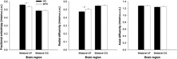
A series of mixed-design ANOVAs (group×hemisphere) was performed to investigate whether HC and BPD participants showed different FA, AD and RD in the primary ROI, the bilateral UF (see Figs 1 and 2). BPD, as compared to HC participants, showed lower FA (effect of group: F 1,50= 4.85, p = 0.03, η p 2 = 0.09; all other effects involving group or hemisphere: F < 2.74, p > 0.10, η p 2 < 0.05) and higher RD (effect of group: F 1,50 = 4.91, p = 0.03, η p 2 = 0.09; all other effects involving group or hemisphere: F < 2.72, p > 0.14, η p 2 < 0.04) in the bilateral UF. By contrast, AD did not differ in the bilateral UF of HC and BPD participants (all effects involving group or hemisphere: F < 0.10, p > 0.89, η p 2 < 0.001). To further investigate how FA and RD in the UF were related to one another, FA or RD were entered as covariates in the above-mentioned mixed-design ANOVAs (group×hemisphere). Controlling for individual differences in FA or RD, eliminated FA and RD differences in the bilateral UF of HC and BPD participants (all effects involving group or hemisphere for FA or RD: F < 1.42, p > 0.24, η p 2 < 0.03). Moreover, correlation analyses revealed a negative relationship between FA and RD in the bilateral UF (all r > |0.78|, p < 0.001), suggesting a common pathophysiological process underlying structural alterations in the bilateral UF of BPD participants. Of note, excluding BPD participants with co-morbid PTSD from the above analyses revealed a similar pattern of results (see Supplementary Material S1).
Structural integrity of the bilateral uncinate fasciculus (UF) and bilateral cingulum (CG). Bars represent mean fractional anisotropy (FA), mean radial diffusivity (RD) and mean axial diffusivity (AD) in the bilateral UF and bilateral CG of healthy control (HC) participants and participants with borderline personality disorder (BPD). Error bars indicate s.e.m. *p < 0.05.

Secondary analyses
Another series of mixed-design ANOVAs (group× hemisphere) was run to investigate whether HC and BPD participants showed different FA, AD and RD in the secondary ROI, the bilateral CG (see Figs 1 and 2). This analysis revealed no differences in FA, AD or RD in the bilateral CG of HC and BPD participants (all effects involving group or hemisphere for FA: F < 2.89, p > 0.10, η p 2 < 0.06; all effects involving group for AD and RD: F < 3.34, p > 0.07, η p 2 < 0.06; all effects involving hemisphere for AD and RD: F > 5.60, p < 0.02, η p 2 > 0.101), indicating the specificity of the structural alterations in the bilateral UF of BPD participants. As above, excluding BPD participants with co-morbid PTSD from the analysis did not change the results (see Supplementary Material S1)
Associations between clinical characteristics and structural integrity
Primary analyses
Several correlation analyses were run to investigate whether FA, RD and AD in the bilateral UF of HC and BPD participants were related to participants’ anxious, depressive or borderline symptoms. None of these analyses revealed a relationship between structural alterations in the UF and self-reports of anxious, depressive or borderline symptoms, neither among BPD nor among HC participants (all r < |0.33|, all p > 0.10).
Secondary analyses
Further correlation analyses were run to investigate whether FA, RD and AD in the bilateral CG of HC and BPD participants were related to participants’ anxious, depressive or borderline symptoms. Mirroring the findings of the above-mentioned correlation analyses, structural alterations in the CG were unrelated to HC and BPD participants’ self-reported symptoms (all r < |0.35|, all p > 0.08).
Discussion
The aim of the present study was to investigate the structural integrity of the UF, the major white-matter tract connecting (para-)limbic to prefrontal brain regions, in BPD. In consideration of previous studies revealing structural alterations in the UF in mental disorders that share common features with BPD (e.g. Versace et al. Reference Versace, Almeida, Hassel, Walsh, Novelli, Klein, Kupfer and Phillips2008; Phan et al. Reference Phan, Orlichenko, Boyd, Angstadt, Coccaro, Liberzon and Arfanakis2009; Fani et al. Reference Fani, King, Jovanovic, Glover, Bradley, Choi, Ely, Gutman and Ressler2012), we expected to find similar alterations in BPD. In addition, we expected these alterations to be related to emotion dsyregulation in BPD.
We revealed distinct structural alterations in the UF of BPD but not HC participants as indicated by multiple diffusion measures. Compared to HC participants, BPD participants showed increased FA and decreased RD but normal AD in the UF. Whereas differences in FA indicate structural alterations, differences in RD and AD help to depict and quantify the causes for these alterations (Mori & Zhang, Reference Mori and Zhang2006). Differences in AD reflect alterations due to axonal injury (Song et al. Reference Song, Sun, Ramsbottom, Chang, Russell and Cross2002, Reference Song, Sun, Ju, Lin, Cross and Neufeld2003; Sun et al. Reference Sun, Liang, Trinkaus, Cross, Armstrong and Song2006) and differences in RD reflect alterations due to myelin degeneration (Song et al. Reference Song, Sun, Ramsbottom, Chang, Russell and Cross2002, Reference Song, Sun, Ju, Lin, Cross and Neufeld2003; Sun et al. Reference Sun, Liang, Trinkaus, Cross, Armstrong and Song2006). Since differences in FA were closely related to differences in RD but not AD, the observed alterations were probably due to demyelination. Demyelination has been shown to be associated with inflammation (Merrill & Benveniste, Reference Merrill and Benveniste1996), which often occurs in the aftermath of stressful life events (Garcia-Bueno et al. Reference Garcia-Bueno, Caso and Leza2008). Considering that BPD patients frequently experience stressful life events (e.g. Pagano et al. Reference Pagano, Skodol, Stout, Shea, Yen, Grilo, Sanislow, Bender, McGlashan, Zanarini and Gunderson2004; Lobbestael et al. Reference Lobbestael, Arntz and Bernstein2010; McGowan et al. Reference McGowan, King, Frankenburg, Fitzmaurice and Zanarini2012), it may be possible that these experiences lead to inflammation. A dysregulation of pro- and anti-inflammatory cytokines, presumably as a consequence of stress-dependent alterations of the hypothalamus-pituitary-adrenal axis (Wingenfeld et al. Reference Wingenfeld, Spitzer, Rullkotter and Lowe2009), has already been reported in BPD patients (e.g. Kahl et al. Reference Kahl, Bens, Ziegler, Rudolf, Dibbelt, Kordon and Schweiger2006; Diaz-Marsa et al. Reference Diaz-Marsa, Macdowell, Guemes, Rubio, Carrasco and Leza2012). Studies are now needed that further investigate the relationship between stress-induced inflammation and myelin degeneration in BPD. Of note, demyelination appeared to be specific for the UF because similar alterations were not evident in the CG, another white-matter tract connecting (para-)limbic brain regions (Wakana et al. Reference Wakana, Jiang, Nagae-Poetscher, van Zijl and Mori2004).
Although we found, for the first time, white-matter alterations in a tract connecting BPD patients’ (para-)limbic and prefrontal brain regions, white-matter alterations in tracts connecting prefrontal brain regions have already been shown (e.g. Grant et al. Reference Grant, Correia, Brennan-Krohn, Malloy, Laidlaw and Schulz2007; Rusch et al. Reference Rusch, Luders, Lieb, Zahn, Ebert, Thompson, Toga and van Elst2007a , Reference Rusch, Weber, Il'yasov, Lieb, Ebert, Hennig and van Elst b ; Carrasco et al. Reference Carrasco, Tajima-Pozo, Diaz-Marsa, Casado, Lopez-Ibor, Arrazola and Yus2012). Moreover, grey-matter alterations in BPD patients’ (para-)limbic and prefrontal brain regions have also been reported (e.g. Schmahl et al. Reference Schmahl, Vermetten, Elzinga and Bremner2003; Tebartz van Elst et al. Reference Tebartz van Elst, Hesslinger, Thiel, Geiger, Haegele, Lemieux, Lieb, Bohus, Hennig and Ebert2003; Hazlett et al. Reference Hazlett, New, Newmark, Haznedar, Lo, Speiser, Chen, Mitropoulou, Minzenberg, Siever and Buchsbaum2005; Weniger et al. Reference Weniger, Lange, Sachsse and Irle2009; Bruehl et al. Reference Bruehl, Preissler, Heuser, Heekeren, Roepke and Dziobek2013; Niedtfeld et al. Reference Niedtfeld, Schulze, Krause-Utz, Demirakca, Bohus and Schmahl2013), implying structural alterations on the microscopic and macroscopic level in BPD. These alterations are most pronounced in prefrontal regions (e.g. Tebartz van Elst et al. Reference Tebartz van Elst, Hesslinger, Thiel, Geiger, Haegele, Lemieux, Lieb, Bohus, Hennig and Ebert2003; Hazlett et al. Reference Hazlett, New, Newmark, Haznedar, Lo, Speiser, Chen, Mitropoulou, Minzenberg, Siever and Buchsbaum2005; Grant et al. Reference Grant, Correia, Brennan-Krohn, Malloy, Laidlaw and Schulz2007; Rusch et al. Reference Rusch, Luders, Lieb, Zahn, Ebert, Thompson, Toga and van Elst2007a , Reference Rusch, Weber, Il'yasov, Lieb, Ebert, Hennig and van Elst b ; Carrasco et al. Reference Carrasco, Tajima-Pozo, Diaz-Marsa, Casado, Lopez-Ibor, Arrazola and Yus2012; Bruehl et al. Reference Bruehl, Preissler, Heuser, Heekeren, Roepke and Dziobek2013), such as the ACC and OFC, and (para-)limbic regions, such as the amygdala (e.g. Schmahl et al. Reference Schmahl, Vermetten, Elzinga and Bremner2003; Weniger et al. Reference Weniger, Lange, Sachsse and Irle2009; Niedtfeld et al. Reference Niedtfeld, Schulze, Krause-Utz, Demirakca, Bohus and Schmahl2013). Of note, BPD patients often show abnormal activity (e.g. Herpertz et al. Reference Herpertz, Dietrich, Wenning, Krings, Erberich, Willmes, Thron and Sass2001; Koenigsberg et al. Reference Koenigsberg, Fan, Ochsner, Liu, Guise, Pizzarello, Dorantes, Guerreri, Tecuta, Goodman, New and Siever2009a , Reference Koenigsberg, Siever, Lee, Pizzarello, New, Goodman, Cheng, Flory and Prohovnik b ; Niedtfeld et al. Reference Niedtfeld, Schulze, Kirsch, Herpertz, Bohus and Schmahl2010; Schulze et al. Reference Schulze, Domes, Kruger, Berger, Fleischer, Prehn, Schmahl, Grossmann, Hauenstein and Herpertz2011; Hazlett et al. Reference Hazlett, Zhang, New, Zelmanova, Goldstein, Haznedar, Meyerson, Goodman, Siever and Chu2012) and abnormal connectivity (e.g. New et al. Reference New, Hazlett, Buchsbaum, Goodman, Mitelman, Newmark, Trisdorfer, Haznedar, Koenigsberg, Flory and Siever2007; Silbersweig et al. Reference Silbersweig, Clarkin, Goldstein, Kernberg, Tuescher, Levy, Brendel, Pan, Beutel, Pavony, Epstein, Lenzenweger, Thomas, Posner and Stern2007; Niedtfeld et al. Reference Niedtfeld, Kirsch, Schulze, Herpertz, Bohus and Schmahl2012) in these regions, indicating a dysfunctional interplay between (para-)limbic and prefrontal brain regions in BPD (Mauchnik & Schmahl, Reference Mauchnik and Schmahl2010; Krause-Utz et al. Reference Krause-Utz, Winter, Niedtfeld and Schmahl2014). Since these regions are implicated in the experience and regulation of emotions (Ochsner et al. Reference Ochsner, Silvers and Buhle2012), emotion dysregulation may be due to structural and functional alterations in these regions (Mauchnik & Schmahl, Reference Mauchnik and Schmahl2010; Krause-Utz et al. Reference Krause-Utz, Winter, Niedtfeld and Schmahl2014). Accordingly, structural and functional alterations in BPD patients’ (para-)limbic and prefrontal regions have been related to abnormalities in emotional sensitivity and abnormalities in emotion regulation (e.g. Hazlett et al. Reference Hazlett, New, Newmark, Haznedar, Lo, Speiser, Chen, Mitropoulou, Minzenberg, Siever and Buchsbaum2005; Rusch et al. Reference Rusch, Weber, Il'yasov, Lieb, Ebert, Hennig and van Elst2007 b; Niedtfeld et al. Reference Niedtfeld, Schulze, Kirsch, Herpertz, Bohus and Schmahl2010, Reference Niedtfeld, Schulze, Krause-Utz, Demirakca, Bohus and Schmahl2013; Hazlett et al. Reference Hazlett, Zhang, New, Zelmanova, Goldstein, Haznedar, Meyerson, Goodman, Siever and Chu2012; Bruehl et al. Reference Bruehl, Preissler, Heuser, Heekeren, Roepke and Dziobek2013). Nonetheless, it remains open whether structural alterations, in fact, account for functional alterations in the same regions as suggested by the present and previous findings (e.g. Tromp et al. Reference Tromp, Grupe, Oathes, McFarlin, Hernandez, Kral, Lee, Adams, Alexander and Nitschke2012). Further studies combining structural and functional imaging procedures may help to elucidate the interdependence of structural and functional alterations in BPD.
Although we assume that structural alterations in the UF account for abnormalities in emotional sensitivity and abnormalities in emotion regulation, we have to admit that we were unable to show a specific relationship between structural alterations and emotion dysregulation in BPD participants. BPD participants’ scores on the self-report questionnaires assessing psychopathological symptoms (BDI, STAI, BSL) indicated abnormalities in emotional sensitivity and abnormalities in emotion regulation, but none of these scores was related to structural alterations in BPD participants’ UF. We did not expect to find a relationship between structural alterations and anxiety or depression scores because the BDI and STAI do not explicitly assess symptoms related to emotion dysregulation (Laux et al. Reference Laux, Glanzmann, Schaffner and Spielberger1981; Hautzinger et al. Reference Hautzinger, Bailer, Worall and Keller1995). A relationship between structural alterations and borderline scores, on the contrary, was expected because symptoms related to emotion dysregulation are, amongst others, assessed by the BSL (Bohus et al. Reference Bohus, Kleindienst, Limberger, Stieglitz, Domsalla, Chapman, Steil, Philipsen and Wolf2009). However, we employed a shortened version of the BSL, which may be less sensitive to capture abnormalities in emotional sensitivity and abnormalities in emotion regulation than the original version (Bohus et al. Reference Bohus, Limberger, Frank, Chapman, Kuhler and Stieglitz2007, Reference Bohus, Kleindienst, Limberger, Stieglitz, Domsalla, Chapman, Steil, Philipsen and Wolf2009). Notably, the BSL assess emotion dysregulation as typically displayed by BPD patients. The non-existent relationship between structural alterations and borderline symptoms, may, thus, indicate that these alterations are not specific for BPD. Similar alterations have already been found in other mental disorders, such as BD, SAD or PTSD (e.g. Versace et al. Reference Versace, Almeida, Hassel, Walsh, Novelli, Klein, Kupfer and Phillips2008; Phan et al. Reference Phan, Orlichenko, Boyd, Angstadt, Coccaro, Liberzon and Arfanakis2009; Fani et al. Reference Fani, King, Jovanovic, Glover, Bradley, Choi, Ely, Gutman and Ressler2012). Considering that these disorders are also characterized by abnormalities in emotional sensitivity and abnormalities in emotion regulation (e.g. Goldin et al. Reference Goldin, Manber, Hakimi, Canli and Gross2009; New et al. Reference New, Fan, Murrough, Liu, Liebman, Guise, Tang and Charney2009; Schulze et al. Reference Schulze, Domes, Kruger, Berger, Fleischer, Prehn, Schmahl, Grossmann, Hauenstein and Herpertz2011; Townsend et al. Reference Townsend, Torrisi, Lieberman, Sugar, Bookheimer and Altshuler2013), it may be possible that structural alterations in the UF reflect emotion dysregulation in general. To determine the specificity of these alterations, further studies are needed that compare these alterations between different disorders known to be associated with emotion dysregulation. Of these disorders, PTSD may be of particular relevance because BPD is often associated with PTSD (e.g. Zanarini et al. Reference Zanarini, Frankenburg, Dubo, Sickel, Trikha, Levin and Reynolds1998, Reference Zanarini, Frankenburg, Hennen, Reich and Silk2004, Reference Zanarini, Horz, Frankenburg, Weingeroff, Reich and Fitzmaurice2011; McGlashan et al. Reference McGlashan, Grilo, Skodol, Gunderson, Shea, Morey, Zanarini and Stout2000; Shea et al. Reference Shea, Stout, Yen, Pagano, Skodol, Morey, Gunderson, McGlashan, Grilo, Sanislow, Bender and Zanarini2004). It may, thus, be possible that structural alterations in the UF of BPD participants were due to the presence of PTSD rather than BPD. BPD participants, however, still showed alterations in the UF after controlling for the presence of PTSD, indicating that these alterations were most likely due to the presence of BPD. Further studies investigating BPD patients with different degrees of trauma exposure and trauma-related symptoms by means of appropriate measures may help to clarify how stressful life experiences lead to structural alterations in the UF. Considering that these alterations are already present in adolescents with BPD features (New et al. Reference New, Carpenter, Perez-Rodriguez, Ripoll, Avedon, Patil, Hazlett and Goodman2013), it may be worthwhile to investigate BPD patients across the life span in these studies, preferably before and after pharmacological or psychotherapeutic interventions to determine the effects of these interventions on the structural integrity of the UF.
Taken together, we found, for the first time, distinct white-matter alterations in the UF of BPD as compared to HC participants. These alterations were due to myelin rather than to axonal degeneration, possibly as a consequence of stress-induced inflammation in the aftermath of stressful life events. These alterations were present in a white-matter tract connecting (para-)limbic and prefrontal brain regions that have been related to abnormalities in emotional sensitivity and abnormalities in emotion regulation. Although we suppose that these alterations account for emotional dysregulation in BPD, it remains to be determined whether these alterations are specific for BPD. It may be possible that these alterations can generally be found in mental disorders that are characterized by emotion dysregulation.
Supplementary material
For supplementary material accompanying this paper visit http://dx.doi.org/10.1017/S0033291715001142.
Acknowledgements
Funding for the study was provided by in-house grants from the Department of Psychiatry and Psychotherapy of the University of Greifswald and from the Center for Diagnostic Radiology and Neuroradiology of the University of Greifswald. The funding source had no further role in study design, in the collection, analysis and interpretation of data; in the writing of the report; and in the decision to submit the paper for publication. The authors thank Anna Buchheim for valuable suggestions regarding the design of the study and Alfons Hamm for valuable suggestions regarding the analysis and interpretation of results of the study.
Declaration of Interest
None.