Takotsubo cardiomyopathy is a transient myocardial disease named after its characteristic findings of hypocontraction of the apex and hypercontraction of the basal segment of the left ventricle. Reference Dawson1 It is mostly seen in post-menopausal women and rarely in the paediatric population, Reference Rozema and Klein2 particularly in those with single ventricle disease. Reference Derk, Aboulhosn and Reardon3,Reference Watanabe, Shiraishi, Takahashi and Tsuchida4 We report a case of paediatric takotsubo cardiomyopathy with right single ventricle disease.
Case report
A 4-year-old boy underwent Glenn surgery at 3 months of age for asplenia, single right ventricle disease, and pulmonary atresia. Following intrapulmonary-artery septation and left aortopulmonary shunting at 17 months for left pulmonary artery development, he developed symptomatic epilepsy at 19 months. Despite levetiracetam therapy, he had occasional seizures. On the day of takotsubo cardiomyopathy onset, he suffered a 40-minute seizure. His previous doctor administered midazolam, and the convulsions aborted. However, immediately, atrial tachycardia (250 beats/minute) appeared and persisted despite antiarrhythmic drug administration. During endotracheal intubation, cardiopulmonary arrest occurred; cardiopulmonary resuscitation was performed for 4 minute. Once cardiac rhythm resumed, sinus rhythm was maintained by defibrillation, and he was transferred to our hospital.
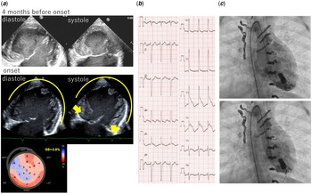
At presentation, he had hypotension (60/40 mmHg) and sinus tachycardia (160 beats/minute). Since echocardiography showed diffuse hypocontractility, catecholamines (adrenaline 0.2 μg/kg/minute and dobutamine 5 μg/kg/minute) were used to stabilise the blood pressure. There was no cardiac enlargement on chest radiography and no ST-T changes on electrocardiography. On day 2, he had prolonged disturbance of consciousness, and electroencephalography showed generalised low amplitude. Echocardiography showed akinesis at the apex and hypercontraction at the base of the heart (Fig 1a). Electrocardiography showed ST-segment elevation in leads V2 to V6, II, III, and aVF (Fig 1b). Blood test results were as follows: creatinine kinase, 565 U/L (upper limit: 197 U/L); creatine kinase myocardial band, 46 U/L (upper limit: 25 U/L); and troponin T, 373 pg/ml (upper limit: 26.2 pg/ml).
(a) Transthoracic echocardiogram showing no ventricular dysfunction before onset. At onset, and apical ballooning of the right ventricle, with hypokinesis of the apical segment (arrowheads) in the diastolic phase and normo-to-hyperkinesis of the basal segment (arrow) in the systolic phase. Longitudinal strain polar maps show abnormal values in the mid-wall and apical wall segments. (b) Twelve-lead electrocardiogram at the onset of takotsubo cardiomyopathy showing ST-segment elevation in leads V2 to V6, II, III, and aVF. (c) Right ventriculogram showing hypokinesis with the basal contraction at end-systole and end systole. The ejection fraction was estimated to be 35%. GS, global strain; ANT, anterior; LAT, lateral; POST, posterior; SEPT, septal; INF, inferior.

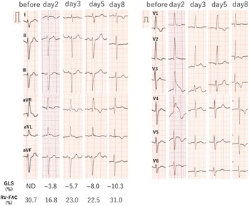
Based on the echocardiogram, takotsubo cardiomyopathy was diagnosed. Catecholamine doses were reduced; a switch was made to vasopressin and olprinone to stabilise the circulation. On day 5, ventriculography showed morphology similar to that of takotsubo (Fig 1c); the coronary angiography was normal. The electrocardiography almost normalised over time (Fig 2a); on day 8, cardiac contraction recovered to normal, and catecholamines could be terminated. However, the patient had a prolonged disturbance of consciousness; electroencephalography showed low amplitude, and head MRI showed global cerebral oedema and obscured cortical-medullary boundaries, leading to the diagnosis of hypoxic-ischemic encephalopathy.
Twelve-lead electrocardiographic changes. ST-segment changes were not seen before the onset of takotsubo cardiomyopathy. On day 2, ST-segment elevation was seen in leads V2 to V6. On day 3, a T-wave inversion was seen. Five days after onset, T-wave normalisation occurred. Subsequently, giant T-wave inversion was observed on day 8. ND, no data; GLS, global longitudinal strain; RV-FAC, right ventricle-fractional area change.

Discussion
We identified two important clinical findings in this case. First, serial electrocardiographic findings and speckle-tracking echocardiographic changes in the paediatric case of takotsubo cardiomyopathy with single right ventricle is similar to those in adults. Second, seizures can induce takotsubo cardiomyopathy in children.
Takotsubo cardiomyopathy is rare in children, Reference Rozema and Klein2 particularly in those with a single ventricle. Reference Derk, Aboulhosn and Reardon3,Reference Watanabe, Shiraishi, Takahashi and Tsuchida4 Previous paediatric reports did not detail serial electrocardiographic findings and speckle-tracking echocardiographic changes. Reference Rozema and Klein2–Reference Watanabe, Shiraishi, Takahashi and Tsuchida4 A previous adult study Reference Mitsuma, Kodama and Ito5 showed serial findings in women with takotsubo cardiomyopathy and reported that the clinical course comprised four electrocardiographic phases, with phase 1 characterised by ST-segment elevation immediately after onset. T-wave inversion was observed on days 1–3 (phase 2), and inverted T-waves improved transiently on days 2–6 (phase 3). Subsequently, giant inverted T-waves appeared and persisted for more than 2 months (phase 4). Finally, all electrocardiographic abnormalities normalised over several months. Similarly, the present case showed ST-segment elevation in leads V2 to 6 after onset and T-wave inversion 2 days later. On day 5, T-wave normalisation occurred, and giant T-wave inversion was observed on day 8. Gradually, negative T-waves normalised by day 40. Echocardiographic findings showed similar changes. Immediately after onset, the right ventricle-fractional area change was 16.8% (30.8% before onset) and the global longitudinal strain was −3.8%, which gradually improved to 31.0% and −10.3%, respectively, when the electrocardiogram showed giant negative T-waves on day 8. In adults, left ventricular strain has been reported to improve before the systolic function, while right ventricular strain improves simultaneously with systolic function, Reference Tibrewala, Freed and Akhter6 suggesting that takotsubo cardiomyopathy with single right ventricle disease follows the same course as takotsubo cardiomyopathy in the right ventricle.
The triggers for takotsubo cardiomyopathy in adults and children are almost the same: psychological stress in 52% and physical stress in 48% (comprising acute respiratory failure in 26%, central nervous system disease in 18%, and malignant disease in 11%). Reference Sharkey, Windenburg and Lesser7 In adults Reference Stöllberger, Wegner and Finsterer8 and children, Reference Yamaguchi, Nagase and Yoshida9 seizures inducing takotsubo cardiomyopathy have been noted. According to the latter report, takotsubo cardiomyopathy occurred within 72 hours after seizure, and in the present case, the disease onset was 36 hours after seizure. In the present case, it is thought that the severe seizure caused physical stress, leading to the onset of takotsubo cardiomyopathy. However, the previous physician did not evaluate cardiac function, and the exact mechanism of onset was unclear because the tachycardia that occurred after the severe seizure may have triggered takotsubo cardiomyopathy. In any case, seizures in patients with single ventricular disease can lead to takotsubo cardiomyopathy; therefore, epilepsy should be strictly controlled.
There is no specific treatment for takotsubo cardiomyopathy, and unnecessary inotropic drugs should be avoided because its pathogenesis is associated with excessive sympathetic nervous system activity. Reference Paur, Wright and Sikkel10 Excessive sympathetic nervous system activity is thought to be the main pathogenesis because there are more β2-adrenergic receptors in the apex than in the base of the heart, and more sympathetic nerve endings are distributed in the basal segment. Accordingly, sympathetic overactivity suppresses the signal switch mechanism, causing contractile dysfunction in the apex, while sympathetic stimulation causes hypercontraction in the base segment. In the present case, catecholamines were used because of the marked decrease in cardiac contraction at disease onset; however, the clinical course and echocardiographic findings strongly suggested takotsubo cardiomyopathy; therefore, early termination of treatment was possible.
In conclusion, serial electrocardiogram findings in a paediatric single right ventricle can be the same as in adult takotsubo cardiomyopathy. To our knowledge, this is the first report to show serial electrocardiogram and two-dimensional speckle tracking changes in a child with single-ventricle disease and takotsubo cardiomyopathy.
Acknowledgements
None.
Financial support
This research received no specific grant from any funding agency, commercial, or not-for-profit sectors.
Conflicts of interest
None.
Ethical standards
Informed consent was obtained from the patient for publication of this case report.