INTRODUCTION
The cercozoan class Imbricatea Cavalier-Smith, 2003 (in Cavalier-Smith & Chao, Reference Cavalier-Smith and Chao2003), emend. Howe et al. (Reference Howe, Bass, Scoble, Lewis, Vickerman, Arndt and Cavalier-Smith2011) contains two flagellate and amoeboid orders of heterotrophic protists, the Thaumatomonadida Shirkina, 1987 and the Euglyphida Copeland, 1956, emend. Cavalier-Smith, 1997, wherein cells are encased in a layer of endogenously-produced silica scales.
Within the Thaumatomonadida, the genus Thaumatomastix Lauterbourn, 1896 was thought, until recently, to contain about 20 species (see e.g. Thomsen et al., Reference Thomsen, Kosman and Ikävalko1995; Thomsen & Ikävalko, Reference Thomsen and Ikävalko1997; Mikrjukov, Reference Mikrjukov2002). It had been redefined by Beech & Moestrup (Reference Beech and Moestrup1986) to include the monospecific genus Reckertia Conrad, 1920 as well as some other scaled protists previously thought to be members of the photo-autotrophic genus Chrysosphaerella. Species are distinguished on the basis of the ultra-structure of the siliceous scales which cover the cell. Only three species are known from freshwater: T. triangulata (Balonov) Beech & Moestrup, from Russia (and a few other locations discussed later in this paper), T. bipartita discovered in a New Zealand tidally influenced lake (Wujek & O'Kelly, Reference Wujek and O'Kelly1992), and T. nigeriensis known from Nigeria (Wujek et al., Reference Wujek, Pershon and Kadiri2008) and from South Carolina, USA (Wujek, Reference Wujek2010).
Howe et al. (Reference Howe, Bass, Scoble, Lewis, Vickerman, Arndt and Cavalier-Smith2011) reinstated and amended the description of Reckertia and made eight new combinations resulting from the transfer of species of Thaumatomastix that lacked spine scales. Included among these transfers to Reckertia was the freshwater species T. nigeriensis, because the previous two reports of this species had included no description of spine-scales. Howe et al. (Reference Howe, Bass, Scoble, Lewis, Vickerman, Arndt and Cavalier-Smith2011) also believed that Balonov's C. triangulata should not have been transferred to Thaumatomastix based on the information provided by Balonov (Reference Balonov1980).
The purpose of this paper is to: (i) describe six new marine species of Thaumatomastix and one new freshwater species of Reckertia; (ii) provide the first re-description of whole cells with both spine-scales and plate-scales of T. triangulata, demonstrating its likely identity with Balonov's Chrysosphaerella triangulata and thus provide support for the transfer to Thaumatomastix made by Beech & Moestrup (Reference Beech and Moestrup1986); and (iii) provide an amended description of T. nigeriensis based on electron microscopy of scale structure and light microscopy of living cells (thus invalidating ‘Reckertia nigeriensis').
MATERIALS AND METHODS
Samples containing the freshwater Thaumatomastix specimens were collected by filling 500 ml polycarbonate bottles near the water–sediment interface of several lakes and ponds in Ontario Canada, between 1983 and 2011. Specific conductance and pH measurements were made at the time of collection with a portable meter (DspH Pocket Pal, Presto Tech Corp.) with automatic temperature compensation to 25°C. Accuracy with laboratory standards was ±5 µS cm−1 and ± 0.2 pH units.
Samples were fixed at the time of collection with Lugol's solution and concentrated by sedimentation, or were kept alive as unpreserved material in the original collection vessels for several days while observations were made with a light microscope (LM). Drops of concentrated sample were dried at room temperature on copper location grids coated with a support film of Butvar B98 (J.B. EM Services, Dorval P.Q.). Preparations of previously fixed material were washed with distilled water to remove iodine crystals (fixative residue) and re-dried. Some preparations of Thaumatomastix were shadowed with Pd/Pt alloy at an angle of about 20° in a high vacuum evaporator before examination at 80 Kv with a transmission electron microscope (TEM). For scanning electron microscopy (SEM), individual cells were isolated with a micro-pipette and transferred to No. 2 coverslip chips and glued to standard aluminium SEM stubs in preparation for coating with atomized Au in an evaporator–sputter coater.
Marine sampling methods are described in Nicholls Reference Nicholls(2009a, Reference Nichollsb) and locations of sampling on the Canadian Pacific and Atlantic coasts are summarized under the ‘type locality' headings for each of the new marine species described here. Salinity was measured with a refractometer (model FG-211, Sun Instruments Corp., Torrance CA) with automatic temperature compensation, when the samples arrived at my Ontario laboratory a few days after collection (shipped unpreserved by air mail express). Triangular plate-scale sizes follow those of Wujek et al. (Reference Wujek, Pershon and Kadiri2008), based on length from one apex of the triangle to the mid-point on the opposite side.
RESULTS
SYSTEMATICS
Phylum CERCOZOA Cavalier-Smith, 1998
Class IMBRICATEA Cavalier-Smith & Chao, Reference Cavalier-Smith and Chao2003
Order THAUMATOMONADIDA Shirkina, 1987
Family THAUMATOMONADIDAE Holland, 1934; emend. Howe, Bass, Scoble, Lewis, Vickerman, Arndt & Cavalier-Smith, Reference Howe, Bass, Scoble, Lewis, Vickerman, Arndt and Cavalier-Smith2011
Genus Thaumatomastix Lauterborn, 1899; emend. Howe, Bass, Scoble, Lewis, Vickerman, Arndt & Cavalier-Smith, Reference Howe, Bass, Scoble, Lewis, Vickerman, Arndt and Cavalier-Smith2011
Marine species
NEW SPECIES
Thaumatomastix asymmetrica sp. nov.
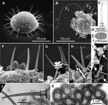
(Figure 1A–K)
Although several living Thaumatomastix cells from the sample containing T. asymmetrica were examined by light microscopy, there were none that I could unequivocally state could be assigned to this species, as opposed to some other, owing to the resolution limitations imposed by LM. No details on internal structure, movement, swimming behaviour, etc. can therefore be provided at this time for this species.
DIAGNOSIS
Cells (dried) 11–21 µm in diameter. Spine-scales 3.5–5.0 µm long, including basal structure. Spine-scale basal structure consists of a circular base-plate 0.7–0.9 µm in diameter and a secondary plate 0.4–0.6 µm in diameter joined together by three arched and fully separated band-like struts 0.50–0.55 µm long. Shaft of the spine attached at its base to the secondary plate; spine shaft hollow, slightly curved, tapering from 0.4 µm above the basal structure to 0.25 µm at the apex. Apex with 2–3 small denticles. Plate-scales of an upper (distal) elliptical plate 1.0–1.3 µm wide and 1.5–2.0 µm long, separated from a lower (proximal) plate that is up to 15% larger by a hollow goblet-shaped pillar near each end of the scale, about 0.5 µm high, with the wide end attached to the underside of the distal plate and the narrow stem-end attached to the proximal plate. The point of attachment to the upper (distal) plate is revealed on the distal surface as a large hole in the surface of the plate. This hole is larger at one end of the scale than the other, apparently as a result of the fusion of two pillars, revealed in TEM images as two electron-dense ‘eyes' in close proximity. The inter-hole space on the upper surface of the scale is marked by a prominent ridge oriented across the width of the scale.
ETYMOLOGY
The specific epithet (asymmetrica) refers to the dissimilar size of the holes in the upper surface of the plate-scales located near the ends of the scale; the large hole at one end indicating the fusion of two pillars and the smaller hole at the opposite end of the scale, a single pillar serving to join the upper and lower plates of the scale.
TYPE SPECIMEN
Thaumatomastix asymmetrica sp. nov: (A) whole cell with complete investiture of plate-scales and spine-scales; (B) whole cell with incomplete scale covering; F1 and F2 are the long and short flagella, respectively; (C–E) diagrammatic representation of the structure of a spine-scale (C) and a plate-scale (D—oblique, lateral view; E—plan view); (F–I) edges of cells with both plate-scales and spine-scales; (F) well-developed secondary base-plate (arrow) forming part of the basal structure of a spine-scale; (H) arrow shows the collar-like rim on the underside of the distal plate of a plate-scale that surrounds the pillars that join the two plates of the scale; (I) arrows point to plate-scales revealing the underside of the proximal plate of four plate-scales; (J) spine-scale; (K) appearance of plate-scales in transmission electron microscope.

TYPE LOCALITY
Sand beach, south shore of Sable Island, Atlantic Ocean, Nova Scotia, Canada; 43°55′25″N 59°57′52″W. Salinity = 33 ppt.
MATERIAL FROM THE TYPE LOCALITY
Collected at low tide, 21 August 2007; retained by K. Nicholls as formalin-preserved aqueous sample No. V-2079.
ADDITIONAL REMARKS
This species was also found in samples collected from the north shore of Sable Island, 20 August 2007. The prominent side-to-side ridge through the middle of the scale is a major distinguishing feature of this species. This ridge is clearly revealed in SEM images, especially those scales viewed obliquely (e.g. Figure 1A—those scales oriented obliquely around the edge of the cell), but is not revealed easily in TEM images; this suggests that the ridge consists of an elevated fold rather than a thickening of the scale surface. A thickening of scale material would be revealed in a TEM image as a zone of higher electron-opacity. There are several features of the spine-scales of T. asymmetrica that resemble those found in T. multipora (see below), notably they are hollow, slightly curved and taper throughout their length to an apex with 2–3 small teeth. The differences include a wider, better developed secondary plate above the base plate in T. asymmetrica and an inter-base-plate structure that is more completely separated into three supporting struts anchoring the spine shaft to the base-plate (Figure 1J). The most important differences though, relate to the presence of the porous central area in the plate-scales of T. multipora that was not present in any of the many specimens of T. asymmetirca examined by EM. Also, the marginal rim in the plate-scales of T. asymmetrica is narrower than the comparable structure in T. multipora (compare Figure 2H for T. multipora and Figure 1K for T. asymmetrica).
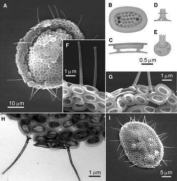
Thaumatomastix multipora sp. nov.: (A, I) whole cells with complete investitures of plate-scales and spine-scales; (B, C) diagrammatic representation of the structure of a plate-scale; (B) plan view; (C) lateral view; (D, E) diagrammatic representation of the basal structure of a spine-scale; (D) lateral view; (E) oblique lateral view; (F–H) edges of cells with spine-scales and plate-scales; (F, G) scanning electron microscope; (H) transmission electron microscope.

Thaumatomastix multipora sp. nov.
(Figure 2A–I)
Many cells were examined with TEM and SEM, but the identities of many other cells examined by LM only, that possibly were T. multipora, could not positively be assigned to this taxon.
DIAGNOSIS
Cells (dried) about 20–30 µm in diameter covered with two types of scales: plate-scales nearly elliptical, consisting of two elliptical plates of nearly identical size (1.3–2.1 µm long × 1.0–1.4 µm wide) and separated by a hollow columnar structure (‘pillar') about 0.4 µm high near one end of the scale and two fused pillars near the opposite end of the scale (or much less frequently, a single pillar at each end). The distal surface of the scale consists of a broad (0.4 µm wide) convex marginal zone devoid of surface ornamentation. The elliptical region inside the marginal zone consists of a depressed nearly elliptical plate ornamented with 4 or 5 rows of pit-like depressions about 0.06–0.08 in diameter. Near one end of this region is a single larger pore (0.10–0.12 µm in diameter) that is open to the interior of the pillar that joins the upper and lower plates of the plate-scales. The opposite end of the scale has usually two such large pores which are manifested on the underside of the upper plate as a pair of fused pillars. Spine-scales are far fewer in number than the plate-scales and consist of a nearly circular base plate about 0.6–0.8 µm in diameter to which the base of the hollow shaft of the spine is attached. Near the base plate are three or four large holes in the wall of the shaft, above which is a secondary (smaller) base plate, about 0.4 µm in diameter. Spine scales are slightly curved over their 4–5 µm total length and taper in width from 0.27 µm near the base to about 0.17 µm near the distal end where there is a slight flare with 2–4 small tooth-like distal projections.
ETYMOLOGY
The specific epithet (multipora) refers to the pattern of pit-like depressions ornamenting the central area of the upper (distal) surface of the plate-scales.
TYPE SPECIMEN
TYPE LOCALITY
Sand beach, south shore of Skidegate Inlet, Haida Gwaii (British Columbia, Canada), opposite Gillat Island, east of Onward Point at 53.2410°N 131.8980°W.
MATERIAL FROM THE TYPE LOCALITY
Collected at low tide, 8 December 2008; retained by K. Nicholls as formalin-preserved aqueous sample No. V-2111.
ADDITIONAL REMARKS
The main distinguishing features of this species include the plate-scales with their broad unpatterned borders and the punctate central elliptical zone. The punctae are easily seen in SEM images (Figure 2F, G), but difficult to see in TEM images (Figure 2H), suggesting that these pits or punctae are not pores, but are circular depressions on the distal surface of the scale. Occasionally the broad borders of the plate-scales were found to curve upward, imparting a general concave shape to the distal plate of the scale, despite the convex shape of the border itself. The spine-scales all have a slight curvature and taper over their length (Figure 2A, I). The base-plates of the spine-scales are of a size that allows them to ‘lock' into position between adjacent plate-scales, with the base plate of the spine-scale located between the two plates of a plate-scale. The unique scale structure and ornamentation allows this species to be distinguished from all other known species of Thaumatomastix.
Thaumatomastix inornata sp. nov.
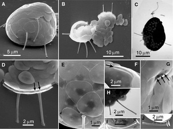
(Figures 3A–J)
Several whole cells were examined with both SEM and TEM.
DIAGNOSIS
Cells 12–25 µm in diameter. Plate-scales of the double plate structure typical of the genus, rounded triangular, 2.2–3.4 µm (outer edge of one corner to mid-point of opposite side). Distal surface of the upper plate and the proximal surface of the lower plate are smooth, unornamented with pits, pores or ridges. Connection points on the plate where the three pillars join the upper and lower plates together were visible in both SEM and TEM as small electron-dense patches spaced about 1 µm apart in a triangular pattern. The pillars are about 0.3 µm wide and about 0.3 µm high (i.e. the distance between the plates). Spine-scales 5–7 µm long including a basal structure consisting of a primary base-plate 0.8–0.9 µm in diameter and a secondary plate reduced to a collar-like swelling about 0.6 µm in outside diameter encircling the spine shaft about 0.6 µm above the primary base-plate. The connective material between the two plates appears hollow and undivided into separated struts (as is common in some other species). The spine shaft is widest (0.30–0.35 µm in diameter) just above the secondary plate and tapers and bends slightly through most of it length, but flares slightly near the apex of the spine, remaining hollow and open-ended at the apex with 2–3 small denticles around the apical rim.
ETYMOLOGY
The specific epithet (inornata) refers to the predominantly smooth, unornamented surface of the plate-scales.
TYPE SPECIMEN
Thaumatomastix inornata sp. nov.: (A–C) whole cells with complete investitures of plate-scales and spine-scales; (A, B) scanning electron microscope; (C) transmission electron microscope (TEM); (D) group of plate-scales in lateral view showing pillars (arrows) joining the two plates of a scale; (E) distal surfaces of a group of interlocking plate-scales; (F) the short inter-plate distance between the two plates of a plate-scale (arrow); (G) three pillars (arrows) separating the two plates of a plate-scale; (H) TEM image showing the hollow and slightly flared apices of two spine-scales; (I, J) spine-scales with slight differences in their basal structures.

TYPE LOCALITY
Sand beach, south shore of Skidegate Inlet, Haida Gwaii (British Columbia, Canada), opposite Gillat Island, east of Onward Point at 53.2410°N 131.8980°W.
MATERIAL FROM THE TYPE LOCALITY
Collected at low tide, 8 December 2008; retained by K. Nicholls as formalin-preserved aqueous sample No. V-2111.
ADDITIONAL REMARKS
Included here is a specimen (Figure 3B, D & J) for which the plate-scales conform to the description given above, but includes spine scales that are also nearly identical to those described above, but for a differently structured secondary base-plate. The secondary base-plate on this specimen is better developed into a distinctly wider plate that is angled downward towards the primary base-plate, resembling more a sloping shelf than the thickened collar described above for this taxon. More specimens need to be found to determine if this spine structure is consistent over space and time. In the meantime, because the other characteristics of its spine-scales and its plate-scales are so much like those described here for T. inornata, I propose to include this ‘variant' with T. inornata for taxonomic purposes.
Thaumatomastix gwaii sp. nov.
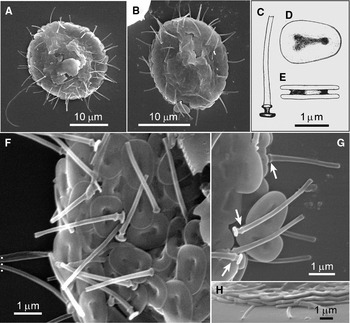
(Figure 4A–H)
DIAGNOSIS
Cells 18–22 µm in diameter, covered in interlocking plate-scales and spine-scales. Plate-scales 1.5–1.9 µm long by 1.3–1.5 µm wide; the upper and lower plates about 0.19 µm thick. The distal surface of the upper plate contains an elongated depression of variable shape and degree of development occupying the middle 1/3–1/2 of the total length of the scale. At each end of the depression may be one or two holes leading to the interior of the pillar material that bonds the two plates together. Distance between the two plates is about 0.1 µm. Spine-scales 2.4–4.4 µm long, including a basal structure consisting of a primary base-plate about 0.09 µm thick and 0.45–0.55 µm in diameter. The secondary base-plate is reduced to a thickened collar 0.27–0.32 µm in diameter about 0.14 µm above the primary base-plate. The connective material is hollow with one or two minor clefts or small holes and 2–3 curved but weakly developed ribs, oriented longitudinally. The spine shaft is widest (0.18–0.23 µm in diameter) immediately above the secondary base-plate and tapers very little throughout the remainder of its length. The apex of the spine shaft is open to the hollow interior and ornamented with 2–3 small denticles around the apical rim.
ETYMOLOGY
The specific epithet (gwaii) refers to the type locality of this species (Haida Gwaii).
TYPE SPECIMEN
Thaumatomastix gwaii sp. nov.: (A, B) whole cells with complete investitures of plate-scales and spine-scales; (C–E) diagrammatic representation of the structure of a spine-scale (C) and the two views (D, E) of a plate-scale; (F) edge of a cell showing several spine-scales and the distal surfaces of many plate-scales; (G) secondary base-plate on spine-scales reduced to a collar-like rim (arrows); (H) the thickness of the plates of plate-scales relative to the diameters of two spine shafts.

TYPE LOCALITY
Sand beach, south shore of Skidegate Inlet, Haida Gwaii (British Columbia, Canada), opposite Gillat Island, east of Onward Point at 53.2410°N 131.8980°W.
MATERIAL FROM THE TYPE LOCALITY
Collected at low tide, 8 December 2008; retained by K. Nicholls as formalin-preserved aqueous sample No. V-2111.
ADDITIONAL REMARKS
The spine-scales of T. gwaii are of similar structure to those of T. inornata, but T. inornata spine-scales are about twice as long. The plate-scales of these two species are very different and cannot be easily confused; the plate-scales of T. inornata are clearly equilateral triangular with no depression in the cental area of the distal plate, while those of T. gwaii are weakly elliptic-isosceles triangular shape with a strongly developed but irregularly-shaped depression in the central area of the distal plate.
Thaumatomastix sablensis sp. nov.
(Figure 5A–E)
DIAGNOSIS
Cells about 13 µm in diameter (but only one whole cell measured in an SEM image) covered with interlocking plate-scales and spine-scales. Plate-scales of the double plate structure in the shape of an equilateral triangle with rounded corners; 2.1–2.7 µm in length (corner to mid-point of opposite side). Proximal plate separated from the distal plate by three pillars about 0.75 µm in height. Pillars anchored at the corners of a triangular area in the middle of scale marked by a well-defined triangular depression. The size of this depression is 30–35% of the scale size. A small depression or pit, about the same size as the pits marking the anchor points of the three pillars, is located in the middle of the triangular depression, about equidistant from each of the attachment points of the three pillars. Spine-scales 42–50 µm long; primary base-plate about 0.8 µm in diameter; secondary base-plate about 0.6 µm in diameter, separated from the primary base-plate by a hollow column about 0.2 µm in diameter and about 0.25 µm high, some of which appear to have 3–5 small holes at the base of the cylinder above its connection with the primary base-plate. Base of the shaft 0.4–0.6 µm in diameter, tapering over the proximal ½ of its length, and in some shafts expanding slightly near the apex. Spine shafts, in the distal ½ of their lengths, about 0.18 µm in diameter.
ETYMOLOGY
The specific epithet (sablensis) refers to the type locality of this species (Sable Island, Atlantic Ocean, Canada).
TYPE SPECIMEN
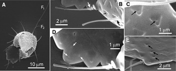
Thaumatomastix sablensis sp. nov.: (A) whole cell with its investiture of triangular siliceous plate-scales and spine-scales; F1 and F2 are the long and short flagella, respectively; (B) two spine-scales showing difference is diameter of the base of the spine shaft (large arrows) and several plate-scales; (C) the small depressions in the centres of the triangular depressions of two plate-scales (arrows: see also small arrow in B); (D) a single spine-scale and the undersides (proximal surfaces) of several plate-scales showing the attachment points (arrows) of the three inter-plate pillars joining the two plates of a scale; (E) the proximal surfaces of plate-scales showing the central depression in three adjacent scales (arrows).

TYPE LOCALITY
Sand beach, south shore of Sable Island, Atlantic Ocean, Nova Scotia, Canada; 43°55′25″N 59°57′52″W. Salinity = 33 ppt.
MATERIAL FROM THE TYPE LOCALITY
Collected at low tide, 21 August 2007; retained by K. Nicholls as formalin-preserved aqueous sample No. V-2079.
ADDITIONAL REMARKS
Although only one specimen was observed with electron microscopy (Figure 5A), the cell was complete, showing both the flagella, several spine-scales and plate-scales in various planes of view, including the underside (proximal surface) of a large patch of imbricate plate-scales. This species could possibly be confused with only one other species (T. curvata), owing to the presence of the distinctively large triangular depression on the distal surface of the outer plate of the plate-scales of both of these species (but see Remarks under T. curvata, below for a summary of major differences between these two species). The small circular central depression in the outer plate of plate-scales (Figure 5B, C) was only seen in SEM images of scales oriented angled off the right-angle plane; scales viewed straight-on (plan view) did not reveal this feature (Figure 5D). This suggests that it is a depression or dimple that is not accompanied by any significant thickening of the scale material and contrasts with the appearance of the connection points of the pillars connecting the two plates which are revealed clearly in plan view (Figure 5D). A corresponding depression apparently occupies the centre of the proximal (lower) plate also (Figure 5E). Although there was considerable variation in the diameter of the spine shaft above the secondary base-plate, there was limited variation seen in the structure of plate-scales.
Thaumatomastix curvata sp. nov.
(Figure 6A–E)
DIAGNOSIS
Cells (dried) about 12 µm in diameter (only one seen in EM) covered in plate-scales and spine-scales. Plate-scales rounded triangular, 1.1–1.4 µm (outside corner to mid-point of opposite side) with a wide and thick boarder surrounding a triangular depression in the centre of the outer surface of the scale 0.35–0.61 µm (outside corner to mid-point of opposite side). Scale boarder strongly convex 0.35–0.41 wide. Spine-scales 4–7 µm long, strongly curved throughout. The basal structure of the spine-scale is reduced to a primary basal disc 0.54–0.62 µm in diameter; the secondary plate reduced to a nearly imperceptible swelling on the spine shaft about 0.2 µm above the basal disc. Spine shaft immediately above basal structure 0.2–0.4 µm in diameter and tapers strongly throughout its length to an apex of about 0.05 µm in diameter.
ETYMOLOGY
The specific epithet (curvata) refers to the well-developed curvature of the spine-scales.
TYPE SPECIMEN
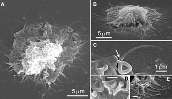
Thaumatomastix curvata sp. nov.: (A) whole cell (collapsed on drying) with investiture of triangular plate-scales and curved spine-scales; (B) same cell as in (A), but tilted; (C) the much reduced secondary basal plate of a spine-scale (arrow) with plate-scales; (D) the nail-head appearance of the primary basal plates of two spine-scales; note also the large difference in the size of the central triangular depressions of some of the plate-scales; (E) several spine-scales with their characteristic highly tapered and curved structure. The scale bars in D and E are both 1 µm.

TYPE LOCALITY
Sand beach, south shore of Skidegate Inlet, Haida Gwaii (British Columbia, Canada), opposite Gillat Island, east of Onward Point at 53.2410°N 131.8980°W.
MATERIAL FROM THE TYPE LOCALITY
Collected at low tide, 8 December 2008; retained by K. Nicholls as formalin-preserved aqueous sample No. V-2111.
ADDITIONAL REMARKS
The triangular depression in the central area of the outer surface of the plate-scales of this species is reminiscent of the plate-scales of T. sablensis; the size of T. sablensis scales, however, is about 2 times greater. The other major distinguishing features of T. curvata are the strongly curved and tapered spine-scales. Its very distinctive spine-scale structure is like that of no other known Thaumatomastix species.
Freshwater species
Thaumatomastix nigeriensis Wujek, Pershon & Kadiri, Reference Wujek, Pershon and Kadiri2008.
Plate-scales from this species appear identical to those described from Nigeria and from South Carolina, USA by Wujek et al. (Reference Wujek, Pershon and Kadiri2008) and Wujek (Reference Wujek2010). This species was described as possessing plate-scales only and was the reason for its transfer to Reckertia as R. nigeriensis (Wujek, Pershon & Kadiri) Scoble & Cavalier-Smith (in Howe et al., 2011). Several whole cells have been found in Ontario with complete investitures of scales; spine-scales were present in all cases and included the characteristic double plate structure at the base of the spine, typical of many species of this genus. It is therefore necessary to provide an amended description of T. nigeriensis.
Thaumatomastix nigeriensis emend. Nicholls
(Figure 7A–G)
(non-Reckertia nigeriensis, sensu Howe et al. (Reference Howe, Bass, Scoble, Lewis, Vickerman, Arndt and Cavalier-Smith2011).)
AMENDED DIAGNOSIS
Cells 20–28 µm in diameter with a large nucleus (7–10 µm in diameter); 1–3 contractile vacuoles of various size depending on the stage of diastole. Both bi-flagellate and amoeboid stages known. Cells covered in two types of scales. Plate-scales consist of outer (distal) and inner (proximal) rounded-triangular plates measuring 1.7–2.1 µm from the rounded outer edge of a corner to the mid-point of the opposite edge of the scale. The two plates are separated by three columnar structures about 0.5 µm high, fused at the underside of the distal plate, but nearly (or completely) separated where they attach to the proximal plate. The distal outer surface of the distal plate is ornamented with a floral-like, tri-cornered opening to an inner cavity, leaving a smooth, broad (0.5–0.9 µm) and moderately convex margin surrounding the central cavity. Spine-scales consist of a basal structure similar to a plate-scale, but often smaller in size. The shaft of the spine is anchored at a point on the rim of the central ornamentation. The spine shaft is circular in cross-section, nearly of uniform width (0.3 µm) throughout most of its length of 6–15 µm, narrowing near the distal end to a sharp-pointed apex.
NEOTYPIC IMAGE
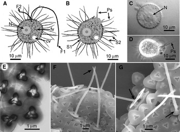
Thaumatomastix nigeriensis (Wujek, Pershon & Kadiri, Reference Wujek, Pershon and Kadiri2008) emend.: (A, B) diagrammatic representation of the flagellate stage (A) and the amoeboid stage (B) showing the long and short flagellum (F1 and F2), food vacuole (fv), nucleus (N), contractile vacuole (cv), plate-scale (S1), spine-scale (S2) and pseudopodia (Ps); (C, D) living specimens (light microscope) showing nucleus and pseudopodia; (E) a group of plate-scales (transmission electron microscope), showing floral-like central ornamentation; (F, G) edges of whole cells (scanning electron microscope) showing some plate-scales and the shafts of two spine-scales tapering to pointed apices (arrow in F) and the attachment of the spine shaft to the basal structure of the spine-scales (arrows in G: see also F).

NEOTYPIC HABITAT
Shallow backwater area of the Cinder Lake outflow; Hindon Township, Haliburton County, Ontario Canada (45°03′49″N 78°56′55″W).
MATERIAL FROM THE NEOTYPE LOCALITY
Retained by the author as a formalin-preserved aqueous sample (No. V-2093) collected 10 October 2007.
ADDITIONAL REMARKS
I collected loose scales of this taxon from Ontario as early as 1984 from three freshwater sites in Ontario, Canada as follows: 1. Pincher Lake, McCraney Township, 45°33′N 78°54′W (12 September 1984; pH = 5.8, sp. cond. = 20 µS cm−1); 2. Pond No. 18, Draper Township, 45°02′37″ N 79°14′0″W (4 September 1987; pH = 5.6; sp. cond. = 30 µS cm−1 3. Pond No. 19, Hindon Township, 45°03′43″N 78°57′38″W (25 October 1988; pH = 5.5; sp. cond. = 20 µS cm−1). Not until 2007 did I find abundant specimens in a wild population wherein two life history stages of this species were observed—a bi-flagellated stage (Figure 1A) and an amoeboid stage lacking visible flagella (Figures 1B, D). In the Cinder Lake outflow collection, the amoeboid stage was especially abundant and was the source of cells upon which measurements were made in their living state (N = 24): mean, median and standard deviation cell diameter = 23.6, 23.0 and 2.04 µm, respectively. The amoeboid stage appeared to be a trophic adaptation for exploring particles for their potential ingestion as food items.
The plate-scale structure agrees well with that described by Wujek et al. (Reference Wujek, Pershon and Kadiri2008) for Nigerian specimens, except that the Ontario specimens were slightly larger. The TEM image (Figure 1E) clearly shows the floral-like pattern of electron-dense siliceous material surrounding central cavity and corresponds to the vertical walls of the pillars that separate the upper and lower plates of the scale. The three-dimensional construction of this region of the scale is most easily visualized in TEM images that reveal also the three points of attachment of the columns to the lower plate and their linkage by the three ridges that radiate from a central point on the lower plate (Figure 1E). These images are convincingly identical to the Nigerian material (compare with plates 1–3 in Wujek et al. (Reference Wujek, Pershon and Kadiri2008)). The spine-scales of this species have a basal structure that is structurally like a plate-scale (double plate with similar surficial features and infrastructure), but is usually smaller by up to about 30%. It is the solid rim of electron-dense material surrounding the floral-like cavity in the distal plate of the scale upon which the shaft of a spine-scale is anchored (Figure 1F, G). Spine shafts rarely extend from the basal plate at right-angles to it. More often the spine shaft is eccentrically attached along an edge or corner of the cavity of the distal plate and extends upward at an angle of about 30° off the vertical, pointing over the far side, rather than the near side of the base plate. A similar orientation of the spine shaft was found for T. triangulata (see below).
The lack of spine-scales reported by Wujek et al. (Reference Wujek, Pershon and Kadiri2008) and Wujek (Reference Wujek2010) should now, in my opinion, be discounted as a taxonomic descriptor; it is more likely that their absence from those collections was a function of the paucity of material examined and/or the loss of spine-scales from the specimen observed. Whole cells of very many Ontario specimens all possessed both types of scales reported here (none lacked spine-scales).
Thaumatomastix triangulata (Balonov) Beech & Moestrup, Reference Beech and Moestrup1986; emend. Nicholls
(Figures 8A–N)
(non-Chrysosphaerella triangula Balonov, Reference Balonov1980sensu Howe et al. (Reference Howe, Bass, Scoble, Lewis, Vickerman, Arndt and Cavalier-Smith2011).)
AMENDED DIAGNOSIS
Cells 15–22 µm in diameter. Plate-scales rounded-triangular in outline with a distance of 1.1–1.4 µm between a rounded corner and the mid-point of the opposite side. Plate-scales consist of a double plate structure separated by three separated, hollow columnar pillars about 0.3 µm high. The pillars are of larger diameter (about 0.15 µm) closer to the outer plate. In TEM, the most distinctive feature is the pattern of three small circular electron-dense rings about 0.13 µm in diameter, marking the fusion points of the plates to the inter-plate pillars. Spine-scales consist of a hollow shaft, straight or slightly curved, 5–8 µm long, tapering to a simple sharp-pointed apex. The shaft diameter through most of its length is 0.18–0.23 µm. The shaft is attached near the centre of the upper plate of a double-plated base that has a structure similar to that of a plate-scale. Differences include a more closely spaced arrangement of the inter-plate columns and a sturdier but smaller lower plate.
NEOTYPIC IMAGE
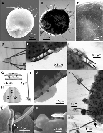
Thaumatomastix triangulata (Balonov, Reference Balonov1980) Beech & Moestrup, Reference Beech and Moestrup1986, emend.: (A, B) whole cells with investitures of plate-scales and spine-scales; (C) the distal surface of a patch of interlocking plate-scales showing their rounded triangular outlines; (D) apices of several spine-scales; (E, F) lateral views of plate-scales showing the three pillars in each (arrow); (G–I) diagrammatic representation of the plate-scale (G, H) and spine-scale (I) structure; (J) plate-scales; (K) plate-scales and two spine-scales; (L) shaft of the spine-scale anchored at an angle to the upper base-plate (see also I and N); (M) plate-scales in plan view (on the left) and in oblique lateral view (upper right); (N) edge of a cell with plate-scales and a single spine-scale showing the angle of attachment of the spine shaft to the basal structure.

NEOTYPIC HABITAT
Benthic sediment–water interface, west arm of Riley Lake, Ryde Township, Muskoka District (44°50′15″N 79°12′30″W).
MATERIAL FROM THE NEOTYPE LOCALITY
Retained by the author as a formalin-preserved aqueous sample (No. V-2096) collected 10 October 2007.
ADDITIONAL REMARKS
Five (in TEM) and six (in SEM) different whole cells from the Ontario collection (Riley Lake) were examined for their scale structure; all revealed the presence of two scale types of typical Thaumatomastix morphology. Many whole cells in living and formalin-fixed states were examined by light microscopy (100 × oil-immersion phase contrast and differential interference contrast), and no chloroplasts were found; neither were any colonies like those reported by Balonov (Reference Balonov1980) found. Balonov (Reference Balonov1980) described the cells as obovate, 15–22 × 18–25 µm; the plate-scales as rounded-triangular 1.1–1.4 µm ‘long' (it is possible that this was a measurement along one side of the triangle, which is difficult to define owing to the rounded corners, and perhaps the reason Wujek et al. (2008) advocated measurement from the outer edge of one corner to the mid-point on the opposite side of the scale). Balonov described the spine-scales as hollow, 4.8–8.1 µm long, tapering to a sharp-pointed apex; the diameter of the spine ‘trunk' not greater than 0.22 µm and attached to a triangular base-plate, 1.1–1.2 µm wide. Aside from the mention of the 'colonies', and the chloroplast, cell size and scale structure is in excellent agreement with the specimens described here from Ontario.
Clearly, this organism is a Thaumatomastix, not a Chrysosphaerella, and the evidence presented here suggests strongly that the Ontario specimens are identical to those described by Balonov (Reference Balonov1980), thus providing support for Beech & Moestrup's (1986) transfer of this taxon from Chrysosphaerella to Thaumatomastix and challenging the conclusion by Howe et al. (Reference Howe, Bass, Scoble, Lewis, Vickerman, Arndt and Cavalier-Smith2011) that Beech & Moestrup (Reference Beech and Moestrup1986) ‘were wrong'. It now appears much more likely that Balonov (Reference Balonov1980) made errors in interpretation of his light microscope observations that led him to assign this taxon to Chrysosphaerella (see also the Discussion section, below).
Additional freshwater unidentified Thaumatomastix
The Ontario freshwater collections include three plate-scales (Figure 9) whose structures are not in accordance with those of either T. triangulata or T. nigeriensis, although the well-defined border surrounding the triangular central areas of these scales suggests some similarity to the marine species T. formosa Thomsen et al. (Reference Thomsen, Hällfors, Hällfors and Ikävalko1993) (= Reckertia formosa Cavalier-Smith, in Howe et al., 2011). Because whole cells or spine-scales were not found in association with these scales they are simply included here as unidentified Thaumatomastix (or perhaps, but less likely, Reckertia) species, owing to similarities in scale structure.
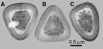
Three unidentified Thaumatomastix (Reckertia ?) plate-scales. The three different scales (A–C) may represent a single species or more than one species.

SYSTEMATICS
Phylum CERCOZOA Cavalier-Smith, 1998
Class IMBRICATEA Cavalier-Smith & Chao, Reference Cavalier-Smith and Chao2003
Order THAUMATOMONADIDA Shirkina, 1987
Family THAUMATOMONADIDAE Holland, 1934; emend. Howe, Bass, Scoble, Lewis, Vickerman, Arndt & Cavalier-Smith, Reference Howe, Bass, Scoble, Lewis, Vickerman, Arndt and Cavalier-Smith2011
Genus Reckertia Conrad, 1920; emend. Howe, Bass, Scoble, Lewis, Vickerman, Arndt & Cavalier-Smith, Reference Howe, Bass, Scoble, Lewis, Vickerman, Arndt and Cavalier-Smith2011
NEW SPECIES
Reckertia hindoni sp. nov.
(Figure 10A–F)
DIAGNOSIS
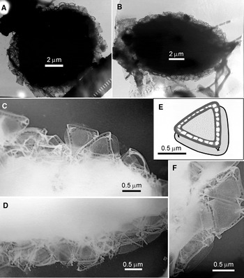
Cells 10–13 µm in diameter covered plate-scales of a single type. Plate-scales consist of a rounded-triangular baseplate, 0.7–1 µm in size, separated from a more sharply tri-angled secondary plate by a short branched strut located at each of the three corners. The secondary plate has straight or convex edges with a thickened margin and a submarginal border of elongate perforations.
ETYMOLOGY
The specific epithet (hindoni) refers to the administrative locality (Hindon Township) within which the type collection was made.
TYPE SPECIMEN
Reckertia hindoni sp. nov.: (A, B) transmission electron microscope (TEM) images of whole cells with investitures of scales; (C, D & F) TEM images of scales; (E) diagrammatic representation of a single scale.

TYPE LOCALITY
A shallow pool in a small intermittent stream in Hindon Township, Ontario, Canada (45°03′44″N 78°57′37″W).
MATERIAL FROM THE TYPE LOCALITY
Collected 25 October 1988 and retained by K. Nicholls as a formalin-preserved aqueous sample (No. V-826).
ADDITIONAL REMARKS
A second whole cell (Figure 10B) with scales identical to those on the type specimen was found in a different sample (V-823) collected 25 October 1988 from a marshy stream, 0.5 km north-east of Clear Lake, Oakley Township, 45°0′55″N 79°0′35″W (pH = 5.3, sp. cond. = 20 µS cm−1). There is some resemblance of the scales of Reckertia hindoni to those of Thaumatomastix ‘sp. #5′ of Thomsen & Ikävalko Reference Thomsen and Ikävalko(1997; now considered to be an unidentified Reckertia species per Howe et al. (Reference Howe, Bass, Scoble, Lewis, Vickerman, Arndt and Cavalier-Smith2011)), or those of R. igloolika (Thomsen & Ikävalko) Cavalier-Smith (in Howe et al. Reference Howe, Bass, Scoble, Lewis, Vickerman, Arndt and Cavalier-Smith2011), but R. igloolika scales have convex margins and lack the submarginal row of perforations in the secondary plate.
Reckertia hindoni represents the only freshwater member of this genus; all others being known from marine sediments or sea-ice.
DISCUSSION
Until now, the marine species of the genus Thaumatomastix were known to have either elliptical plate-scales (T. salina (Birch-Andersen) Beech & Moestrup, Reference Beech and Moestrup1986; T. bipartita Beech & Moestrup, Reference Beech and Moestrup1986), or triangular plate-scales (T. formosa Thomsen G. Hällfors, S. Hällfors & Ikävalko, 1993; T. patelliformis (Takahashi & Hara) Beech & Moestrup, Reference Beech and Moestrup1986; T. salina (Birch-Andersen) Beech & Moestrup, Reference Beech and Moestrup1986; T. tripus (Takahashi & Hara) Beech & Moestrup, Reference Beech and Moestrup1986; T. tauryanini Mikrjukov, Reference Mikrjukov2002). To this latter group can now be added the new species described here, T. inornata, T. sablensis and T. curvata. The plate-scales of these species all have well-defined tri-radial symmetry with a separate pillar located near each corner of the scale joining the two plates of the scale.
The other three new marine species described in this paper (T. asymmetrica, T. multipora and T. gwaii) do not truly belong in the aforementioned elliptical scale group. Instead, they represent an intermediate morphology with characteristics of both the elliptic group and the triangular group. All have bilateral symmetry and a strong tendency to display two pillars at one end (these may be fused into a single structure) and a single pillar at the other end of the nearly elliptical scales. The outline of the T. gwaii plate-scales is closer to a triangular form than that of either T. asymmetrica or T. multipora. Whereas the other 10 marine triangular scaled species listed above and the two freshwater species (T. triangulata and T. nigeriensis) have plate-scales in the shape of equilateral triangles, the plate-scales of T. gwaii are well-defined isosceles triangles with rounded corners, and could be imagined as the product of a blending of an ellipse and an equilateral triangle. It is therefore likely that all of these species had a common ancestor and the differences in scale shape of the extant species today reflect different degrees of genetic isolation and change over time. Searches in other parts of the world for the six new marine species reported here would help to define the stability over large geographical distances of these distinctly different morpho-species. It is interesting to note that the freshwater T. nigeriensis, although perhaps rare (Wujek et al., Reference Wujek, Pershon and Kadiri2008; Wujek, Reference Wujek2010; to which can be added the Ontario, Canada findings reported here) appears to be morphologically stable over its known range (Africa and North America), but is perhaps restricted to soft water, acidic habitats.
This paper reports for the first time the ultra-structure of whole cells of T. triangulata, since Balonov's (1980) initial discovery. Four loose scales from Sri Lanka (Dürrschmidt, Reference Dürrschmidt1989; her figure 53) and attributed to T. triangulata by her reveal scale size of under 1 µm and a considerable zone of electron-dense material surrounding the ‘eyes' and so may represent a different species or subspecies. No spine scales or whole cells were reported, however, so further judgement about the taxonomic status cannot be made of those and other specimens reported by Dürrschmidt (Reference Dürrschmidt1989) as being common in samples from Chile, New Zealand and Malaysia, presumably with scale structure similar to those illustrated from the Sri Lankan collection. Other reports with EM images of plate-scales that appear very similar to T. triangulata (but did not include whole cells or associated spine-scales) include Vørs (Reference Vørs1992) from the Gulf of Finland, Thomsen et al. (Reference Thomsen, Hällfors, Hällfors and Ikävalko1993) from Isefjorden Denmark and Jocobsen (Reference Jocobsen1985) from Greenland. Additionally, a large cluster of plate-scales with sizes ranging between 1.1 and 1.3 µm and attributed to T. triangulata from a collection in eastern Bohemia (Czech Republic) by Štephanová & Kalina (1992) appear to be identical in size and structure to those reported here from Riley Lake in Ontario. Thomsen et al.'s (1993) group of plate-scales (their figure 28) from a Danish brackish-water locality reveals scales identical in structure to the Ontario and Czech specimens, but of a larger size (1.4–1.5 µm).
Unfortunately, images of the lateral views of the plate-scales were not included in the Czech or Danish illustrations mentioned above; the pillars were separate in the Sri Lankan material (but again, these scales may not belong to T. triangulata—see reference to Dürrschmidt (Reference Dürrschmidt1989) above). Because no other published images of spine-scales existed, Thomsen et al.'s (1993) drawing of a spine-scale of T. triangulata must have been based on Balonov's images, and would appear to be an accurate interpretation of the structure that includes a relatively large triangular upper basal plate separated from the lower (proximal) plate by three separated pillars. The spine shaft appears to be anchored near the centre of the upper plate and tapers to a sharp point. The scale structure of the Ontario specimens is in agreement with this interpretation.
Thaumatomastix triangulata is similar in some ways to the marine species T. formosa Thomsen, Hällfors, Hällfors & Ikävalko, Reference Thomsen, Hällfors, Hällfors and Ikävalko1993. The differences between them include: (i) T. formosa apparently lacks spine-scales; (ii) its plate-scales include a wider separation of the columnar structures (pillars) separating the two plates of the scale (the pillars are much closer to the edges of the scale in T. formosa than in T. triangulata); and (iii) the scales of T. formosa have a prominent marginal zone easily distinguished from the central region of the scale (such a marginal zone is not delimited in T. triangulata).
It is not clear why Thomsen et al. (Reference Thomsen, Hällfors, Hällfors and Ikävalko1993) concluded that the three pillars separating the two plates of a plate-scale of T. triangulata were fused (they cited this as a major feature distinguishing T. triangulata from T. formosa, in which the pillars are separated). In their figure 52, Thomsen et al. (Reference Thomsen, Hällfors, Hällfors and Ikävalko1993) show the pillars in T. triangulata as separate structures (certainly not fused to the degree they are in T. tripus and T. patelliformis, which they also illustrate for comparison purposes). In the Ontario specimens, they clearly are separate structures, as they are in the type material from Rybinsk, Russia (Balonov's plate 2, figure 3). This is important, because Thomsen et al. (Reference Thomsen, Hällfors, Hällfors and Ikävalko1993) state that this difference (fused or not) is a major difference between T. formosa and T. triangulata. Assuming their statement is in error (i.e. the correct conclusion is that pillars between the plates are separate in both species), the differences between the two species are still well-defined, given the four differences listed here (above) and are, in my opinion, adequate to keep the freshwater T. triangulata taxonomically distinct from the marine T. formosa.
Howe et al. (Reference Howe, Bass, Scoble, Lewis, Vickerman, Arndt and Cavalier-Smith2011) gave several reasons to justify the invalidity of Beech & Moestrup's (1986) transfer of Chyrsosphaerella triangulata Balonov to Thaumatomastix. I have presented (above) evidence to show that Balonov's Chrysosphaerella triangulata is very likely the same organism as the Ontario specimens described here (and amended) under Thaumatomastix triangulata (Balonov) Beech & Moestrup, Reference Beech and Moestrup1986. The rejection of the Beech & Moestrup transfer to Thaumatomastix by Howe et al. (Reference Howe, Bass, Scoble, Lewis, Vickerman, Arndt and Cavalier-Smith2011) must itself be rejected in light of the present evidence. Still, there needs to be an accounting of the source of the confusion associated with Balonov's publication.
The silica-scaled autotrophic heterokont freshwater genus Chrysosphaerella is well-circumscribed morphologically (Nicholls, Reference Nicholls1980; Wujek, Reference Wujek1985/1986). Balonov's (1980) description of his ‘Chrysosphaerella triangulata' included mention (and a drawing—his plate 1a) of a 4-celled ‘colony' 54 µm in diameter. Chrysosphaerella species are colonial, but Thaumatomastix is not known to form colonies. It is possible that Balonov observed two cells in close proximity that were in the late stages of binary fission (= 4 cells grouped together, but not forming a true colonial structure). He also mentioned the presence of a chloroplast, a stigma, and a ‘flagellum pinnatum' (presumably the mastigonemes of the longer flagellum known for Chrysosphaerella species (Wujek, Reference Wujek1968)). None of these organelles is known for Thaumatomastix and, together with an argument about scale structure (see below), was the reason Balonov's taxon was reinstated by Howe et al. (2011) as Chrysosphaerella, thus rejecting the combination proposed by Beech & Moestrup (Reference Beech and Moestrup1986) who had transferred this taxon to Thaumatomastix.
Of course, these are all good reasons for the transfer back to Chrysosphaerella, had the original description been more believable. Evidence in the form of camera images was not provided by Balonov to support his claim of a tinsellated flagellum, a chloroplast or a stigma. Others have made similar mistakes in describing the presence of these organelles when, in fact, they were absent. For example, Takahashi & Hara (Reference Takahashi and Hara1984) described the presence of ‘a single yellowish or pale-green chloroplast and a stigma …' when they described Chrysosphaerella tripus and C. patelliformis. The same mistake was apparently made by Birch-Andersen (Reference Birch-Andersen1973) in describing ‘two parietal chloroplasts, or possibly one deeply lobed, yellowish-brown …' for his ‘Chrysosphaerella salina'. All three of the above ‘Chrysosphaerella' species are now considered to be ‘good' species of Thaumatomastix. Similarly, Štefanová & Kalina (Reference Štefanová and Kalina1992) erroneously described ‘Spiniferomonas pectinata' as a new species of the chloroplast-bearing genus Spiniferomonas, obviously unaware of the proper classification of this organism as the non-chloroplast-bearing Pterocystis fortesca (Nicholls, 1983) Siemensma & Roijackers, 1988. In conclusion, in the absence of critical microscopic images, verbal descriptions alone of organelles like chloroplasts, stigmata, and mastigonemes must not be afforded a high degree of authenticity in this case.
The other main argument put forward by Howe et al. (Reference Howe, Bass, Scoble, Lewis, Vickerman, Arndt and Cavalier-Smith2011) for rejection of the Beech & Moestrup transfer of Balonov's C. triangulata to Thaumatomastix centred on the structure of the spine-scales. Howe et al.'s (2011) concept of a ‘typical' Thaumatomastix spine-scale was patterned after the marine species that have ribbed spine shafts, bi- or tri-furcate apices and a basal structure consisting of a primary base-plate and a smaller ledge-like secondary plate or collar around the base of the shaft above the primary base-plate. Correctly, they identified more similarities between the spines of Balonov's C. triangulata and those of the two most common freshwater colonial Chrysosphaerella species. Now, however, with the amended descriptions of both T. nigeriensis and T. triangulata given in this paper, it is apparent that the concept of spine-scale morphololgy in Thaumatomastix must be expanded to include those with hollow spine shafts tapering to a simple sharp-pointed apex and a secondary base plate that is much more prominent than that known previously. This new model of Thaumatomastix spine-scale structure clearly supports Balonov's taxon. Furthermore, the plate-scales of Chrysospharella are not of the double plate structure with three interconnecting pillars commonly found in Thaumatomastix (including Balonov's and the Ontario specimens reported here), so the scale morphology argument used by Howe et al. (Reference Howe, Bass, Scoble, Lewis, Vickerman, Arndt and Cavalier-Smith2011) must be discounted. These reasons, and those given above relating to the high degree of similarity between the EM images provided by Balonov (Reference Balonov1980) and the non-colonial, non-chloroplast-bearing cells described here from Ontario, must lead to the reinstatement of this taxon as Thaumatomastix triangulata (Balonov) Beech & Moestrup, Reference Beech and Moestrup1986.
It is possible that further studies of the spined thaumatomonads will find a reason to separate (in a new genus) the hollow-spined taxa reported here from those with ribbed spines (e.g. T. bipartita and T. salina). I believe this to be premature at this time, however, owing to the great degree of similarity among the plate-scales and the spine bases of both groups. Certainly, there exist gradients among those taxa with triangular plate-scales and others with near-elliptical shapes. Clearly, the diversity of form is large in this group and needs further investigation based on much more collecting from both marine and freshwater habitats. Expanded study of this group must lead to clearer lines of taxonomic separation based on morphology and genetics before any useful and stable revision of the classification of Thaumatomastix can be undertaken.
Interestingly, the validation of the genus Reckertia by Howe et al. (Reference Howe, Bass, Scoble, Lewis, Vickerman, Arndt and Cavalier-Smith2011) may have been on the minds of other protistologists for some time. Wujek et al. (Reference Wujek, Pershon and Kadiri2008) described Thaumatomastix as ‘… an unwieldy, artificial conglomeration of forms …'. In 2001, I reviewed a draft of a paper by K. Mikrjukov in which he proposed to reinstate Reckertia for reasons (scale morphology) similar to those put forward by Howe et al. (Reference Howe, Bass, Scoble, Lewis, Vickerman, Arndt and Cavalier-Smith2011). Unfortunately, Mikrjukov's paper was not published (he died in 2002), but it is likely that the changes formally introduced by Howe et al. (Reference Howe, Bass, Scoble, Lewis, Vickerman, Arndt and Cavalier-Smith2011) will be welcomed by students of this branch of protistology.
ACKNOWLEDGEMENTS
I appreciate the efforts of Zoe Lucas and Doug Burles for sampling on the Atlantic and Pacific coasts, respectively. John Wainwright and David Fung, Ontario Ministry of the Environment, assisted with electron microscopy and developed film during the 1980s. More recently, Karen Rethoret, Biology Department, York University, Ontario, assisted with the scanning electron microscope and transmission electron microscope operations.