INTRODUCTION
The flagellated trypanosomatids of the genus Phytomonas and some Herpetomonas spp. are parasites of plants. In some cases they parasitize plants without apparent pathogenicity but they can also cause diseases of economic significance in plantations of coconut, oil palm, cassava and coffee (Camargo et al. Reference Camargo, Kastelein and Roitman1990; Redman et al. Reference Redman, Schineider, Mehlert and Ferguson1995). These trypanosomatids have also been detected in various edible fruits, such as pomegranates, peaches, guavas and tangerines and in their insect vectors (Redman et al. Reference Redman, Schineider, Mehlert and Ferguson1995). The parasites live mostly in the xylem and phloem of the infected plants and are thought to be transmitted through the bite of phytophagous insects (Camargo et al. Reference Camargo, Kastelein and Roitman1990; Redman et al. Reference Redman, Schineider, Mehlert and Ferguson1995). In the biological cycle of these pathogens, several plant-sucking insects act as intermediate hosts and the plant acts as the main host (Jankevicius et al. Reference Jankevicius, Jankevicius, Campaner, Conchon, Maeda, Teixeira, Freymuller and Camargo1989). These parasites including Herpetomonas spp. have ultrastructural features typical of the Trypanosomatidae family. They contain a kinetoplast, endoplasmic reticulum, acidocalcisomes and single tubular mitochondria (with few cristae) (Attias et al. Reference Attias, Roitman, Camargo, Dollet and De Souza1988; Freymuller et al. Reference Freymuller, Milder, Jankevicius, Jankevicius and Camargo1990; Redman et al. Reference Redman, Schineider, Mehlert and Ferguson1995; Sodré et al. Reference Sodré, Moreira, Nobrega, Gadelha, Meyer-Fernandes, Dutra, Vercesi, Lopes, Scofano and Barrabin2000; Moysés and Barrabin, Reference Moysés and Barrabin2004; Medeiros et al. Reference Medeiros, Moreira, Kildare, de Souza, Plattner, Hentschel and Barrabin2005).
It is well established that calcium ions play a crucial role in controlling a large number of cellular functions, conveying signals received at the cell surface to inside the cells. The free cytosolic Ca+2 concentration is maintained in different cells from diverse organisms at a very low level (10−7m), as compared to the concentration in the extracellular fluid (about 10−3m). This gradient is sustained by the active transport of the ion by specific Ca2+ pumps present in the plasma membrane, endoplasmic reticulum, and mitochondria (Clapham, Reference Clapham1995; Berridge et al. Reference Berridge, Bootman and Roderick2003). In order to exert such functions, Ca2+ distribution across the plasma membrane and the membranes of other organelles must be regulated precisely and presents the capacity for fast fluctuations (Clapham, Reference Clapham1995; Berridge et al. Reference Berridge, Bootman and Roderick2003).
In other trypanosomatids such as Trypanosoma cruzi, Trypanosoma brucei and Leishmania spp, it has been shown that mitochondria, endoplasmic reticulum and acidic vacuoles participate in the Ca2+ homeostasis in these cells (Docampo and Vercesi, Reference Docampo and Vercesi1989; Moreno et al. Reference Moreno, Docampo and Vercesi1992; Docampo, Reference Docampo1993; Vercesi et al. Reference Vercesi, Moreno and Docampo1994; Docampo et al. Reference Docampo, Scott, Verscesi and Moreno1995; Lu et al. Reference Lu, Zhong, De Souza, Benchimol, Moreno and Docampo1998; Scott and Docampo, Reference Scott and Docampo1998; Rodrigues et al. Reference Rodrigues, Catisti, Uyemura, Vercesi, Lira, Rodrigues, Urbina and Docampo2001). The study of the mechanisms by which a plant parasite (Herpetomonas sp.) regulates intracellular Ca2+ distribution to maintain cell viability could provide new insights into the physiology of these parasites and, in addition, could be useful for the design of new drugs.
Our group investigated the Ca2+ transport and the mitochondrial bioenergetics of digitonin-permeabilized Herpetomonas sp. promastigotes (Sodré et al. Reference Sodré, Moreira, Nobrega, Gadelha, Meyer-Fernandes, Dutra, Vercesi, Lopes, Scofano and Barrabin2000). The response of the promastigote mitochondrial membrane potential to ADP, oligomycin, Ca2+ and antimycin A indicated that it behaves similarly to vertebrate and T. cruzi mitochondria regarding the properties of their electrochemical proton gradient. Ca2+ transport by permeabilized cells appears to be performed mainly by the mitochondria and a vanadate-sensitive Ca2+ transport system, attributed to the endoplasmic reticulum (Sodré et al. Reference Sodré, Moreira, Nobrega, Gadelha, Meyer-Fernandes, Dutra, Vercesi, Lopes, Scofano and Barrabin2000). Thapsigargin and IP3 were not able to affect the vanadate-sensitive Ca2+ transport. Nigericin, FCCP or bafilomycin A1 also had no effect on the vanadate-sensitive Ca2+ transport, indicating the absence of a Ca2+ transport mediated by a Ca2+/H+ exchanger coupled to an H+-ATPase. No evidence of a third Ca2+ compartment with the characteristics of the acidocalcisomes described by Vercesi et al. (Reference Vercesi, Moreno and Docampo1994) was observed in those experiments. However, under the same conditions, H+ uptake stimulated by pyrophosphatase (Ppase) and structures like acidocalcisomes had already been identified (Medeiros et al. Reference Medeiros, Moreira, Kildare, de Souza, Plattner, Hentschel and Barrabin2005). The proton gradient generated by PPi can be dissipated by the addition of 20–50 μm Ca2+, suggesting the presence of a Ca2+/H+ exchanger (Medeiros et al. Reference Medeiros, Moreira, Kildare, de Souza, Plattner, Hentschel and Barrabin2005). Discrepancies between experiments have been attributed to an undesirable effect of digitonin solubilizing the membranes rather than the cytoplasm. Such an effect of digitonin has been attributed, in some cases, to the cell growth medium composition (Zhang et al. Reference Zhang, McDonald, Tanowitz, Wittner, Weiss, Bilezikian and Morris1998; Miranda et al. Reference Miranda, Vercesi, Catisti, de Souza, Rodrigues and Docampo2005).
To overcome possible alterations in the membrane composition caused by digitonin, we studied the Ca2+ transport systems in a subcellular fraction from Herpetomonas sp. promastigotes isolated after mechanical cell disruption. The results showed a Ca2+ uptake promoted by ATP, or other nucleotide except ITP, which is not affected by H+ ionophores but is inhibited by vanadate.
MATERIALS AND METHODS
Chemicals
Sodium orthovanadate, antimycin A, thapsigargin, oligomycin, carbonylcyanide p-trifluoromethoxiphenylhydrazone (FCCP), nigericin, bafilomycin A1, ATP, GTP, arsenazo III, digitonin, and calcium ionophore (A23187) were purchased from Sigma Chemical Co. All other reagents were analytical grade.
Culture method
Herpetomonas sp. promastigotes (CT.IOC 13; kindly provided by Dr Reginaldo Brazil, Instituto Oswaldo Cruz, FIOCRUZ, RJ, Brazil) were grown at 25°C in a medium containing 20 g/l sucrose, 20 g/l KCl, 3 g/l peptone, 1 mg/l folic acid, supplemented with 10 mg/l haemin and 10% (v/v) fetal bovine serum. At 2–3 days after inoculation, cells were harvested by centrifugation, washed twice with 5 mm phosphate buffer, pH 7·0, containing 150 mm NaCl (PBS) and resuspended in the same solution. Protein concentration was determined by the biuret assay (Gornall et al. Reference Gornall, Bardawill and David1949) in the presence of 0·2% deoxycolate.
Subcellular preparation
Cells (approx. 5×10⁹ for each preparation) were resuspended in a minimal volume of ice-cold lysis buffer: 125 mm sucrose, 50 mm KCl, 4 mm MgCl2, 0·5 mm EGTA, 20 mm HEPES, pH 7·2, 5 mm DTT, 0·1 mm phenylmethanesulphonyl fluoride (PMSF), 10 μm pepstatin A, 10 μm leupeptin, 10 μm quimostatin and 10 μm E64. Then cells were mixed with glass microspheres (0·1 μm) at a 1:2 mass proportion and disrupted in a mortar with gently rotary movements of a pestle at 0–4°C, until most of the cells were disrupted as confirmed by microscopic observation (2–4 min). The homogenate was diluted with 5 volumes of lysis buffer and submitted to a 1 min centrifugation at 100 g to remove the glass particles and then once more at 1000 g for 10 min to remove debris, live cells and most of the nuclei and mitochondria. The supernatant fraction was centrifuged at 15000 g for 15 min and the pellet resuspended in 1 ml of lysis buffer for use in the assays. Protein was measured by the method of biuret (Gornall et al. Reference Gornall, Bardawill and David1949).
Ca2+ uptake
Calcium uptake was measured by incubating the subcellular fraction (0·3 mg.ml−1) in medium containing 75 mm HEPES-Tris, pH 7·0, 120 mm KCl, 2 mm mercaptoethanol, 1 mm ATP, 1 mm MgCl2, 0·15 mm EGTA, 0·2 mm CaCl2, 4 mm K+-oxalate, 1 mm ouabain, 10 mm sodium azide, 50 mm KNO3 and trace of 45Ca-CaCl2 to a final concentration of 2×10³ dpm.nmol−1. We did not perceive any significant effect of ouabain (1 mm) or KNO3 (50 mm) neither in the Ca2+ uptake nor in the ATPase activities under the conditions described in this study; however, they were included in the standard medium because, under different conditions of culture, some Na,K–ATPase or phosphatase can eventually contribute to increasing ATP hydrolysis and challenging the sarcoplasmic reticulum Ca2+-ATPase (SERCA) activity measurements. After incubation for 1 h aliquots of 0·5 ml were withdrawn, filtered through Millipore filters (HPWS 0·45 μm) under vacuum and washed 3 times with 5 ml of a ice-cold solution of 1 mm LaCl3, pH 7·0. The filters were dried and the radioactivity measured in a scintillation counter.
Free calcium concentrations were calculated as described by Fabiato and Fabiato (Reference Fabiato and Fabiato1979), as modified by Sorenson et al. (Reference Sorenson, Coelho and Reuben1986), using the dissociation constants for the Ca-EGTA complexes reported by Schwartzenbach et al. (Reference Schwarzenbach, Senn and Anderegg1957).
ATPase activity
The Ca2++Mg2+-ATPase activity was measured by incubating the subcellular fraction (0·3 mg.ml−1) in medium containing 75 mm HEPES-Tris, pH 7·0, 120 mm KCl, 2 mm mercaptoethanol, 2 mm MgCl2, 0·2 mm CaCl2, 10 μm calcium ionophore A23187, 1 mm ouabain, 10 mm sodium azide, 50 mm KNO3 and 2 mm (γ-32P)ATP (2×10⁵ dpm.μmol−1). After incubation for 1 h the reactions were arrested by the addition of 4 volumes of activated charcoal resuspended in 0·1 m HCl. The samples were centrifuged at 1200 g during 5 min and aliquots of the supernatant were withdrawn to measure 32Pi by scintillation counting. The Mg2+-dependent ATPase was measured in the same medium except that CaCl2 was substituted by 0·15 mm EGTA.
RESULTS
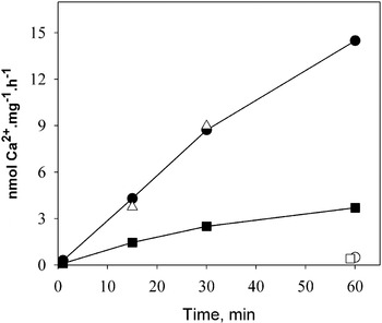
The Herpetomonas sp. subcellular fraction was able to actively transport Ca2+ using ATP as hydrolysable substrate (Fig. 1). This fraction is expected to contain different compartments such as endoplasmic reticulum, mitochondria and acidocalcisomes, all with the capability of sustaining Ca2+ uptake in the presence of the appropriate substrate. In fact, sodium azide (10 mm) was able to inhibit about 30% of the total uptake (result not shown) indicating that the presence of this inhibitor was important to avoid any mitochondrial contribution in the uptake. Using acridine orange as a probe, we observed that PPi, but not ATP, strongly stimulates H+ uptake in our preparations denoting the presence of H+-PPase from acidocalcisomes. Indeed, PPi was able to sustain a measurable Ca2+ uptake, sensitive to FCCP, of about 3–5 nmol of Ca2+.mg−1/h−1 (not shown). However, the Ca2+ uptake promoted by ATP studied here was FCCP insensitive (see ahead and Fig. 6) and so not related to acidocalcisomes.
Oxalate stimulation of Ca2+-uptake in the subcellular fraction. Ca2+-uptake in the subcellular fraction from Herpetomonas sp. was measured as described in the Materials and Methods section, at increasing incubation time in medium containing CaCl2 to have 50 μm free calcium in the presence (•) and in the absence (▪) of 5 mm oxalate. Open triangles represent uptake in the presence of oxalate and 2 μg/ml of calmodulin. Uptake in the presence of 10 μm ionophore A23187 in the presence (○) and absence (□) of oxalate are also indicated. The data represent a typical experiment that was repeated 3 times with an identical pattern.

The transport shown in Fig. 1 was strongly stimulated by the presence of oxalate in the reaction medium that presumably prevents calcium accumulation inside the vesicles by precipitation of Ca2+. In both conditions the Ca2+ ionophore A23187 was able to prevent the uptake indicating that the process was against a concentration gradient. In the presence of oxalate the active transport was approximately linear for at least 60 min.
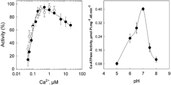
The Ca2+-uptake gradually increased with the calcium concentration, showing an apparent affinity near 0·1 μm and a maximal rate of 20±3 nmol of Ca2+.mg−1.h−1 at 0·5–1 μm Ca2+ (Fig. 2A, open circles). The subcellular fraction hydrolysed ATP in the absence of Ca2+ at a rate of 0·84±0·10 μmol.mg−1.h−1(n=9). Calcium stimulated the hydrolysis with Km=0·083±0·003 μm and nH=2·9±0·4 (Fig. 2A, closed circles). However the observed maximal Ca2+-dependent hydrolytic activity (0·49±0·09 μmol.mg−1.h−1) was much higher than the Ca2+ transport. This result suggests that the Ca2+-activated ATPase may be responsible for the Ca2+ transport observed in Fig. 1. The low levels of transport observed, as compared to the Ca2+-ATPase activity, may be due to some vesicles not being sealed. This Ca2+-dependent ATPase activity was critically dependent on pH, with an optimum at pH 7·0 (Fig. 2B).
(A) Ca2+ dependence of subcellular Ca2+-uptake (○) and Ca2+-ATPase activities (•). Uptake in the presence of oxalate and ATPase activities was measured as indicated in the Materials and Methods section. Values represent the mean of 4 experiments −/+s.e. (standard error). Maximal values representing 100% were 19 nmol of Ca2+.mg−1.min−1 (○) and 0·51 μmol of Pi.mg−1.min−1 (•). Continuous line is the non-linear regression fit of ATPase data using the function activity=100/((1+(Km/[Ca2+])nH).(1+([Ca2+]/Ki)m)), where Km and n H are the Michaelis-Menten constant and Hill number for the activation by Ca2+ and Ki and m are the apparent inhibition constant and the Hill number for the inhibition process. (B) pH dependence on Ca2+-ATPase activity. Ca2+-ATPase activity was measured using HEPES-Tris buffer at pH 7–8 and MES-Tris buffer for the pH range 5–6.5. Values represent mean of experiments −/+s.e.

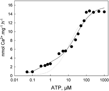
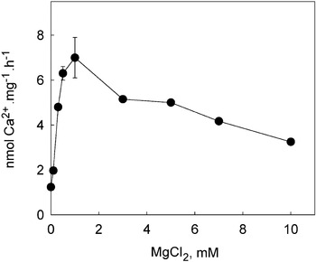
As was shown for the SERCA and plasma membrane Ca2+-ATPase (PMCA) of mammals, the Ca2+ transport was stimulated by ATP by a complex mechanism that involves an activation in the 0·1–5 μm range and a second activation in the 5–1000 μm range of ATP (Fig. 3). This behaviour could be simulated by considering the sum of two Michaelis-Menten curves (Fig. 3, continuous line) with Km1=0·25±0·19 μm, Km2=29·6±6·8 μm and Vm1=3·3±0·8 and Vm2=12·4±0·8 nmol.mg−1.h−1. A poor fitting was obtained by using a single Michaelis-Menten curve (Fig. 3 dotted line). Fig. 4 shows that Mg2+ is necessary for Ca2+ transport; however, concentrations higher than equimolar with ATP resulted in progressive inhibition. This result suggests that the Mg-ATP complex is the true substrate of the ATPase and that free Mg2+ acts as a mild inhibitor.
ATP dependence of Ca2+-uptake. Uptake was measured as described in the Materials and Methods section, in medium containing 1 mm Mg2+, 50 μm Ca2+ and 2 mm phosphoenol-pyruvate plus 2 IU of pyruvate kinase as ATP regenerator system. The traced line represents the best fit using a single Michaelis-Menten function. The continuous line is the non-linear regression fit obtained by using the function v=V 1/(1+Km 1/[ATP])+V 2/(1+Km 2/[ATP]).

MgCl2 dependence of Ca2+-uptake. Uptake was measured as described in the Materials and Methods section, in medium containing 1 mm ATP+ and 50 μm Ca2+. Values represent mean of 3 experiments −/+s.e.

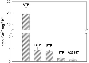
The SERCA and PMCA active Ca2+-transport systems that have been described in the literature were distinguished by their specificity for nucleotides. The former accepts not only ATP but also GTP or ITP while the latter could only use ATP as substrate for Ca2+ transport. We found that GTP and UTP were able to sustain the Ca2+ uptake in the subcellular fraction of Herpetomonas sp. though with a lower rate than ATP (Fig. 5). ITP was almost ineffective. GTP-dependent Ca2+ uptake by endoplasmic reticulum was previously observed in Herpetomonas sp. permeabilized with digitonin (Sodré et al. Reference Sodré, Moreira, Nobrega, Gadelha, Meyer-Fernandes, Dutra, Vercesi, Lopes, Scofano and Barrabin2000).
Substrate specificity of Ca2+ uptake. Uptake was measured as described in the Materials and Methods section, in medium containing 1 mm Mg2+, 50 μm Ca2+ and 1 mm ATP or 3 mm GTP, UTP or ITP. Residual uptake in the presence of ATP as substrate and 10 μm ionophore A23187 is indicated. Values represent mean of 3 experiments −/+s.e.

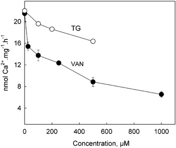
To better characterize the transport of calcium the effects of cyclopiazonic (1 mm) acid and thapsigargin (up to 0·5 mm, see Fig. 7), inhibitors of SERCA, and bafilomycin (50 nm), an inhibitor of V-ATPases, were tested. Both types of inhibitor were ineffective as inhibitors of the Ca2+ uptake. However, it should be mentioned that in other trypanosomatids, including permeabilized Herpetomonas sp thapsigargin was also found to be almost ineffective as an inhibitor of vanadate-sensitive Ca2+ uptake. Vanadate itself is a potent inhibitor of P-type ATPases of mammals: however, it inhibited the Ca2+ uptake with low affinity (Fig. 7). It seems that the structure of the ATPase in Herpetomonas sp. has important differences that confer insensitivity towards the typical inhibitors of SERCA.
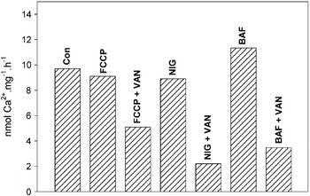
Acidocalsisomes are organelles found in some parasites of the Tripanosomatidae family that actively accumulates Ca2+ and H+. It was previously shown that collapsing the H+ gradient initiates a Ca2+ efflux through an H+/Ca2+ exchanger (Medeiros et al. Reference Medeiros, Moreira, Kildare, de Souza, Plattner, Hentschel and Barrabin2005). Our results show that the H+ ionophore FCCP and the H+/Na+ exchanger nigericyn were ineffective in inhibiting the Ca2+ uptake (Fig. 6) indicating that acidocalcisomes were not involved in the Ca2+ uptake of the subcellular fraction. Bafilomycin, a specific inhibitor of V-H+-ATPases, had also no effect on transport, supporting the idea of a primary active Ca2+ transport system.
Effects of inhibitors and H+ ionophores on Ca2+ uptake. The uptake of Ca2+ in the subcellular fraction was measured in medium containing either 50 nm bafilomycin (BAF), 1 μm FCCP, 1 μm nigericin (NIG) or 1 mm sodium vanadate (VAN). The first column (CON) is the uptake in the absence of inhibitors. Values represent mean of 3 experiments −/+s.e.

As mentioned before, vanadate was the only effective inhibitor of Ca2+ transport. In Fig. 7 it is show that this inhibition displayed two Ki values, one of less than 20 μm, which inhibited 20–30% of the uptake, and a second Ki of approximately 500 μm, which resulted in almost complete inhibition of the uptake. Thapsigargin barely inhibits Ca2+ uptake at concentrations much higher than those used to inhibit mammalian SERCA.
Vanadate and thapsigargin inhibition of Ca2+ uptake. Uptake was measured as described in the Materials and Methods section, in the presence of vanadate or thapsigargin. Values represent mean of 3 experiments −/+s.e.

DISCUSSION
It was reported that digitonin, used to solubilize the cells, can also interfere with the measurements of uptake by causing an undesired permeabilization of membranes other than the cellular one. To overcome this problem, here we used a subcellular fraction prepared by mechanical rupture of Herpetomonas sp. and radioactively traced Ca2+ for the uptake measurements. Our results showed that this subcellular fraction presents a calcium uptake with properties similar to that observed in rabbit sarcoplasmic reticulum (SR) preparations. The uptake is energized by ATP hydrolysis and by other nucleotides like UTP and GTP, albeit with some less efficiency than in SR. In contrast with SR (Makinose and The, Reference Makinose and The1965), ITP was either not a substrate or the hydrolytic rate was negligible. It is important to mention that ATP is the only effective substrate for calcium uptake by plasma membrane Ca-ATPase in mammals. The uptake was highly stimulated by oxalate, which is typical for sarcoplasmic reticulum vesicles (Makinose and Hasselbach, Reference Makinose and Hasselbach1965; Tada et al. Reference Tada, Yamamoto and Tonomura1978). According to the kinetic data of SR uptake, high calcium concentrations in the lumen of the reticulum (mm range) inhibit the ATPase by blocking the enzyme dephosphorylation. The Ca2+ inside vesicles goes up to millimolar levels in a few seconds slowing down the uptake; oxalate can precipitate the Ca2+ inside the vesicles maintaining it below 0·5 mm and so precluding the inhibition (Tada et al. Reference Tada, Yamamoto and Tonomura1978). Our results indicate that oxalate is freely permeable to the subcellular fraction and must act as described in the SR preparations. The conditions of our experiments (presence of oxalate and absence of calmodulin) must eliminate the possibility of any major contribution from plasma membrane Ca2+-ATPase.
The dependence of the Ca2+-dependent ATPase activity on pH exhibited a bell-shaped profile, with optimal activity at pH 7·0. The decrease in enzyme activity was found to be more evident at alkaline pH, as described also in sarcoplasmic reticulum (SR) assays (Tada et al. Reference Tada, Yamamoto and Tonomura1978). MgCl2 dependence showed biphasic behaviour with an activation that encompasses the formation of the complex Mg-ATP, the true substrate, and a weak inhibition that suggests binding of a second Mg2+ to the enzyme. Structural studies on the crystal of Ca-ATPase from SR showed 2 Mg2+ bound at the active site of the enzyme. One interacts directly with 2 phosphates of ATP and is believed to be complexed with ATP, the second one has its function to be defined (Sorensen et al. Reference Sørensen, Møller and Nissen2004; Picard et al. Reference Picard, Jensen, Sørensen, Champeil, Møller and Nissen2007).
The same profile was observed for Ca2+ activation of ATPase activity and Ca2+ uptake, with a K0·5 close to 0·1 μm and Hill number (nH) 2·8±0·4. This apparent affinity for Ca2+ is expected for a system involved in maintenance of the physiological Ca2+ level close to 50–60 nm, as observed in most living cells. An nH higher than unity suggests that activation of the ATPase requires more than 1 Ca2+ ion. The value observed suggests 2 or 3; however, we were unable to measure the equilibrium of Ca2+ binding to the ATPase. So, any conjecture of stoichiometry must be taken with caution. Ca2+-ATPase from SR but not from PM shows nH values higher than 1, usually between 1·8 and 2·2 (Tada et al. Reference Tada, Yamamoto and Tonomura1978; Silva and Verjowski-Almeida, Reference Silva and Verjowski-Almeida1981), reflecting the well-established stoichiometry of 2 Ca2+ transported by ATP hydrolysed. Values greater than 2 can be expected for association of at least 2 ATPases, transporting 2 Ca2+ each, with positive interaction between monomers. Such an interaction has been proposed for the pig kidney Na,K-ATPase (Yokoyama et al. Reference Yokoyama, Kaya, Abe, Taniguchi, Katoh, Yazawa, Hayashi and Mârdh1999) and possibly occurs in the SR Ca2+-ATPase (Silva and Verjowski-Almeida, Reference Silva and Verjowski-Almeida1983). The sequences of the Ca2+-ATPases from SR annotated in the genomes, as well as the cloned and sequenced ones, of Trypanosoma brucei (AAX79576.1), Trypanosoma cruzi (AAD08694.1) (Furuya et al. Reference Furuya, Okura, Ruiz, Scott and Docampo2001), Leishmania major (XP_888512.1) and Leishmania donovani (AAV65111.1) show 60–63% total homology and near 48% identity with rabbit SERCA and conserve all of the amino acids involved in binding of 2 Ca2+ ions. Considering a similar homology for the Ca2+-ATPase of Herpetomonas sp. the nH observed may indicate that in fact there are interactions between ATPase peptides.
The results obtained with mechanically disrupted Herpetomonas sp. are similar to that obtained in digitonin-permeabilized cells monitored with arsenazo (Sodré et al. Reference Sodré, Moreira, Nobrega, Gadelha, Meyer-Fernandes, Dutra, Vercesi, Lopes, Scofano and Barrabin2000) suggesting that digitonin was not able to interfere with the results. The relatively low apparent affinity for vanadate and the insensivity towards thapsigargin and ciclopiazonic acid are distinctive features of the endoplasmic reticulum Ca2+-ATPase of Herpetomonas sp., probably due to some differences in the primary structure of the protein when compared with the mammalian SR. Accordingly, in the absence of digitonin we did not find any evidence for the presence of a Ca2+-ATPase in acidocalcisomes of Herpetomonas sp. ATP-driven Ca2+ uptake from acidocalcisomes should be dependent on the H+ gradient. Neither FCCP, nor nigericin and bafilomycin change the Ca2+ uptake rate.
We thank Rosângela Ferreira for her excellent technical assistance, Dr Leopoldo de Meis for providing the SERCA membranes. This work was supported by grants from the Conselho Nacional de Desenvolvimento Científico e Tecnológico (CNPq) and from Fundacão de Amparo à Pesquisa Carlos Chagas Filho do Estado do Rio de Janeiro (FAPERJ). C. L. M. M., C. L. S. and P. L. M. D. had a fellowship from Conselho Nacional de Desenvolvimento Científico e Tecnológico (CNPq).