INTRODUCTION
Many invertebrates (sponges, worms, cnidaria and molluscs) often exist in symbiosis with diverse groups of eukaryotic and prokaryotic microorganisms (Smith, Reference Smith1991; Zaika, Reference Zaika1991; Bavestrello et al., Reference Bavestrello, Cerrano, Cattaneo and Sarà1996; Maruyama et al., Reference Maruyama, Ishikura, Yamazaki and Kanai1998; Carpenter & Foster, Reference Carpenter, Foster, Rai, Bergman and Rasmussen2002; Ishikura et al., Reference Ishikura, Hagiwara, Takishita, Haga, Iwai and Maruyama2004; Apprill & Gates, Reference Apprill and Gates2007; Loram et al., Reference Loram, Trapido-Rosenthal and Douglas2007; Taylor et al., Reference Taylor, Radax, Steger and Wagner2007). Oxygen-evolving phototrophic microorganisms (OPM)—microalgae and/or cyanobacteria—constitute much of the invertebrates symbionts. Most host animals of photomicrosymbionts live within the limits of the photic zone and have transparent covers or organs open for sunlight (Zaika, Reference Zaika1991). A special group comprises the invertebrates containing functionally active algal chloroplasts in their tissues (Mujer et al., Reference Mujer, Andrews, Manhart, Pierce and Rumpho1996; Rumpho et al., Reference Rumpho, Summer and Manhart2000).
Symbiotic associations between marine cnidarians and different microorganisms are known for the most part from tropical and sub-tropical regions. The best known cases are the symbiotic zooxanthellae in corals (Le Tissier, Reference Le Tissier, Cornelius, Hughes and Robson1991; Achituv et al., Reference Achituv, Benayahu and Hanania1992; Carricart & Torres, Reference Carricart and Torres1993; Leletkin et al., Reference Leletkin, Nechai and Titlyanov1994) and scyphozoans (Hofmann & Brand, Reference Hofmann and Brand1987; Hofmann & Henninf, Reference Hofmann and Henninf1991; Hofmann et al., Reference Hofmann, Fitt and Fleck1996). Only few species of colonial hydroids harbour endosymbiotic algae (Muller-Cale & Kruger, Reference Muller-Cale and Kruger1913; Svoboda & Cornelius, Reference Svoboda and Cornelius1991; Marques et al., Reference Marques, Cantero and Vervoort2000; Gravier-Bonnet & Bourmand, Reference Gravier-Bonnet and Bourmand2005). The symbiotic associations of cnidarians from temperate seas are much more poorly investigated. Several papers describe the epibiotic community of several Mediterranean colonial hydroids (Bavestrello et al., Reference Bavestrello, Cerrano, Cattaneo and Sarà1996, Reference Bavestrello, Cerrano, Di Camillo, Puce, Romagnoli, Tazioli and Totti2008; Di Camillo et al., Reference Di Camillo, Puce, Romagnoli, Tazioli, Totti and Bavestrello2005; Romagnoli et al., Reference Romagnoli, Bavestrello, Cucchiari, De Stefano, Di Camillo, Pennesi, Puce and Totti2007) with analysis of spatial and temporal distribution of diatom algae and bacteria in general.
Colonial hydroids constitute a noticeable part of the benthic community mostly in temperate and high latitudes seas (Naumov, Reference Naumov1969; Cornelius, Reference Cornelius1975, Reference Cornelius1979). Branched shoots of the colonies covered with chitinous skeleton rise up from the substrate into surrounding water and present additional substrate for different sessile organisms (Bavestrello et al., Reference Bavestrello, Cerrano, Cattaneo and Sarà1996; Di Camillo et al., Reference Di Camillo, Puce, Romagnoli, Tazioli, Totti and Bavestrello2006). The skeleton of hydroids differs from the abiotic substrate being not so inert for it is permeable for low molecular substances produced by the tissues of hydroid (Beloussov et al., Reference Beloussov, Labas, Kazakova and Zaraisky1989). Colonies of most long living hydroid species are covered to a greater or lesser extent by different epibiotic organisms (Gorelova et al., Reference Gorelova, Kosevich, Baulina, Fedorenko, Torshkhoeva and Lobakova2009b). In appearance the basis of such a community consists of diverse groups of attached (sessile) microalgae with less numerous representatives of sessile or settled micro-invertebrates.
The main functions of photomicrosymbionts of different invertebrates are: (1) production of nutrients; (2) protection by slimes production, pigmentation and synthesis of specific chemical agents; (3) mutual synthesis of specific biologically active metabolites; and (4) mineralization of external animal covering (Zaika, Reference Zaika1991; Trench, Reference Trench1993; Lee et al., Reference Lee, Lee and Lee2001; Carpenter & Foster, Reference Carpenter, Foster, Rai, Bergman and Rasmussen2002; Taylor et al., Reference Taylor, Radax, Steger and Wagner2007; Usher, Reference Usher2008; Venn et al., Reference Venn, Loram and Douglas2008; Yellowlees et al., Reference Yellowlees, Rees and Leggat2008; Rumpho et al., Reference Rumpho, Pelletreau, Moustafa and Bhattacharya2011). The functions of hydroid microsymbionts are only partially known.
The present paper deals with the characterization of microorganisms associated with the colonial thecate hydroid Dynamena pumila (L., 1758) (family Sertulariidae, subclass Leptomedusae, class Hydroidomedusa) from the White Sea. The soft body of the hydroid colony is equivalent to the branching two-layer tube. Part of this tube—the stolons of the hydrorhiza—attaches the colony to the substrate. The shoots of the colony bearing numerous zooids (hydranths) settle on the upper side of the stolons in regular distances. The rigid chitinous outer skeleton—the perisarc—covers the colony soft tissue from the outside. The rigid perisarc acts as extra substrate for diverse fouling organisms. We analysed the spatial and temporal organization of the epibiotic community upon the hydroid colony in relation to the mode of colony growth and season. The compound biofilm of the diverse microorganism has a definite structural organization that develops in a certain order correlated with the age of the colony part, its morphology and season. The main attention of this paper focuses on the diversity of epibionts and their potential role in development of multicomponent symbiosis.
MATERIALS AND METHODS
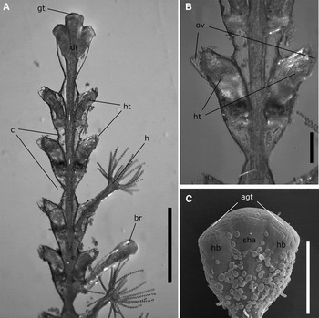
The object of the recent study is the colonial thecate hydroid Dynamena pumila (L., 1758) characterized by monopodial shoot growth with terminal growth zones (Figure 1A) (Kuhn, Reference Kuhn and Spengel1914; Berrill, Reference Berrill1949).
Organization of Dynamena pumila shoot. Only the perisarc (outer skeleton) is visible: (A) general view of the upper part of the shoot; (B) details of the internode organization; (C) distal part of the shoot (scanning electron microscopy). Agt, apical surface of the growing tip; br, lateral branch; c, constrictions between internodes; di, developing internode; gt, growing tip; h, hydranth; hb, hydranth rudiment; ht, hydrotheca; i, internode; ov, operculum valves; sha, shoot axis rudiment. Scale bar: 300 μm.

The perisarcal protective housings—hydrothecae—surround the feeding zooids constituting the shoots. The hydrotheca orifice has two valves of the operculum that are the continuation of the hydrothecae sidewalls. However, the valves thickness is less than the sidewalls at the level of the orifice therefore the valves are relatively flexible (Figure 1B) (Naumov, Reference Naumov1969).
In D. pumila the hydrothecae form two opposing longitudinal rows along the shoot stem (Figure 1A, B). The shoot part with two opposite hydrothecae constitutes flattened shoot internode. The stem constrictions free of hydrothecae often with circular furrows separate the consecutive internodes lying in one plane. The internode length is about 0.3–0.8 mm. The shallow furrows with smooth rounded bottom mark the points of the hydrothecae fusion with the stem (Figure 1C) (Kosevich & Fedosov, Reference Kosevich and Fedosov2008).
Elongation of the hydrorhiza stolons, shoot stem and branches takes place at their termini—the so-called growing tips. The primary perisarc substance secreted at the apical surface of the growing tip is elastic at the time of secretion (Figure 1C). A short distance from the apex along the tip sides, the newly laid perisarc hardens in 15–20 minutes and does not change its shape any more (Kossevitch et al., Reference Kossevitch, Herrmann and Berking2001).
So new, the youngest parts of the shoot stem or branches are located at their distal ends. Under the common summer temperature in the White Sea at the locations of mass D. pumila distribution—about 12–16°C—the development of the new terminal internode takes about 24–36 hours. Under sufficient nutrition the shoot growth occurs without delay—ending of the terminal internode formation denotes the beginning of the next internode development.
In favourable environments, all colony parts—hydrorhiza stolons and shoots—may function without any changes in appearance during a prolonged period—one season (2–4 months at the intertidal zone) or more than a year in the case of development in the subtidal zone deeper than 2 m.
Specimens of D. pumila were collected at the vicinity of N.A. Pertsov White Sea Biological Station of M.V. Lomonosov Moscow State University (Kandalaksha Bay) (66°34′N 33°08′E) from the upper subtidal zone (0–2 m) in June, August and at the end of February to beginning of March (2006–2009).
Light microscopy
Fragments of freshly collected or stained with Indian ink or Lughole solution colonies were studied using light (MBI-2 (Russia), Leitz Laborlux D (Ernst Leitz GmbH D 6330 Wetzlar, Germany)) and epifluorescent microscopy (Axioskop 40 FL (Carl Zeiss, Germany)). The filtration of excitation light was made using an interference filter with the transmission maximum at 365 and 546 nm and the half-width of transmission band of 25 and 12 nm correspondingly. Recording of fluorescence emission was through a boundary filter blocking the radiation with wavelength less than 420 and 590 nm.
Electron microscopy
For electron microscopy parts of the colonies were fixed by isotonic (osmolarity 830 mOsm) solution of 2.5% glutaraldehyde on phosphate buffer with addition of NaCl, рН 7.4 (Millonig, Reference Millonig and Buffa1964) for 1 hour at 4°C, post-fixed with 1% ОsО4 in the same buffer for a period of 0.5–1 hour. For transmission electron microscopy (TEM) fixed samples were dehydrated in the grade series of ethanol, including absolute ethanol saturated with uranyl acetate, and embedded in Araldite® resin. Ultrathin sections were cut on LKB-8800 (Sweden) ultra-microtome, contrasted with lead citrate after Reynolds (Reference Reynolds1963) and studied under JEM-100B and JEM-1011 (JEOL, Japan) transmission electron microscopes.
For scanning electron microscopy (SEM) fixed samples were dehydrated in the grade series of ethanol, then transferred into absolute acetone and dried at the critical points in Dryer HCP-2 (Hitachi, Japan). After mounting upon holders, the samples were sputtered with gold and palladium in IB-3 Ion Coater (Eiko, Japan) and examined under a JSM-6380LA microscope (JEOL, Japan).
Quantitative analysis
The quantity of individuals of the epibiotic microorganisms (cells, their clusters or trichomes) was calculated using the SEM frontal images of the 1st–5th shoot internodes of the hydroid. It was impossible to calculate the quantity of epibiotic microorganisms over the internodes older than the sixth one due to the formation of a multi-layer fouling community. The diatom cell number was checked on the frontal side of the internode including the perisarc of the stem and adjacent hydrothecae. The prokaryotic microorganisms were calculated over the surface free of diatoms at about 500 µm2; the total number of such fragments upon four shoots was 40, 14, 17, 6 and 6 from 1st to 5th apical internode correspondingly.
RESULTS
General characteristics of epibiotic microorganism community
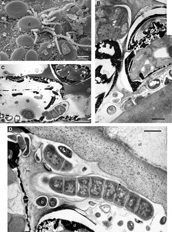
Light microscopy and SEM reveal different microorganisms upon the surface of all hydroid samples. Spacious regions with dense epibiotic fouling community characterize samples of Dynamena pumila collected during the summer period. According to the SEM and pigment autofluorescence the microorganisms possessing chlorophyll and phycobilins—that is exogenous phototropic pro- and eukaryotic microorganisms (OPM)—cyanobacteria and microalgae constitute the major part of such community. The diatom Cocconeis scutellum Ehr. and trichome cyanobacteria dominate among the OPM epibiotic community (Figure 2А). Red filamentous algae, coсcoid unicellular and short filamentous (2–3 cells) microalgae without rigid shell more than 3 µm in diameter, and diatoms belonging to 9 genera (Achnanthes sp., Amphora sp., Grammatophora sp., Licmophora sp., Nitzschia sp., Rhabdonema sp., Rhoicosphenia curvata (Kütz.) Grun., Synedra sp. and Hyalodiscus scoticus (Kütz.) Grun) represent the minor eukaryotic OPM.
Association of epibiotic microorganisms upon the surface of growing shoots in Dynamena pumila colonies: (A) general view (scanning electron microscopy); (B) bacterial cushion between diatoms and perisarc (transmission electron microscopy (TEM)); (C) bacteria penetration into the diatom cells (TEM); (D) structural integration of microorganisms in the biofilm (TEM). b, bacterium; c, cyanobacterium; d, diatom; ds, diatom shell; im, intercellular matrix; ma, microalga; p, perisarc; st, soft tissue. Scale bars: A, 10 µm; B, D, 1 µm; C, 2 µm.

The diatom C. scutellum constitutes about 85–90% of the overall number of the diatom community upon most of the hydroid shoots. At the shoot base Rh. curvata sometimes forms the dense layer over the layer of C. scutellum. Non-numerous solitary unicellular heterotrophic eukaryotic microorganisms without chloroplasts happen in such epibiotic communities.
The morphological characters revealed with the help of SEM and light microscopy showed that the prokaryotic OPM (cyanobacteria) belong to four subsections of the phylum BX cyanobacteria. Unicellular rounded and stick-form cyanobacteria displaying binary division belong to Subsection I (order Chroococcales). Filamentous cyanobacteria with trichomes formed by the cells of the same type dividing in one plain represent Subsection III (order Oscillatoriales). Filamentous cyanobacteria with trichomes formed by the vegetative cells dividing in one plain and specialized cell forms belong to Subsection IV (order Nostocales). Filamentous cyanobacteria with binary division in more then one plain and forming specialized cells correspond to Subsection V (order Stigonematales). Filamentous cyanobacteria of different morphotypes—long and short chains distinguished by the presence of the sheath, by the size and the cell form—constitute the main bulk. The cell can be oval, barrel-shaped or cylindrical. Some of the cyanobacteria form heterocysts (specialized cells responsible for molecular nitrogen fixation) and akinetes (spore-like cells).
Transmission electron microscopy investigation allows characterizing the ultrastructure of certain cyanobacteria morphotypes. They differ in the sheath and cell wall structure and cytoplasm features (Gorelova et al., Reference Gorelova, Baulina and Lobakova2009a).
Together with cyanobacteria, the great number of other bacteria lacking structural features of phototrophs is present on the hydroid surface. Among them are coccus of different size, long and short rods, twisted cells (vibrions and spirillas), thin (less than 1 µm) branching and non-branching micellar forms (presumably actinomycetes and/or trichome bacteria), rods of irregular (sometimes V- and Y- shape) form and constituting also flat rosette-like aggregates, and bacteria with appendages (prosthecobacteria and stalk bacteria). Morphological and ultrastructural peculiarities allowed identifying the planctomycetes, prosthecobacteria belonging to the genus Caulobacter and actinomycetes among non-phototrophic epibiotic bacteria. The actinomycetes were identified as the representatives of the genus Streptomyces by means of isolation in the enrichment culture and phagotyping (Omarova et al., Reference Omarova, Gorelova, Zenova and Lobakova2008). Certain epibiotic forms of cyanobacteria and other prokaryotes display defective rigidity of the cell wall, and partial or complete reduction of the cell wall (forms with defective cell wall—FDCW) (Gorelova et al., Reference Gorelova, Kosevich, Baulina, Fedorenko, Torshkhoeva and Lobakova2009b).
Within all examined samples microalgae live in intimate contact with cyanobacteria and non-phototrophic prokaryotes (Gorelova et al., Reference Gorelova, Kosevich, Fedorenko, Baulina and Lobakova2008) presenting the core of such aggregations. Many C. scutellum individuals upon the D. pumila shoots, settle over the dense bacterial matt embedded into the perisarc matrix to different degree (Figure 2B). The fascicles of filamentous cyanobacteria are mostly associated with such aggregates (Figure 2A). Transmission electron microscopy reveals also bacteria (including cyanobacteria) penetration into the cells of C. scutellum (Figure 2C). Most of such diatoms have the signs of destruction while their protoplasts look sometimes structurally complete. Therefore, it is impossible to determine the viability of the C. scutellum individuals containing bacteria. Investigation of the autofluorescence of the native D. pumila samples pointed out that many of the C. scutellum cells contain 3–7 coccoid bodies (0.7–1.2 µm in diameter) and displaying orange fluorescence. Such bodies are often situated close to the chloroplast displaying dark-red fluorescence. It is quite possible that the phycobilin pigments characteristic for cyanobacteria caused the orange fluorescence of these bodies (Lobakova et al., Reference Lobakova, Georgiev, Fedorenko and Gorelova2008).
The characteristic feature of the majority of D. pumila prokaryotic epibionts is the presence of thick mucous sheaths and capsules. Moreover, secretion of the labile mucous into surrounding in a form of slime fibrils or amorphous mass characterizes most of them, including cyanobacteria. The mucous envelopes cover the separate cells and trichomes of cyanobacteria, together with eukaryotic cells, uniting them by mutual extracellular matrix (Figure 2D). The result is the formation of heterogeneous epibiotic biofilm.
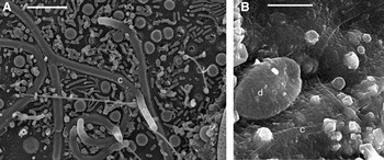
Epibiotic microbial associations upon D. pumila shoots collected at the end of February–beginning of March (those that survived the polar night) also have organization of the heterogeneous biofilm. However, compared to the ebibiotic biofilm of the summer samples less volume, reduced number and species diversity of the components characterize the biofilm after the winter. Thus, there were no filamentous red algae, unicellular eukaryotic OPM were extremely rare, and only few cells of C. scutellum present the diatoms. The prokaryotic components—diverse coccoid forms of different size, short and long rods, vibrions, thin micellar forms, unregular rod organized in V- and Y- shape figures in rosette-like aggregates, prosthecobacteria and filamentous cyanobacteria—reduced to less degree compared to eukaryotic ones (Figure 3A). The morphotype of the filamentous cyanobacteria is similar to the dominating cyanobacteria of summer samples but with more pronounced sheaths. The spatial distribution of cyanobacteria trichomes differs too. In summer samples the trichomes mostly settle over the biofilm surface immersing only the basal portions into the biofilm and attaching to the perisarc (Figure 2A). On the contrary, in the winter samples major parts of trichomes lay along the perisarc within the biofilm matrix displaying outside only the terminal points (Figure 3A).
Association of epibiotic microorganisms upon the surface of survived shoots during the polar night or dead shoots of Dynamena pumila colonies (scanning electron microscopy): (A) diversity of prokaryotic microorganisms within the biofilm of survived shoots during the polar night shoots; (B) part of the embalmed biofilm on the dead shoots. c, cyanobacterium; d, diatom. Scale bars: A, 5 µm; B, 10 µm.

We analysed the perisarc of the dead colonies of D. pumila attached to the stones or brown alga thalloms. The results reveal that the spatial organization of the epibiotic microsymbiocenosis remains after the hydroid death. At the same time the OPM undergo decoloration that is the evidence of their death. The SEM detects the surface structures that are interpreted as more or less lengthy parts of ‘embalmed’ biofilm with microorganisms immobilized in it (Figure 3B). There were no signs of secondary colonization of the dead hydroid colonies by the microorganisms.
The dynamics of the ebibiotic community development
The SEM investigation of D. pumila shoots shows different distribution of the microorganisms over the perisarc of the main stem, lateral branches and hydrothecae. They can be found either as solitary cells or groups of cells of one or several morphoptypes; or they can form continuous covering over spacious surfaces or local and extensive biofilms. Even the perfunctory inspection of the samples starting from the apex towards the shoots bases leads to the conclusion that the density of the epibiotic community and diversity of its components increases with the age of the hydroid colony part.
The unitary coccoid- and rod-like bacteria, diatoms C. scutellum and filamentous cyanobacteria appear on the apices of D. pumila shoots at the age of an hour after its formation. The majority of the microorganisms accumulate in the region of the furrows at the point of the hydrothecae fusion with the stem perisarc; perhaps the colonization of the hydroid skeleton starts from the first hours of its formation that is before its sclerotization.
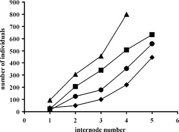
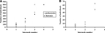
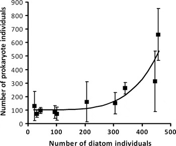
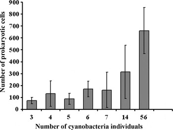
Calculations of the diatoms and prokaryotes number upon the perisarc of the frontal side of the first 4(5) distal internodes showed quick increasing in colonization density proximally to the shoot apex (Figures 4, 5A). It is more difficult to estimate the number of cyanobacteria colonizing the perisarc as it is possible only to count the number of trichomes or even clasters formed by one wriggle or several trichomes covered by common sheath. Yet it is clear that there is disto-proximal gradient of cyanobacteria number correlated with the age of the hydroid colony part (Figure 5B). It makes sense to specify that we register only the microorganism number within the surface monolayer of the community that is the ‘horizontal’ increasing of the epibiotic population number. In reality, the microorganisms' reproduction leads to formation of the three-dimensional aggregations and biofilm of ten and more cells in thickness. Moreover, we count the prokaryotes over the surface free of diatoms while the majority of bacteria including cyanobacteria tend to form aggregations with diatoms. At the same time, the presence of diatoms also leads to increasing of prokaryotes over the ‘free’ areas since the prokaryotes number upon the hydroid fragments of the same age is statistically higher in the presence of more diatoms (Figure 6). Correlation coefficient between the number of prokaryotes and diatoms is equal to 0.81, between the number of cyanobacteria and diatoms it is 0.62. That can be the evidence that the epibiotic microorganism community develops as a balanced system of phototrophic and non-phototrophic components. The cyanobacteria dominate as phototrophs over the areas free of diatoms. Increasing of cyanobacteria number correlates with the increasing of the non-phototrophic prokaryotes (R2 = 0.94) (Figure 7).
Correlation between the ages of the Dynamena pumila shoot internode and the number of associated diatoms. Data present the number of diatoms on the frontal side of the apical part of four different shoots.

The number of diatoms and prokaryotes (A) and cyanobacteria (B) on the perisarc of the frontal side of the apical fragment of individual Dynamena pumila shoot. For diatoms the data present the number of cells for whole frontal side of the internode including stem and hydrothecae surfaces. For prokaryotes, the average number of individuals (cells, trichomes and clasters) is given for topologically similar surfaces equal to 500 µm2 and free of diatoms.

Correlation between the number of prokaryotic individuals (cells, triсhomes and clasters) on the diatom-free space (500 µm2) and the number of diatoms on the frontal side of internode perisarc in Dynamena pumila. The average number and standard deviation for each count are shown. The calculated regression line corresponds to the equation y = 2.17 * x4,62 + 102.5. Correlation R2 = 0.81.

Correlation between cell number of non-phototrophic prokaryotes and number of cyanobacteria individuals (clasters and triсhomes) upon the diatom-free space (500 µm2) on perisarc of internode frontal side in Dynamena pumila. The data show the average number of cells, clasters and triсhomes and standard deviation. Correlation is equal to 0.98.

Microorganisms spatial distributing reflects also the dynamics of the epibiotic microbial community development. Within the youngest shoot parts the microorganism assemblages are mostly found at the points of the natural depressions, namely at the points of the hydrothecae ‘fusion’ with the stem of the shoot marked by shallow furrows. Initially the hydrothcae bases and valves of the operculum remain free of microorganisms and the microorganisms occupy these places later. At that the diatoms and bacteria—solitary or as a member of the local biofilms—can be found on the outer and inner surfaces of the valves, starting from the 3rd–4th distal shoot internodes (Figure 8A). Interestingly the microorganisms we registered upon the operculum valves are less diverse, lacking for example the cyanobacteria.
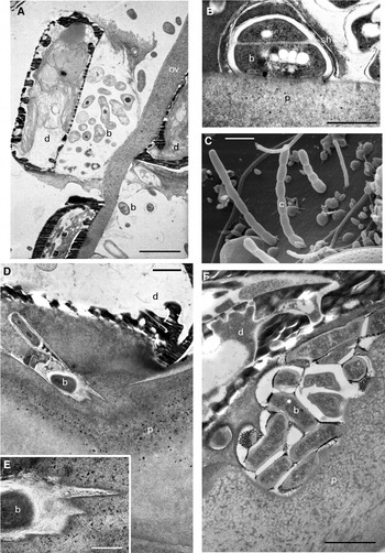
Spatial integration of Dynamena pumila and microorganisms: (A) diatoms and bacteria upon the outer and inner surface of operculum valves (transmission electron microscopy (TEM)); (B) fusion of perisarc and microbial sheath substance (TEM); (C) trichome cyanobacteria adhesion to the perisarc (scanning electron microscopy); (D–F) partial intrusion of bacteria and diatoms into the non-sclerotized material of skeleton (TEM). b, bacterium; c, cyanobacterium; d, diatom; ov, operculum valve; p, perisarc; sh, sheath. Scale bars: A, 2 µm; B, D, F, 1 µm; C, 3 µm; E, 0.5 µm.

Spatial integration of Dynamena pumila and microorganisms
As mentioned above, the microorganisms start hydroid colonization prior to sclerotization of perisarc matrix. The first stage includes adhesion of the solitary microalgae (mostly diatoms) and bacteria cells, and short cyanobacteria trichomes to the surface of the perisarc. At that the diatoms anchor within still soft perisarc by the sharp edges of their shells. The mucous bacterial covering sticks together with the perisarc to the point of microbial sheaths and envelopes fusion with it to the extent of complete disappearance of the border between them and the perisarc (Figure 8B). The filamentous cyanobacteria adhere at first by one terminal cell leaving the rest of trichome free. As a rule, such trichomes have no strongly developed sheaths (Figure 8C). Rarely the apical shoot internodes have cyanobacterial trichomes and their torpedo-like clusters with thick sheaths. Such cyanobacteria lie flat over the perisarc adhering by the maximal space (part) of their surface.
Besides adhesion bacteria and diatoms display partial intrusion (partial embedding) into the material of unsclerotized developing perisarc of the apical parts of D. pumila shoots (Figure 8D–F). Later on intrusion leads to complete embedding of diatoms into the perisarc substance entailing destruction of the alga protoplast.
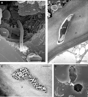
Local mechanical dissection reveals the biofilm components (bacteria, including cyanobacteria, microalgae cells and diatoms shells) inside the perisarc of the elder shoot internodes. It is important that this was found sometimes within the shoot parts free of the biofilm and at different depth levels of the perisarc matrix (Figure 9A). This allows rejecting the probability of the accidental contamination of the perisarc wall with the epibiotic microorganisms and their debris. Transmission electron microscopy displayed the diatom shell fragments without the protoplast but filled with the homogeneous material of medium electron density within the perisarc matrix under the biofilm upon the 4th (from the apex) and elder shoot internodes. Moreover, one can find the algae lacking rigid shells (Figure 9B) and encapsulated solitary bacteria or their microcolonies surrounded by the electron-translucent zone of lysis within the perisarc (Figure 9C). Ultrastructurally these bacteria and algae display no sign of essential degradation compared to diatoms. Embedding of the biofilm components takes place mostly at the points of the hydrothecae fusion with the shoot stem.
Localization of epibiotic microorganisms inside the perisarc: (A) diversity of microorganisms revealed after dissection of shoot parts free of biofilm (scanning electron microscopy (SEM)); (B) microalgae inside the perisarc wall (transmission electron microscopy (TEM)); (C) bacterial microcolonies inside the perisarc wall (TEM); (D) bacteria within the inner space of natural perforations (surface view) (SEM). b, bacterium; bf, biofilm; c, cyanobacterium; ds, diatom shell; ma, microalga; p, perisarc; st, soft tissue. Scale bars: A, 5 µm; B, C, 2 µm; D, 10 µm.

Sometimes the partial lysis of the perisarc local regions results in formation of rounded microperforations up to several microns in diameter. Such perforations are smooth or occasionally elevated over the perisarc surface edges. The inner space of such perforations can be free or occupied by bacteria and algae (Figure 9D). Some biofilm components penetrate the hydroid chitinous perisarc reaching its coenosarcs (Gorelova et al., Reference Gorelova, Kosevich, Baulina, Fedorenko, Torshkhoeva and Lobakova2009b).
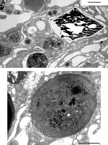
Investigation of the ultrathin sections reveals the vacuoles inside the hydroid coenosarc containing diatom shell fragments and bodies structurally similar to the eu- and prokaryotic epibionts including OPM, i.e. the unicellular algae and cyanobacteria (Figure 10). Such inclusions display more or less expressed manifestations of destruction. Ultrastructure of certain bodies allows supposing their cyanobacterial origin. In such a case the lack of intact cell wall and the presence of the lamellar membrane system resembling photosynthetic thylakoids characterize such bodies. Cyanobacteria-like bodies in D. pumila are present not only in the coenosarc but even in the stolon growing tips, and also in the blastomeres of embryos. We found OPM-like bodies and vacuoles containing diatom shell fragments in epidermal and gastrodermal layers of hydroid.
Coenosarc fragments containing vacuoles with diatom shell wreckage (A) and bodies similar with prokaryotic oxygen-evolving phototrophic microorganisms (OPM) (B) (transmission electron microscopy). ds, diatom shell; opm, OPM-like bodies. Scale bars: А, 5 μm; В, 10 µm.

DISCUSSION
In the present paper we describe the association of the White Sea colonial hydroid Dynamena pumila with its epibiotic microorganisms. That is the first example of such information on the biology of hydroids from the high latitude seas. The works on the associations of hydroids and epibiotic community are not numerous on the whole (Round et al., Reference Round, Sloane, Ebling and Kitching1961; Bavestrello et al., Reference Bavestrello, Cerrano, Cattaneo and Sarà1996; Siqueiros-Beltrones et al., Reference Siqueiros-Beltrones, Serviere-Zaragoza and Argumedo-Hernandez2001; Di Camillo et al., Reference Di Camillo, Puce, Romagnoli, Tazioli, Totti and Bavestrello2006, Reference Di Camillo, Daniel and Brytaev2010; Stabili et al., Reference Stabili, Gravili, Tredici, Piraino, Talà, Boero and Alifano2008). Despite the high species number (Di Camillo et al., Reference Di Camillo, Bavestrello, Valisano and Puce2008) hydroid biomass is low in tropical and subtropical regions and therefore hydroids do not play a significant role in ecosystem functioning. However, in temporal and high latitude regions hydroids constitute sometimes a substantial part of the benthic community of the photic zone (Naumov, Reference Naumov1969; Cornelius, Reference Cornelius1975, Reference Cornelius1979).
Previous studies (e.g. Gorelova et al., 2009) revealed that the epibiotic community upon hydroids is species-specific. Different species of colonial hydroids from the same location support a different set of microorganisms. Dynamena pumila was a focus of great interest for us due to the wide range of occupied depth habitats allowing spatial and temporal comparisons in epibiotic community assemblage.
The epibiotic community upon the D. pumila perisarc includes phototrophic and non-phototrophic eu- and prokaryotic organisms. Diatoms and trichome cyanobacteria dominate among the OPM. Morphological and ultrastructural characters allowed identifying planctomycetes, prosthecobacteria, actinomycetes and cyanobacteria as prokaryotes associated with hydroid. After sterilization we managed to isolate unicellular microalgae and different bacteria (including those capable of nitrogen fixation) from the tissues of the hydroid colony (Omarova et al., Reference Omarova, Gorelova, Zenova and Lobakova2008; Gorelova et al., Reference Gorelova, Baulina and Lobakova2009a, Reference Gorelova, Kosevich, Baulina, Fedorenko, Torshkhoeva and Lobakovab).
The epibiotic microbial association exists mainly in form of the biofilm covering most of the hydroid colony surface. The biofilm constitutes a highly organized community of one or several microorganism species embedded in extracellular mucous matrix produced by themselves and attached to the surface of any object (Davey & O'Toole, Reference Davey and O'Toole2000). According to the current conception the microorganisms within the biofilm can be both in physiologically active state and dormant or persistent state including viable but non-culturable forms. Complex intercellular signals that allow exchange of metabolites and mutual gene expression regulation unite them allowing formation of a cooperative response to the environmental stimuli. During the development of epibiotic microbial biofilm upon D. pumila the quantity of phototrophic and non-phototrophic components displays a certain correlation. Interaction between the biofilm microorganisms of different species is not restricted only to the distant exchange by metabolites through the extracellular matrix. More intimate contacts right up to the intracellular penetration of bacteria into diatoms enrich such interactions. There are data that representatives of several diatom genera may contain intracellular unicellular and heterocyst forming filamentous cyanobacteria (Carpenter & Janson, Reference Carpenter and Janson2000; Janson, Reference Janson, Rai, Bergman and Rasmussen2002; Foster & Zehr, Reference Foster and Zehr2006). During the polar night, in other words during organisms' life in darkness, the spatial connections of microsymbiocenosis with the host organism remain and the reduction of the photosynthetic components mostly takes place owing to the microalgae, while cyanobacteria persist in mucous matrix of the biofilm switching obviously to heterotrophy.
The prokaryotic components of the D. pumila biofilm include also FDCW (Gorelova et al., Reference Gorelova, Kosevich, Baulina, Fedorenko, Torshkhoeva and Lobakova2009b). As mentioned above the cyanobacteria-like bodies within the D. pumila coenosarc lack intact cell walls as was shown for the similar bodies from different hydroid species (Pyataeva et al., Reference Pyataeva, Kosevich and Lobakova2006a, Reference Pyataeva, Lobakova and Kosevichb; Gorelova et al., Reference Gorelova, Kosevich, Fedorenko, Baulina and Lobakova2008). Experiments on cyanobacteria persistence during incubation on the solid medium in darkness at 4°С for the period of 5 months showed that cyanobacteria survival rate couples with the mass production of FDCW in clusters of cells united by extracellular matrix (Gorelova & Baulina, Reference Gorelova and Baulina2009). We suppose that FDCW production by cyanobacteria also serves as an adaptive reaction of the microorganisms that develop under darkening and low temperature conditions and immobilization in mucous matrix of heterogeneous biofilm. The spatial integration of the hydroids with the polycomponent microorganism community remains during the polar night. The partial reduction of the photosynthetic components of the epibiotic microsymbiocenosis mostly takes place owing to the microalgae, while cyanobacteria persist in mucous matrix of the biofilm.
Certain components of the biofilm permeate through the hydroid perisarc and reach coenosarc. That can be due to the microorganism exoenzyme activity in relation to the complex polymers. Such properties are characteristic for many bacteria. In particular the marine actinomycetes belonging to the genus Streptomyces are able to hydrolyse chitin (Hosny et al., Reference Hosny, El-Shayeb, Abood and Abdel-Fattah2010), while some planctomycetes can hydrolyse pectin, xylan, laminarin and chondroitin sulphate (Kulichevskaya et al., Reference Kulichevskaya, Ivanova, Baulina, Bodelier, Sinninghe Damsté and Dedysh2008). And it is important that these microorganisms can ‘work’ effectively in binary associations (‘planctomycetes + actinomycetes’) and in multicomponent communities (Ivanova, Reference Ivanova2008). Several Vibrio species found in associations with colonial hydroids (Stabili et al., Reference Stabili, Gravili, Tredici, Piraino, Talà, Boero and Alifano2008, Reference Stabili, Gravili, Boero, Tredici and Alifano2010) can produce active chitinase and can participate in initiation of hydroid perisarc degradation. According to our electron microscopy data it is quite possible that other bacteria and microalgae can dissolve the perisarc too.
Our results show that association of hydroid D. pumila with the microorganism community has (bears) features of a symbiotic system. Firstly, there is a stable spatial integration of the partners. Its defining facts are: (1) fusion of the microbial envelopes with the perisarc without distinguishable borders; (2) biofilm formation; and (3) embedding of the biofilm elements including ultrastructurally microbial individuals, into the animal's skeleton. Secondly, there is possible metabolic integration of the partners with establishment of the trophic interactions in particular. Although there is no direct evidence yet, our supposition rests on the following observations: (1) local lysis of the perisarc; (2) discovery of the OPM-like bodies within coenosarc cells; (3) lack of dead perisarc colonization by the microorganisms; and (4) temporal conjugacy of perisarc sclerotization and darkening with biofilm development.
The pattern of the hydroid colony growth when new parts develop at the distal points of the branched body allows reconstruction of the epibiotic microorganism community developmental order. The distal portions of the hydroid shoot stem and branches are their youngest parts. The apices of so-called growing tips that occupy the distalmost points of the hydroid colony secrete the primary matter of the perisarc. Newly secreted perisarc is elastic and hardens in 15–20 minutes. New layers of the perisarc are added constantly from the inner side of the skeleton tube (Hughes et al., Reference Hughes, Tardent, Tardent, Tardent and Tardent1980) increasing its thickness. Secondary sclerotization of the perisarc is due to the phenolic substances (dopa-containing proteins and quinones) secreted by the hydroid tissues (Knight, Reference Knight1970; Kossevitch et al., Reference Kossevitch, Herrmann and Berking2001). Secondary sclerotization of the perisarc leads to its darkening (from translucent up to dark-brown) and loss of transparency. The examined ‘summer’ D. pumila samples have three to five terminal internodes that remain translucent and light. The biofilm upon the perisarc surface develops exactly at the level of the 3rd–5th internodes. Since it is known that microorganisms (cyanobacteria and microalgae in particular) are able both to synthesize and secrete, and expose the degradation of various phenolic compounds (e.g. Unson et al., Reference Unson, Holland and Faulkner1994; Semple & Cain, Reference Semple and Cain1996; Duval et al., Reference Duval, Shetty and Thomas2000; Hirooka et al., Reference Hirooka, Akiyama, Tsuji, Nakamura, Nagase, Hirata and Miyamoto2003; Scholz & Liebezeit, Reference Scholz and Liebezeit2006) their participation in the processes of hydroid perisarc sclerotization cannot be ruled out. It is quite possible that the valves of the hydrotheca operculum retain their elasticity due to the lack of cyanobacteria upon their surfaces.
Thus, the development of the heterogeneous epibiotic biofilm upon D. pumila correlates with reorganization of the micro- and microorganism association into the integral system of the higher level–multicomponent symbiosis.
ACKNOWLEDGEMENTS
The authors are grateful to the anonymous referees for their constructive criticism. The work was supported by the Russian Foundation for Basic Research, grant numbers 06-04-48626-а and 09-04-00675-а.