Introduction
Cochlear implantation (CI) is accepted worldwide for the treatment of severe to profound sensorineural hearing loss, and has relatively few reported complications.Reference Jain and Mukherji1
In patients with auditory neuropathy spectrum disorders, CI can provide synchronous neural stimulation to the auditory system, which is not possible acoustically.Reference Runge-Samuelson, Drake and Wackym2
The perimodiolar-positioned electrode array has been developed from the straight electrode array in order to bring the contacts as close as possible to the spiral ganglion cells, which are the target of electrical stimulation. The elastic memory electrode array is straightened by an internal stylet. The stylet is then withdrawn during insertion so that the electrode array curls close to the inner wall of the cochlea.Reference Roland3
Problems related to the electrode array occur in 0.17–2.12 per cent of CI cases.Reference Tange, Grolman and Maat4 The electrode array may occasionally be accidentally positioned in structures adjacent to the cochlea, due to the small and complex anatomy of the inner ear.Reference Jain and Mukherji1 If the surgeon suspects inappropriate positioning of the electrode array, intra-operative X-ray examination can be requested to clarify placement.Reference Copeland, Pillsbury and Buchman5 Intra-operative electrophysiological testing can also be used to confirm or allay the surgeon's suspicions.Reference Pau, Parker, Sanli and Gibson6 Electrode array misplacement can be definitively diagnosed post-operatively by high resolution computed tomography.Reference Jain and Mukherji1
Here, we report a series of pitfalls and complications in a case of electrode array misplacement.
Case report
After extensive counselling, an 11-year-old boy affected by auditory neuropathy was implanted in the right ear with a Cochlear Nucleus Freedom CI24RE Contour implant with Contour Advance electrode (Cochlear Pty Ltd, Sydney, Australia), on July 2009. Pre-operative high resolution computed tomography and magnetic resonance imaging of the temporal bone had excluded gross inner ear malformations.
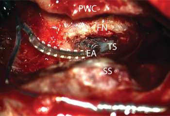
At surgery, the facial nerve was found to lie more anteriorly than normal, concealing the round window niche (Figure 1). Insertion of the electrode array was not possible through the narrow posterior tympanotomy, even though it had been widened as much as possible. Thus, a retro-facial approach was carried out by a skilled surgeon (MAB) familiar with the procedure, in a manner similar to that described by Beltrame et al. Reference Beltrame, Frau, Shanks, Robinson and Anderson7 After identification and sufficient exposure of the round window niche, a cochleostomy was performed antero-inferior to it using a 1-mm burr. The electrode array was inserted completely through the cochleostomy (Figure 1).
Surgical photograph showing electrode array fully inserted into the cochleostomy, via a retro-facial approach. PWC = posterior wall of the external ear canal; FN = facial nerve; TS = tympanic sinus; EA = electrode array; SS = sigmoid sinus

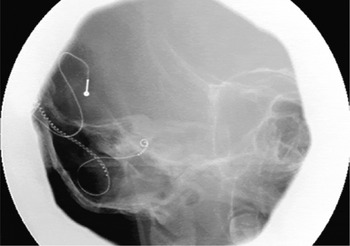
During intra-operative CI testing, impedance results were normal while neural response telemetry was undetectable. The stapedial reflex response could not be evaluated due to the lack of visible anatomical references. Intra-operative plain radiographs were apparently consistent with correct placement of the electrode array (Figure 2).
Intra-operative plain X-ray image.

After activation, vestibular symptoms were elicited at effective electrical stimulation, leading to suboptimal mapping of the processor and unsatisfactory functional results. A device integrity test and a high resolution computed tomography scan of the temporal bone were planned.
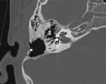
The computed tomography scan (Figure 3) showed that the electrode array was not entering the basal turn of the cochlea because it was entirely coiled within the anterior vestibule.
Axial, high resolution computed tomography scan showing the electrode array self-curling within the vestibule.

Re-implantation was carried out via the same approach, but was complicated by marked cerebrospinal fluid leakage from the reshaped cochleostomy, which probably had notched the lateral aspect of the internal auditory canal. Several attempts were made to insert a straight probe electrode array into the cochlea, under fluoroscopic control, but the tip of the dummy invariably entered the internal auditory canal.
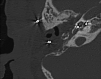
It was decided to convert to a canal wall down procedure. The resulting broad visualisation of the promontorium and round window enabled a second, safe, promontorial cochleostomy. A straight electrode array was inserted correctly into the second cochleostomy, as confirmed by post-operative high resolution computed tomography (Figure 4). Intra-operative neural response telemetry was still absent during revision surgery. The mastoid and tympanic cavities were obliterated with abdominal fat grafts in order to control cerebrospinal fluid leakage and to prevent infection.
Axial, high resolution computed tomography scan taken to follow up re-implantation, showing the straight electrode array correctly positioned within the cochlea. The canal wall down cavity is obliterated with fat grafts.

Facial nerve monitoring was uneventful during all surgical procedures.
The post-operative course was unremarkable.
The device was successfully activated four weeks later. At the time of writing, the patient was doing well.
Discussion
The traditional CI technique uses the transmastoid facial recess approach. This requires a posterior tympanotomy in order to expose the round window adequately. This surgical procedure carries the risk of iatrogenic facial nerve and chorda tympani injury, especially in patients with inner ear malformations.Reference Beltrame, Frau, Shanks, Robinson and Anderson7, Reference Huang, Lin and Wu8 Although congenital deformities of the temporal bone can be demonstrated in most cases by pre-operative high resolution computed tomography, mild facial nerve displacement can go unnoticed on pre-operative imaging, as in the case reported by Huang et al. Reference Huang, Lin and Wu8 However, even mild facial nerve anteriorisation can obscure the round window niche and result in a narrow posterior tympanotomy.
In this scenario, retro-facial air cell dissection is considered a second choice; however, when performed by a skilled surgeon it enables preservation of the chorda tympani and adequate exposure of the round window.Reference Huang, Lin and Wu8 This approach should be carried out with caution, since it is reported to increase the risk of injury to the posterior semicircular canal.Reference Huang, Lin and Wu8
Other procedures to enhance the operative field have been described, such as mobilisation of the bony posterior canal wall, keeping intact the skin lining of the external ear, and careful mobilisation of the exposed facial nerve. Moreover, a suprameatal approach can be performed or a tympanomeatal flap lifted to assist correct electrode array insertion via the round window or cochleostomy. When feasible, some surgeons prefer to avoid mobilisation of the external auditory canal because of the risk of developing an iatrogenic cholesteatoma. A safe but demanding alternative approach is the canal wall down mastoidectomy.Reference Beltrame, Frau, Shanks, Robinson and Anderson7
• The retro-facial approach to the tympanum can be a challenging alternative to a narrow posterior tympanotomy
• Electrode array misplacement is a rare complication of cochlear implantation
• Intra-operative evaluation of electrode array insertion (radiological or electrophysiological) may be unreliable
• Intra-operative fluoroscopy and post-operative high resolution computed tomography may aid diagnosis of electrode array misplacement
Cochlear perimodiolar electrodes have become popular following research in the normal cochlea which compared the straight electrode to the perimodiolar electrode, and which demonstrated that a more reliable and less traumatic insertion can be obtained using the perimodiolar electrode with a soft insertion technique.Reference Roland3 Misdirection of the electrode array occurs more often in cases of deviated cochleostomy.Reference Tange, Grolman and Maat4 Jain and Mukherji reported that the electrode array can be misplaced into the middle-ear cavity, mastoid bowl, cochlear aqueduct, petrous carotid canal or eustachian tube, or may be only partially inserted into the cochlea.Reference Jain and Mukherji1 The electrode array can also be inserted in the vestibular system, e.g. into the superior or lateral semicircular canal.Reference Tange, Grolman and Maat4, Reference Mecca, Wagle, Lupinetti and Parnes9–Reference Sorrentino, Coté, Eter, Laborde, Cochard and Deguine11 Vestibular symptoms have sometimes been reported as complications of CI, and should arouse suspicion of electrode array misplacement.Reference Mecca, Wagle, Lupinetti and Parnes9, Reference Sorrentino, Coté, Eter, Laborde, Cochard and Deguine11
Intra-operative neural response telemetry recordings may suggest CI failure.Reference Sorrentino, Coté, Eter, Laborde, Cochard and Deguine11 However, neural response telemetry results cannot determine whether the electrode array is placed within the cochlea rather than in the vestibule, because cochlear and vestibular action potentials are similar.Reference Pau, Parker, Sanli and Gibson6 On the other hand, the absence of a detectable neural response telemetry threshold is observed in some patients intra-operatively, without precluding successful functional results. In such cases, intra-operative electrically evoked auditory brainstem response testing can be useful to monitor appropriate activation of brainstem auditory pathways; however, responses may be undetectable in some forms of auditory neuropathy.Reference Pau, Parker, Sanli and Gibson6
Intra-operative plain radiographs are not routinely requested in uncomplicated CI cases.Reference Copeland, Pillsbury and Buchman5 Occasionally, X-ray imaging may enable intra-operative repositioning of the electrode array, avoiding the need for revision surgery.Reference Ramalingam, Ramalingam and Padmaja10 However, in Copeland and colleagues' prospective analysis, intra-operative plain radiographs appeared of negligible value in assessing correct electrode array placement.Reference Copeland, Pillsbury and Buchman5 These authors reported that intra-operative plain radiographs changed intra-operative management in only one out of 79 CI cases, despite requests for multiple X-ray examinations in a considerable proportion of cases (23 per cent).
Stenver's transorbital view of the temporal bone is the most common projection performed to study the position of the electrode array during post-operative plain X-ray imaging. This projection is not obtainable in the operating theatre, as the patient needs to lie prone with the sagittal plane of the head at a 45° angle and the side being radiographed close to the film cassette.
Intra-operative radiographs are often obtained at less than optimal angles, given the constraints of patient positioning, the need to avoid contact with the operative field, and the technical limitations of portable equipment.Reference Copeland, Pillsbury and Buchman5
In the intra-operative plain radiographs taken in the presented case, the self-curling electrode array had a radiological appearance consistent with appropriate positioning within the cochlea. Bearing in mind this pitfall, at revision surgery the electrode array was inserted under fluoroscopic guidance. Intra-operative fluoroscopy has been advocated as a simple, safe and effective technique in CI cases with difficult insertion and/or cochlear malformation, because it can provide a real-time view of electrode array insertion.
High resolution computed tomography of the temporal bone is indicated if plain radiographs are unreliable, if the patient experiences post-operative complications or if there is clinical evidence of CI failure.Reference Mecca, Wagle, Lupinetti and Parnes9
Conclusion
The CI surgeon must be aware of temporal bone anatomy and also of the potential pitfalls and complications of CI. Even when performed by a skilled surgeon, the retro-facial approach to the tympanum can be challenging. Considering that intra-operative plain radiography and neural response telemetry are not completely reliable, intra-operative fluoroscopy and post-operative high resolution computed tomography may aid the investigation of possible electrode array misplacement in difficult CI cases.