Introduction
Multifocal fibrosclerosis is a rare systemic disorder of unknown cause characterised by fibrous proliferation involving multiple anatomical sites.Reference Orabi, Negam and Bulman1–Reference Kitano, Shimomura, Okada and Takahashi3 It was first reported by Bartholomew et al. in 1963.Reference Orabi, Negam and Bulman1 The clinical course of the disease is variable and poorly defined.
To date, few cases of multifocal fibrosclerosis involving otolaryngological symptoms have been reported. This paper aims to report a case of multifocal fibrosclerosis with a nine-year follow up, and to discuss its imaging appearance and management.
Case report
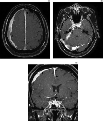
A 50-year-old woman presented with a long clinical history of intermittent fever, headache, anaemia, recurrent sinusitis, 30 kg weight loss and seizures. In May 2001, magnetic resonance imaging (MRI) of the brain showed frontal and temporal pachymeningitis with invasion of the left cavernous sinus, sella turcica and left orbital apex, as well as frontal lobe oedema and pituitary stalk thickening (Figure 1). The patient underwent the first of two meningeal biopsies, which showed fibrous connective tissue. The second biopsy, taken the following year, revealed the presence of chronic pachymeningitis and a fibrous meningioma, and the patient underwent 25 sessions of radiotherapy.
(a) & (b) Axial, postcontrast, T1-weighted magnetic resonance imaging (MRI) scans showing intensive, homogeneous enhancement (arrows) in the temporal, frontal and interhemispheric regions, indicating thickening of the dura mater. (c) Coronal MRI scan showing frontal and temporal pachymeningitis (arrows) with invasion of the sella turcica, pituitary stalk thickening (star) and frontal lobe oedema.

In 2004, computed tomography (CT) scanning of the sinuses showed injury of the apex of the left orbit and thickening of the ipsilateral optic nerve and the lateral rectus and superior rectus muscles.
In 2005, the patient started suffering from abdominal pain and was assessed by CT. The examination was suggestive of retroperitoneal fibrosis, which was confirmed by laparoscopy with multiple biopsies of the peritoneum. Laboratory evaluation revealed anaemia, an elevated erythrocyte sedimentation rate, elevated C-reactive protein level, elevated α1-glycoprotein level, decreased albumin level, and normal levels of serum complement and thyroid-stimulating hormone. Serological testing was negative for lupus anticoagulant and for Paracoccidioides brasiliensis, aspergillosis and histoplasmosis. Specific testing for antibodies such as antinuclear antibodies and anti-neutrophil cytoplasmic antibodies was also negative.
The findings of pachymeningitis and peritoneal and retroperitoneal fibrosis led to the search for a common pathophysiology for these diseases, and the diagnosis of multifocal fibrosclerosis was postulated. Thus, treatment with prednisone 60 mg (almost 1 mg/kg/day) was initiated, with azathioprine subsequently added. This treatment enabled the patient to no longer be hospitalised due to her illness and to gain weight, thus requiring only out-patient treatment.
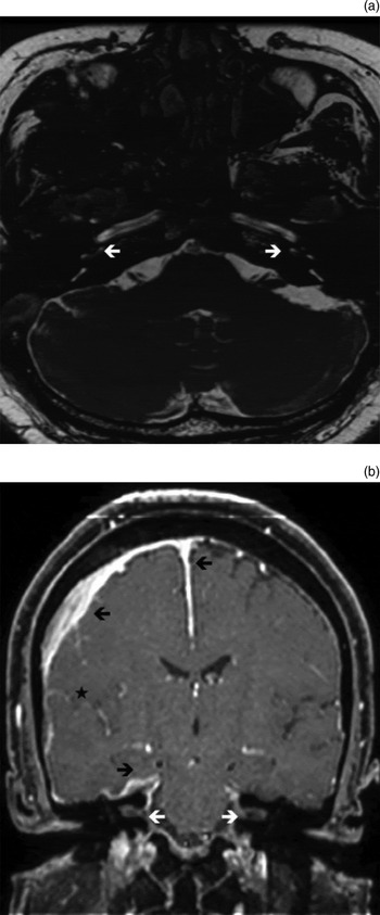
In February 2006, the patient was still experiencing headaches and started presenting with vertigo, visual haze, diplopia, diabetes insipidus, and gradual hypoacusia in the right ear with tinnitus. In May 2006, she complained of deafness in her right ear and hypoacusia in the left ear. A second MRI scan showed cochlear and internal auditory canal involvement (Figure 2) that was worse on the right side, involvement of the posterior fossa, and involvement of the right orbital apex and the ipsilateral optic nerve. However, a reduction in left orbital apex involvement was observed.
(a) Axial 3D-FIESTA magnetic resonance imaging (MRI) scan showing bilateral involvement of the cochlear basal turns (arrows). (b) Coronal, postcontrast, T1-weighted MRI scan showing enhancement suggestive of highly fibrotic tissue involving the meninges (black arrows) and the internal auditory canal (white arrows). Disappearance of the cerebral sulci in the right hemisphere is also shown (star).

In April 2007, the patient presented with significant abdominal pain and underwent an abdominal CT scan, which showed bilateral moderate pyelocalyceal dilatation. In August 2007, she began to suffer from anosmia.
At her most recent visit, in 2010, the patient presented with occasional seizures, bilateral deafness, anosmia and vertigo. Nevertheless, a brain MRI scan did not show any additional abnormalities compared with her 2006 MRI.
Discussion
The term multifocal fibrosclerosis is used to describe a rare disease entity of unknown aetiology, which is characterised by a variable combination of pathologically similar disorders that show a fibrotic process infiltrated with blood vessels, lymphocytes and plasma cells.Reference Kitano, Shimomura, Okada and Takahashi3 The main presentations of multifocal fibrosclerosis include retroperitoneal and mediastinal fibrosis, sclerosing cholangitis, Riedel thyroiditis and orbital pseudotumours.Reference Orabi, Negam and Bulman1 A number of cases have also been described involving intracranial fibrosis in the form of inflammatory pachymeningitis and suprasellar, parasellar and intrasellar lesions resulting in hypopituitarism or cranial nerve palsies.Reference Orabi, Negam and Bulman1
Depending on the site of involvement, different clinical features and radiological findings may appear. To the best of our knowledge, our patient represents the first documented case of involvement of the cochleovestibular nerve in multifocal fibrosclerosis. The patient presented with vertigo and bilateral, severe sensorineural hearing loss and tinnitus. Other otolaryngological manifestations included anosmia and recurrent sinusitis.
The majority of multifocal fibrosclerosis cases seem to be sporadic. However, some rare reports have described familial occurrence amongst siblings, suggesting a recessive form of inheritance.Reference Orabi, Negam and Bulman1
Magnetic resonance imaging clearly displays the fibrotic nature of the lesions found in multifocal fibrosclerosis. On T1-weighted sequences, lesions may appear as gadolinium-enhanced, whereas on T2-weighted sequences they are deeply hypodense.Reference Orabi, Negam and Bulman1, Reference Oguz, Kiratli, Oguz, Cila, Oto and Gokoz218F-fluorodeoxyglucose positron emission tomography imaging can help to establish the diagnosis of multifocal fibrosclerosis and to evaluate disease activity and patient response to corticosteroid therapy.Reference Orabi, Negam and Bulman1
The diagnosis of multifocal fibrosclerosis is confirmed with tissue biopsy and histopathological examination. Lesions show prominent hyalinising collagen bundles, proliferation of myofibroblastic cells, and infiltration by lymphocytes.Reference Orabi, Negam and Bulman1 The differential diagnosis should include malignancy, inflammatory pseudotumour and myofibroblastic tumours.Reference Orabi, Negam and Bulman1
• Multifocal fibrosclerosis is a rare disorder involving fibrous proliferation at multiple sites
• Otolaryngological manifestations and cochleovestibular nerve involvement are possible
• Magnetic resonance imaging clearly shows fibrosclerotic lesions
• Histopathologically, there are prominent hyalinising collagen bundles, myofibroblastic proliferation and lymphocytic infiltration
• Glucocorticoids plus immunosuppressants are beneficial
Pharmacological therapy with glucocorticoids combined with immunosuppressant agents (i.e. azathioprine, cyclophosphamide or cyclosporine) has proven beneficial in the treatment of multifocal fibrosclerosis.Reference Kitano, Shimomura, Okada and Takahashi3 Normalisation of abnormal laboratory data and improvements in symptoms and abnormal CT and MRI findings have been noted in some patients after corticosteroid use, as in the present patient after prednisone and azathioprine treatment.Reference Kitano, Shimomura, Okada and Takahashi3 Our patient gained weight in more recent years and no longer suffered from anaemia; however, her inflammatory marker levels generally remained increased. Surgical removal and/or radiation therapy may be considered in the presence of obstructive or compressive effects of the fibrous masses.Reference Orabi, Negam and Bulman1
The rare association between fibrotic diseases and masses showing different clinical patterns should be kept in mind by otolaryngologists, and imaging should be performed to document the possible existence of multifocal fibrosclerosis as the underlying cause.
Conclusion
Multifocal fibrosclerosis is a rare, systemic disorder of unknown aetiology characterised by a fibrous proliferation involving multiple anatomical sites. Clinical presentation can include retroperitoneal fibrosis, mediastinal fibrosis, sclerosing cholangitis, Riedel thyroiditis and orbital pseudotumours. Less common features are Dupuytren contracture, Peyronie disease, periaortitis, and fibrosis of the subcutaneous tissues, lungs and parotid glands. There can also be involvement of the cochleovestibular nerve, and other otolaryngological manifestations.
Magnetic resonance imaging clearly displays the lesions. On T1-weighted sequences, lesions may appear as gadolinium-enhanced, whereas on T2-weighted images they are deeply hypodense. Diagnosis is confirmed by histopathological examination, which shows prominent hyalinising collagen bundles, proliferation of myofibroblastic cells, and infiltration by lymphocytes. Differential diagnoses include malignancy, inflammatory pseudotumour and myofibroblastic tumours.
Pharmacological therapy with glucocorticoids combined with immunosuppressant agents (azathioprine, cyclophosphamide or cyclosporine) is beneficial.