Introduction
Visceral larva migrans syndrome (VLM) is caused by the L2 of Toxocara canis. Due to its ability to migrate into several tissues, direct diagnosis of this parasitic infection in humans is difficult. Some immunological methods for immunodiagnosis using some parasitic antigens have been described (Gillespie et al. Reference Gillespie, Bidwell, Voller, Robertson and Maizels1993).
In some studies, the products released by larvae cultured in vitro in third-stage L2 were designated as TES antigens (T. canis excretion–secretion antigens). These antigens have been used in immunoenzymatic analyses and indirect immunoassays, such as ELISA, to identify IgGs against Toxocara. These techniques have been shown to be effective diagnostic tools with both high sensitivity and high specificity (Bojanich et al. Reference Bojanich, Marino, López and Alonso2012; Camargo et al. Reference Camargo, Nakamura, Vaz, da Silva, Chieffi and de Melo1992; Magnaval et al. Reference Magnaval, Glickman, Dorchies and Morassin2001; Roldán et al. Reference Roldán, Cornejo and Espinoza2006).
The Western blot (WB) test is proposed as a complementary test to ELISA, considering its specificity and sensitivity when different TES antigens are used in accordance with their molecular weights, allowing for the identification of specific antigens. Some studies suggest that low-molecular-weight proteins are more specific antigens for diagnosing VLM (Pawlowski Reference Pawlowski2001; Smith et al. Reference Smith, Holland, Taylor, Magnaval, Schantz and Maizels2009).
The objective of the present study was to analyse antigens in different developmental stages of T. canis by WB in sera from epileptic Mexican children previously diagnosed by a positive test for L2 T. canis via ELISA.
Materials and methods
Patient sera
Fourteen ELISA-positive positive samples for T. canis were selected from a serum collection of children with cryptogenic epilepsy. Fourteen sera from epileptic children without history of visceral larva migrans and with negative ELISA test with epilepsy, but without a history of visceral larva migrans to T. canis, were used as controls. (Caballero-Garcìa et al. Reference Caballero-García, Simón-Salvador, Hernández-Aguilar, Reyes-López, Nogueda-Torres and Jiménez-Cardoso2019). An informed consent form was signed by the parents or guardians of children under eight years of age, whereas older patients signed informed assent forms drafted with age-appropriate language. The Ethics Review Board of the hospital approved the study.
Obtaining Toxocara canis eggs
T. canis eggs were obtained by dissecting the gravid uteri of female worms. Embryonated eggs were deposited in 0.1 N H2SO4 concentrated by centrifugation at 3500 rpm for 5 min and then placed in culture dishes with 20 ml of 1X PBS at 28°C for 30 days to induce larval formation (Caballero-García et al. Reference Caballero-García, Simón-Salvador, Hernández-Aguilar, Reyes-López, Nogueda-Torres and Jiménez-Cardoso2019).
Obtaining antigen from T. canis eggs
T. canis eggs were homogenized with liquid nitrogen and diluted in 10 mM Tris pH 7.4 (UltraPureTris, Invitrogen® no. 15504-020) with a mixture of protease inhibitors: 10 mM EDTA (Sigma E-9884), 20 mM PMSF (Sigma® P-76-26), 10 mM leupeptin (Sigma® L-2884), and 1 mM pepstatin (Sigma® P-7626). This suspension was centrifuged at 1000 × g at 4°C for 30 min, and the resulting supernatant was used as the source of antigens (Caballero-Garcìa et al. Reference Caballero-García, Simón-Salvador, Hernández-Aguilar, Reyes-López, Nogueda-Torres and Jiménez-Cardoso2019). The protein concentration was determined via the Bradford method (Bradford Reference Bradford1976). The supernatant was stored at −20°C until use.
Collection and in vitro culture of L2 larvae of T. canis
L2 larvae of T. canis were obtained from egg larvae suspended in 0.26% sodium hypochlorite at room temperature (22°C) with constant agitation for 30 min. L2 larvae were washed twice with a 0.8% saline solution under sterile conditions, shaken vigorously until larval eclosion occurred, and then observed under a microscope. The larvae were transferred to tubes with sterile PBS and centrifuged twice at 800 × g. The larvae were collected via the modified technique of Oaks and Kayes (Reference Oaks and Kayes1979) and placed in RPMI 1640 (Sigma® R-0883) medium (containing 1% dextrose and 300 mg/mL L-glutamine supplemented with 100 mg/mL penicillin and 100 mg/mL streptomycin) at 37°C in a 5% CO2 atmosphere (Queu® Incubator no. 2710).
TES antigens from L2 larvae of T. canis
TES antigens were obtained following De Savigny’s (Reference De Savigny1975) method as modified by Bowman et al. (Reference Bowman, Mika-Grieve and Grieve1987). The concentrations of the TES proteins were determined by the Bradford method (Bradford Reference Bradford1976). This material was stored at −70°C until use.
Total antigens from other parasites
Ancylostoma caninum and Dipylidium caninum (adult worms) were collected from the small intestines of euthanized dogs at one canine control centre; we did not sacrifice the animals but collected the intestines of slaughtered dogs in accordance with Mexican Rule NOM-051-ZOO-1995 (‘Humanitarian treatment in the mobilization of animals’). Larvae of T. canis were obtained following the procedure of Oaks and Kayes (Reference Oaks and Kayes1979). Larvae of Ascaris lumbricoides were collected from eggs of human faecal samples obtained from people living in Guerrero State, México, according to the procedure of Caballero-García et al. Reference Caballero-García, Simón-Salvador, Hernández-Aguilar, Reyes-López, Nogueda-Torres and Jiménez-Cardoso2019. Gnathostoma binucleatum larvae were obtained as proposed by Caballero-García et al. Reference Caballero-García, Almeyda-Artigas, Mosqueda-Cabrera and Jiménez-Cardoso2007. Echinococcus granulosus (homogenized hydatid cysts) were collected from the livers of naturally infected pigs at an abattoir in Zacatecas, Mexico. Taenia solium (metacestodes) were collected from the meat of infected pigs at a slaughterhouse in the State of Mexico, Mexico. Trichinella spirallis (muscular larvae) samples were collected from experimentally infected rats in our laboratory. Antigens were obtained following De Savigny’s (Reference De Savigny1975) method as modified by Bowman et al. (Reference Bowman, Mika-Grieve and Grieve1987). The concentrations of the TES proteins were determined by the Bradford method (Bradford Reference Bradford1976). This material was stored at −70°C until use.
Electrophoretic analyses of antigens from different developmental stages, TES, and other parasites
Antigens from the different morphological stages of T. canis (eggs, larvae, adults), TES, and other parasites (5 μg/line) were analysed via 12% SDS-PAGE (Sigma® no. 79-06-1/bisacrylamide Sigma® no.110-26-9 at 30%; UltraPure Tris, Invitrogen® no. 15504-020 pH 8.8; lauryl dodecyl sodium sulfate, Sigma® no. 205-788-1 at 10%; ammonium persulfate, Sigma® no. 7727-54-0; and TEMED, Sigma® no. 203-744-6) under reducing conditions at 100 V in an electrophoretic chamber (Bio-RAD® Mini Protean) via the Laemmli method (Laemmli Reference Laemmli1970). The gels were incubated for 24 h in a fixative solution of silver (methanol, glacial acetic acid, Golden Bell® no. 20100, concentrated enhancer, and distilled water) and stained with a silver stain (Silver Stain PlusTM Bio-RAD® no. 161-0449).
Western blot analysis of different morphological stages of T. canis in sera from epileptic children
Somatic antigens (5 μg/mL) from the egg, larval, and adult worm T. canis stages and 5 μg/mL TES antigens were separated via 12% SDS–PAGE under reducing conditions and then transferred to 0.2-μm nitrocellulose membranes (Bio-RAD® no. 162-0112) via a Bio-RAD® Transblot transfer chamber (Towbin et al. Reference Towbin, Staehelin and Gordon1979). WB analyses were performed following the methods of Magnaval et al. (Reference Magnaval, Fabre, Mauriéres, Charlet and de Larrard1992). Membranes were cut into 3–4 mm strips and incubated with blocking solution (5% milk in PBS) overnight at 4°C. Patient sera (positive for T. canis by ELISA) (Caballero-García et al. Reference Caballero-García, Simón-Salvador, Hernández-Aguilar, Reyes-López, Nogueda-Torres and Jiménez-Cardoso2019) were diluted 1:250 in blocking solution and incubated overnight at 4°C with nitrocellulose strips. The samples were then washed five times with 0.05% Tween-20 in PBS and incubated with an anti-human IgG peroxidase conjugate (Sigma® A-9040) at 1:10,000 for 2 h at 37°C. The strips were then washed with 0.05% Tween-20 in PBS, and immunodetection was performed with 4-chloro-1-naphthol (Sigma C-8890) in PBS with 4% H2O2 as the substrate. The reaction was stopped with distilled water.
Parasites antigen determination by Western blot analysis in sera from children with T. canis-positive ELISA
Somatic antigens (10 μg/mL) from T. canis (TES), Ascaris lumbricoides (L2), Ancylostoma caninum (adult), Dipylidium caninum (adult), Echinococcus granulosus (hydatid cyst), Taenia solium (metacestode), Trichinella spiralis (L1), and Gnathostoma binucleatum (larva) were tested as described above.
Results
Patterns of the total proteins from different T. canis morphological stages evaluated by SDS–PAGE
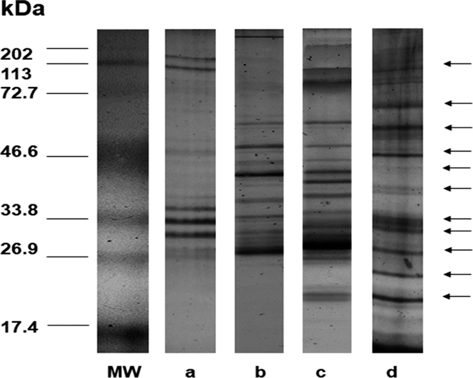
The molecular weight profiles ranged from 20 to 190 kDa. The parasite eggs presented a triplet of proteins between 30 and 36 kDa (Figure 1A). The total antigens from larvae contained proteins of 30, 40, 55, and 70 kDa (Figure 1B), and the TES antigens presented molecular weights ranging from 20 to 90 kDa (Figure 1C), whereas the adult worms presented broader protein profiles ranging from 20 to 195 kDa (Figure 1D).
Comparative SDS-PAGE pattern of antigens in different stages of T. canis. MW = Molecular weights. A, Eggs; B, TES of larva culture in vitro; C, larva; D, adult.

Profile of the proteins from T. canis and other parasites evaluated by SDS–PAGE
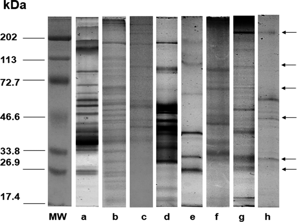
The protein patterns of the TES products from the T. canis larvae cultured in vitro (Figure 2A) and those of nematode parasites A. lumbricoides (Figure 2B), A. caninum (Figure 2C), G. binucleatum (Figure 2D), and T. spiralis (Figure 2E) and tapeworms D. caninum (Figure 2F), T. solium (Figure 2G), and E. granulosus (Figure 2H) showed different molecular weights that ranged from 20 to 210 kDa.
Analysis of total proteins in SDS-PAGE of different parasites and TES of T. canis. MW = Molecular weights. A, TES of T. canis. Total protein; B, Ascaris lumbricoides; C, Ancylostoma caninum; D, Gnathostoma binucleatum; E, Trichinella spiralis; F, Dipylidium caninum; G, Taenia solium; H, Echinococcus granulosus.

Determination by Western blot of specific and nonspecific bands in positive and negative sera
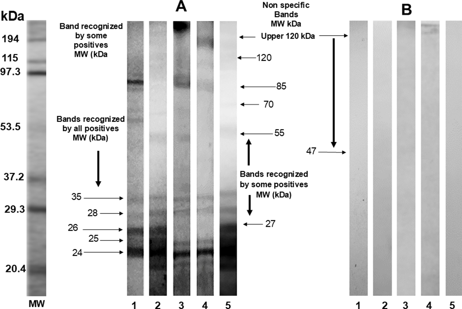
The protein profiles found in positive and negative control sera were bands between 20 and more than 150 kDa; molecular weights of 130 kDa, 150 kDa, and greater were considered nonspecific and were also found in some samples of the negative control group. The bands of interest in the positive control group were those of 35, 30, 28, 26, and 24 kDa, with the 24 kDa protein present in all positive sera (Figure 3).
Specific bands for Toxocara canis in positive and negative sera by Western blot. MW = Molecular mass. A, 1–5. Positive sera; B, 1–5. Negative sera.

Western blot analysis of serum samples from epileptic children in different morphological phases of T. canis antigens
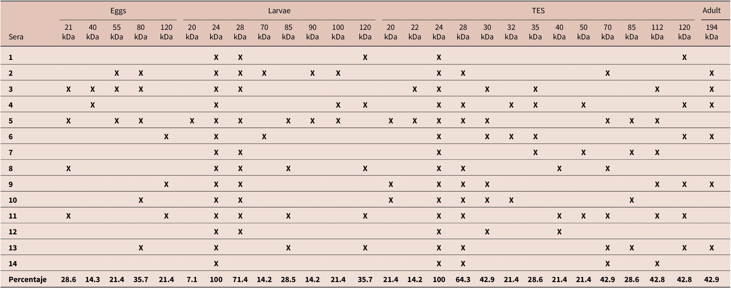
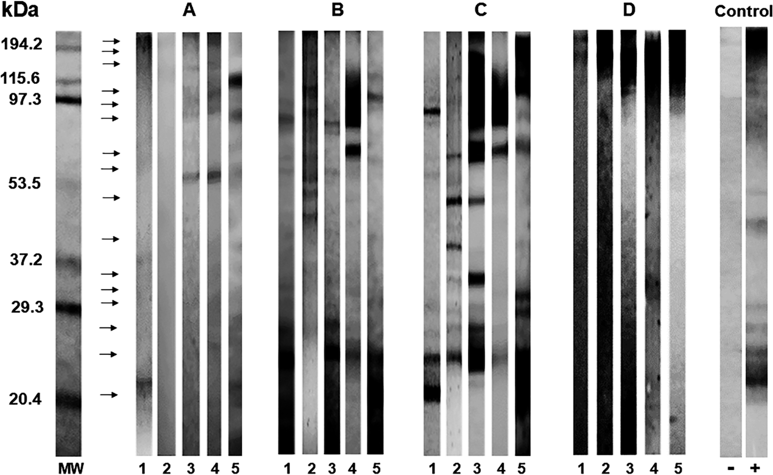
Proteins of 24 kDa were recognized in all the samples. Proteins of 28 kDa were found in 71.4% of the larval-phase samples and in 64.3% of the TES samples. In the TES samples, the recognized proteins were mainly 30 kDa (42.9%), 32 kDa (21.4%), 35 kDa (28.6%), 40 kDa (21.4%), and 50 kDa (21.4%). The 70 kDa protein was recognized by the analysed sera in 14% of the larval-phase samples and 42% of the TES-phase samples. The 120 kDa protein was recognized in 21.4% of the egg-phase samples, in 35.7% of the larval samples, and in 42.8% of the TES samples. In the adult phase, only recognition of the 194 kDa protein was observed; it was found in 42.9% of the samples (Table 1). All the sera from the children with epilepsy showed greater recognition of low, medium, and high-molecular weight proteins in the larval parasite stages and in the TES. In the egg phase, proteins of 21, 40, 55, 80, and 120 kDa were recognized, but not all sera recognized the antigens of this parasite phase. In the adult phase, the sera recognized high-molecular weight proteins (194 kDa) (Figure 4).
Percentage of sera recognition of children with epilepsy toward Toxocara canis antigens of different stages

The protein bands detected in different stages of T. canis infection in serum samples from epileptic children were evaluated by Western blotting. MW = Molecular mass. A, Eggs; B, larvae; C, TES; D, adult. 1–5. Representative positive control serum from epileptic children.

Cross-reactions of anti-T. canis antibodies and different parasite antigens in sera from epileptic children evaluated by Western blot analysis
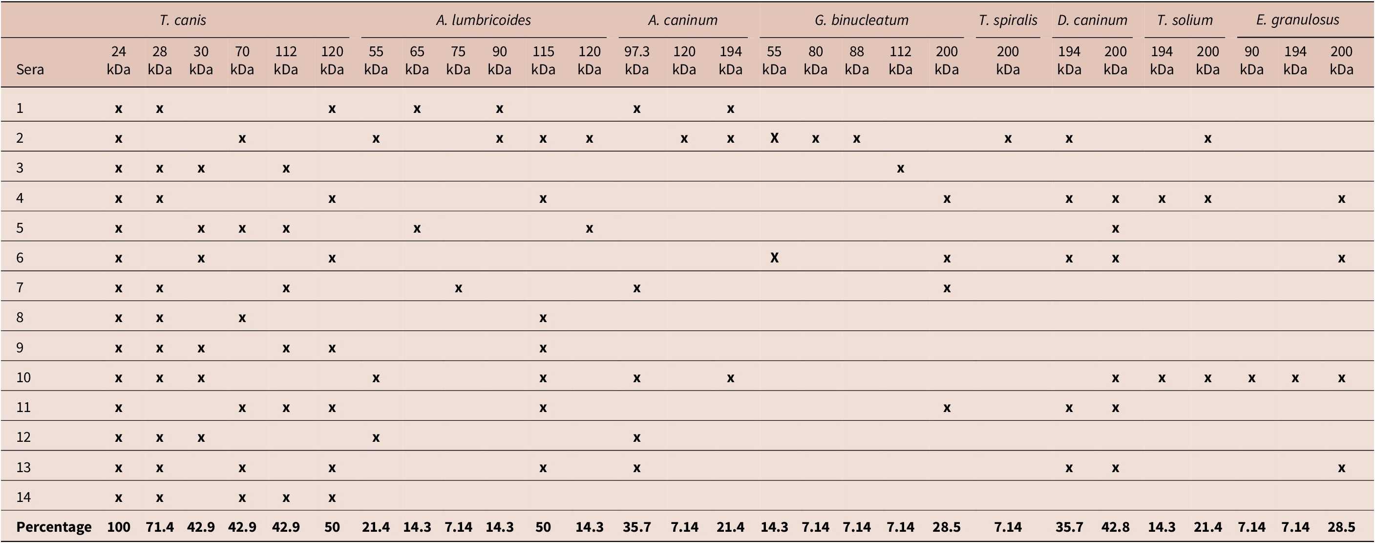
Table 2 shows the percentage of the sera of epileptic children that recognized antigens of different parasites. None of the sera recognized antigens with a molecular weight of less than 55 kDa in parasites other than T. canis. There was greater recognition of proteins of high and medium molecular weights (55, 65, and 75 kDa) in A. lumbriocoides, with percentages of 21.4%, 14.3%, and 7.14%, respectively, as well as high-molecular weight proteins: 90 kDa (14.3%), 115 kDa (50%), and 120 kDa (14.3%). The 55 kDa protein was recognized in G. binucleatum (14.3%); in this same parasite, proteins of 80 (7.14%), 88 (7.14%), 112 (7.14%), and 200 kDa (28.5%) were also recognized. In other parasites, high-molecular weight proteins were detected: one protein of 194 kDa (21.4%) in A. caninum and flatworms D. caninum (35.7%), T. solium (14.3%), and E. granulosus (7.14%); the 200 kDa protein was also detected in T. spiralis (7.14%), D. caninum, (42.8%), T. solium (21.4%), and E. granulosus (28.5%). A protein of 90 kDa was also recognized in E. granulosus (7.14%).
Percentage of sera recognition of children with epilepsy toward antigens different of parasites

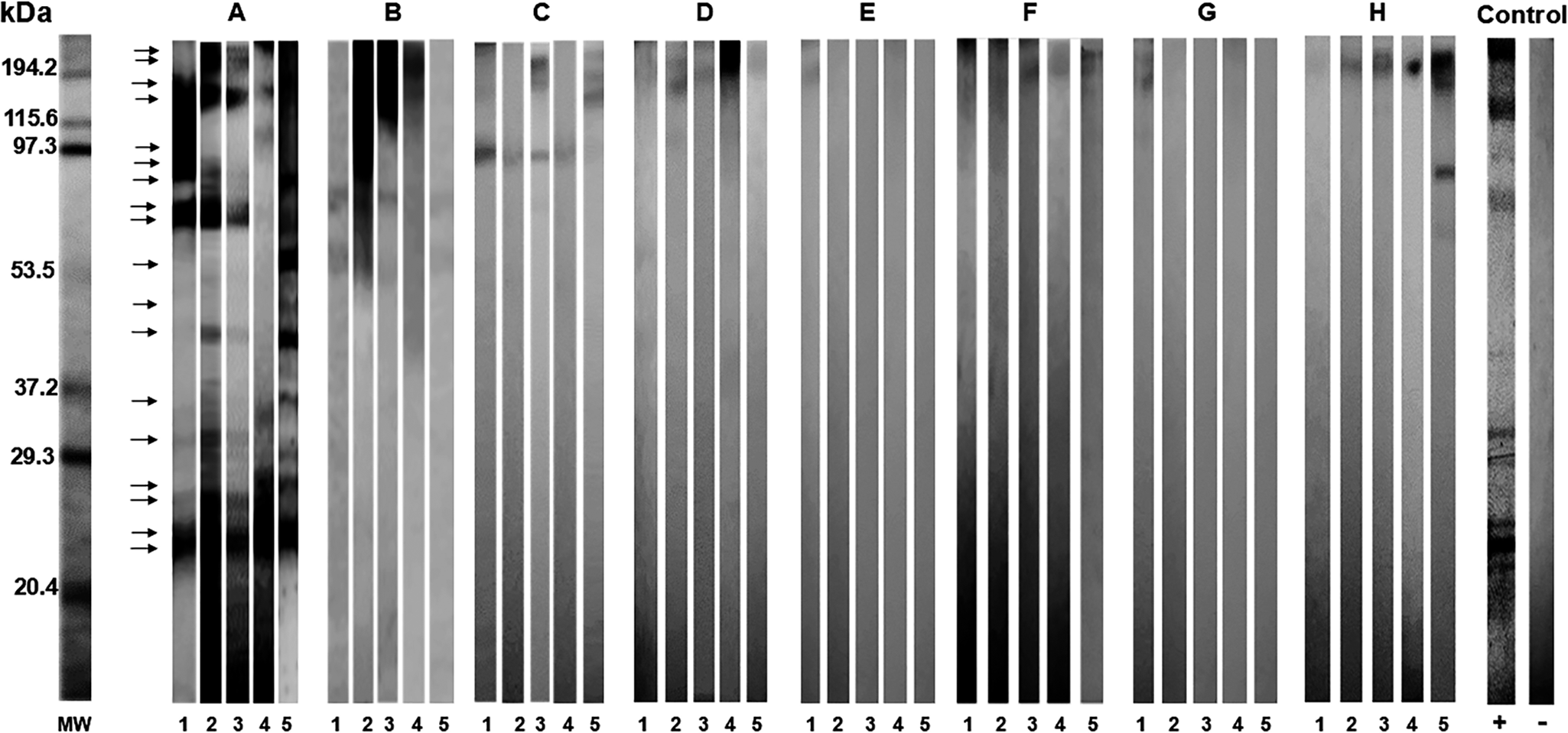
In most of the serum samples, cross-reactivity with antigens of A. lumbricoides proteins of medium and high-molecular weights was detected. In children’s serum, the total antigens of other parasites were detected, mainly high-molecular weight proteins between 90 and 200 kDa. No cross-reaction was detected between low TES molecular weight proteins of T. canis and the analysed parasites (Figure 5).
Western blot cross reaction analyses of the serum of epileptic children with antigens from different parasites. MW = Molecular mass. A, Toxocara canis; B, Ascaris lumbricoides; C, Ancylostoma caninum; D, Gnathostoma binucleatum; E, Trichinella spiralis; F, Dipylidium caninum; G, Taenia solium; H, Echinococcus granulosus. 1–5. Representative positive control serum from epileptic children.

Discussion
In humans, T. canis is diagnosed on the basis of clinical, epidemiological, and serological information. Among immunodiagnostic tests, IgG ELISA is a widely used serodiagnostic method for detecting LMV. The patients selected for this study had samples from children with epilepsy whose IgG ELISA tests against T. canis were positive.
In all included children, WB analysis of the serum was performed to determine the molecular weights of the proteins that were expressed more frequently in the different stages of T. canis infection. Protein-recognition patterns were variable in the different parasite phases. In the adult T. canis stage, only the 194 kDa protein was detected, while five proteins between 21 and 120 kDa were recognized in the egg phase, but those with major recognition were 21 and 80 kDa proteins (28.6% and 35.7%, respectively). In the larval stage, eight proteins with a predominance of 24 and 28 kDa (71.4% and 100%, respectively) were recognized. In the TES, 13 proteins ranging between 20 and 120 kDa were identified, with a predominance of those less than 50 kDa, highlighting the 24, 28, and 30 kDa proteins (100%, 64%, and 42.9%, respectively). It has been argued that, since, in the larval stage, the parasite wanders through the tissues for months or years without apparent interference from the host due to evasion of the immune system, proteins secreted by T. canis are responsible. These findings suggest that the excretion/secretion proteins of the L2 phase are more useful for immunodiagnosis (Despommier Reference Despommier2003). Morales et al. Reference Morales, López, López, Nicholls and Agudelo2002 used a WB of TES T. canis from the sera of rabbits infected with the parasite on different days of infection and found two proteins of 92 kDa and 35 kDa. Roldán and Espinoza (Reference Roldán and Espinoza2009) performed WB and TES analyses of T. canis larvae from patients diagnosed with VLM to detect IgG anti-T. canis antibodies and reported antigens of 24, 28, 30, 35, 56, 117, 136, and 152 kDa, of which the 24 and 35 kDa proteins were considered T. canis specific. The results of the present study agree with the predominance of proteins with a molecular weight less than 50 kDa. According to the analysis of different parasite stages, parasite larvae, specifically their excretion-secretion products, should be used for more specific diagnoses of anti-T. canis antibodies.
It is known that cross-reactivity has a direct influence on the specificity of immunodiagnostic tests based on serology due to the difficulties involved in parasitological confirmation (De Noya et al. Reference De Noya, Colmenares and Noya2001; Muñoz-Guzmán et al. Reference Muñoz-Guzmán, Del Río-Navarro, Valdivia-Anda and Alba-Hurtado2010; Nunes et al, Reference Nunes, Tundisi, Heinemann, Ogassawara and Richtzenhain1999). As shown in Figure 2, all antigens analysed from parasites other than T. canis in this study presented protein patterns between 55 kDa and 200 kDa, and proteins less than 50 kDa were not detected in any parasites other than T. canis. WB analyses revealed that most of the sera recognized specific antigens to T. canis of 24 and 28 kDa in 100% and 71% of the patients, respectively. Proteins <50 kDa could be specific antigens for the diagnosis of VLM, including brain infection.
Proteins with a molecular weight greater than 50 kDa were found in A. lumbricoides in 78.5% of patients, in A. caninum and G. binucleatum in 42.8% of patients and in D. caninum in 50% of patients; proteins that cross-reacted with T. canis were one A. lumbricoides of 120 kDa and one G. binucleatum of 112 kDa. The T. canis antigen cross-reacted with other parasites in the study by Magnaval et al. (Reference Magnaval, Fabre, Mauriéres, Charlet and de Larrard1992) differ from those identified in the present study because they used ELISA and evaluated different parasites, including Fasciola sp., Schistosoma sp., and Strongyloides sp., among others. Ozkoc et al. (Reference Ozkoç, Bayram and Akısü2012), using TES from T. canis, reported a low cross-reaction with Trichinella sp. In WB, the presence of different proteins allowed them to exclude cases that were considered positive by ELISA, suggesting that the 120 kDa protein was not specific to T. canis and had no value for toxocariasis diagnosis. In low-income countries, studies based on immunoassays of TES can be confused by high rates of coinfections with other soil-transmitted helminths, as antibodies to other nematodes may cross-react with T. canis antigens. Knowledge of cross-reactive antigens between different parasites has raised the possibility of using these shared antigens to construct usable targets for generating vaccines and treatments against various similar parasitic agents (De Noya et al. Reference De Noya, Colmenares and Noya2001).
To date, in clinical studies of VLM, immunodiagnostics are the only means of detection because the direct presence of parasites has not been detected in T. canis larvae-disseminated diseases. For humans, in suspected T. canis infection, a diagnosis is made via ELISA. Considering the limitations of ELISA tests for diagnostic confirmation, TES antigens with a low-molecular weight could be evaluated to create more specific immunodiagnostics. In studies combining EIA with TES to improve sensitivity and specificity, some investigators have examined the advantages of measuring IgG subclass antibodies; in one study, the sensitivity could be increased by measuring IgG2 subclass antibodies (Hotez and Wilkins Reference Hotez and Wilkins2009; Watthanakulpanich et al. Reference Watthanakulpanich, Smith, Hobbs, Whalley and Billington2008). Importantly, T. canis excretion-secretion antigens are highly specific for the diagnosis of visceral larva migrans in humans. Low-molecular weight proteins have been shown to be specific for the parasite (Bojanich and López, Reference Bojanich and López2024), while high-molecular weight antigens, greater than 100 kDa, are considered nonspecific and can lead to cross-reactions (Bojanich et al. Reference Bojanich, Marino, López and Alonso2012; Mosquera et al. Reference Mosquera, Medina, Lares, Delgado, Martínez and Ferrer2014). These studies support our findings since the detection of specific antigens of low-molecular weight in positive WB analysis allowed a reliable diagnosis, even in patients exposed to multiple helminths. However, the interpretation of a positive WB should not be based solely on the presence of antibodies, but on the recognition of a characteristic antigenic protein pattern that includes the specific low-molecular-weight components of T. canis. The absence of this pattern of specific proteins, despite the presence of high-molecular weight bands, suggests the possibility of a cross-reaction, which in this case may give an inconclusive result or a false positive. The findings of this study suggest that WB analysis could be a complementary test with high specificity, especially when low-molecular-weight proteins are present in serum samples. For a better diagnosis, it would be convenient to perform both ELISA and WB tests to avoid cross-reactions and obtain a more effective diagnosis. To improve diagnostic specificity and reduce cross-reaction interference, it is essential to use purified antigens whose specificity has been properly validated, which would contribute to reducing false positives and improving the accuracy of serological diagnosis.
Conclusions
The evaluation of T. canis proteins in sera from epileptic children revealed better recognition of TES antigens and those from the larval phase than of antigens from other stages of the parasite. Antigens with a molecular weight <50 kDa are not detected in other parasites, and 24 kDa and 28 kDa are the most frequent proteins in T. canis larvae and TES. WB with low-molecular weight TES could be performed in patients with a positive ELISA test to confirm the diagnosis. Additional studies are needed to define the number of bands of different molecular weights, which could increase the certainty of the diagnosis.
Acknowledgements
Dr. Alejandro Cruz-Reyes of Instituto de Biologia, México, for providing homogenized hydatid cysts of Echinococcus granulosus, Dr. Juan Calixto Hernández-Aguilar of the Neurology Department. Hospital Infantil de México Federico Gómez for providing and cataloguing the sera of the epileptic children used in this work and Dra. Enedina Jimenez Cardoso for your support with the facilities and equipment of Laboratorio de Investigación en Parasitologia, Hospital Infantil de México Federico Gómez.
Author contribution
MLCG, MPCV, and FSS contributed to the original idea and conceived the paper; MLCG, MPCV, and FIGS collected the data; FIGS and MLCG performed laboratory techniques; MLCG, MPCV, LEG, and FSS wrote the initial draft of the paper; MLCG, MPCV, FIGS, LEG, and FSS contributed to the revision of the manuscript; and the final version was reviewed by FSS. All the authors have read and approved the final manuscript.
Financial support
This study was supported by a grant from CONACyT-Mexico (Project SALUD 2009-CO1-115346).
Competing interests
The authors report no conflicts of interest.
Ethical standard
The authors of this paper affirm that all procedures that contributed to this work were reviewed by the Ethics Committee of the Hospital Infantil de Mexico Federico Gómez.