Introduction
The cryopreservation of reproductive cells and embryos is part of a subject that comprises a considerable theoretical knowledge. Since the protective effects of glycerol were discovered (Polge et al., Reference Polge, Smith and Parkers1949), several experiments on freezing and thawing have been performed, not only involving spermatozoa and embryos, but also a wide range of cells and tissues (Watson & Holt, Reference Watson and Holt2001).
While the cryopreservation techniques in mammals have presented consistent results, there are still a lot of problems related to freezing fish genomes and embryos (Ahammad et al., Reference Ahammad, Bhattacharyya and Jana1998). Since the former experiences from Blaxter (Reference Blaxter1953), the trials on cryopreservation of teleostean oocytes and embryos have remained unsuccessful, despite great effort from several researchers (Zell, Reference Zell1978; Whittingham & Rosenthal, Reference Whittingham and Rosenthal1978; Haga, Reference Haga1982; Harvey et al, Reference Harvey, Kelley and Ashwood-Smith1982, 1983; Stoss & Donaldson, Reference Stoss and Donaldson1983; Erdahl et al., Reference Erdahl, Erdahl and Graham1984; Zhang et al., Reference Zhang, Rawson and Morris1993; Zhang & Rawson, Reference Zhang and Rawson1998; Liu et al., Reference Liu, Zhang and Rawson1999; Robles et al., Reference Robles, Cabrita, Real, Álvarez and Hérraez2003).
Fish, reptiles, birds and amphibian embryos have a great amount of yolk and represent a complex multicompartmentalized biological class, whose cryopreservation has constantly failed. Rall (Reference Rall, Cloud and Thorgaard1993) pointed out five features that would hinder the cryopreservation of oocytes and embryos from teleosteans: a final large size, leading to a low surface/volume ratio, cells of great size, the possibility that each embryo compartment presents distinct osmotic properties, the semi-permeability of membranes surrounding the embryo and the high sensitivity to low temperatures.
Most experiments developed on freezing teleostean embryos have been directed towards knowledge about membrane permeability (Adam et al., Reference Adam, Raba and McAndrew1995; Hagedorn, et al., Reference Hagedorn, Hsu, Pilatus, Wildt, RalI and Blackband1996, Reference Hagedorn, Kleinhans, Wildt and Rall1997; Harvey & Chamberlain, Reference Harvey and Chamberlain1982; Zhang & Rawson, Reference Zhang and Rawson1998), the putative barriers to it (Hagedorn et al., Reference Hagedorn, Hsu, Pilatus, Wildt, RalI and Blackband1996), and the precise causes related to the low permeability usually observed (Liu et al., Reference Liu, Zhang and Rawson1999).
Hagedorn et al. (Reference Hagedorn, Kleinhans, Wildt and Rall1997) suggested that the main barrier to a successful freezing of teleostean embryos is the yolk syncytial layer or periblast (YSL) that surrounds the yolk and is impermeable to cryoprotectants.
Another factor able to hinder the freezing of teleostean embryos is their high sensitivity to both low temperatures and exposure to cryoprotectants in the early embryo development stages (cleavage, blastula and gastrula). Some authors suggest that resistance of embryos is reached when it already presents somites and rudimentary organs (Dinnyés et al., Reference Dinnyés, Urbányi, Baranyai and Magary1998; Hagedorn, et al., Reference Hagedorn, Kleinhans, Wildt and Rall1997; Zhang & Rawson, Reference Zhang and Rawson1995), i.e., when it is more complex, hindering a successful cryopreservation.
Neotropical teleostean species used as a model in the present study was Prochilodus lineatus, which is widely distributed over southeastern Brazil (Fowler, 1951) and, according to Corrêa and Castro (1990), it occurs in all Paraná–Paraguay river and Paraíba river basins.
Therefore, this work aimed to develop a procedure for cryopreservation of P. lineatus embryos and to observe, at both structural and ultrastructural levels, the morphologic alterations after application of freezing/thawing techniques.
Materials and Methods
The embryos were obtained through spawning of adult and mature individuals of P. lineatus belonging to the broodstock from Aquaculture Section, at the Animal Production Department at Medicine, Veterinary e Zootecny College/UNESP/Botucatu/São Paulo/Brazil. The material was then processed and analyzed in the Fish Genetics and Biology Laboratory, Morphology Department from IB/UNESP/Botucatu/São Paulo/Brazil. The eggs were incubated in vertical incubators coupled to a closed water system with controlled temperature (28 °C).
In order to test the toxicity of cryoprotectants, chorion-free embryos from three females in two development stages: morula (1.5 h/28 °C) and 4 to 6 somites (7 h/28 °C) were used. Chorion extraction was performed by enzymatic treatment (Pronase – Merck), based on the technique described by Westerfield (Reference Westerfield1993) for zebrafish, and modified for P. lineatus. Embryonated eggs were exposed to a 0.25 mg/ml pronase solution for 30 s at 28 °C, followed by four washes under water up to releasing the enzyme and chorion.
Fifty embryo samples were used for each treatment including two controls, one with free-chorion embryos and another with chorion. The cryoprotectant substances tested were ethylene glycol monomethyl ether and propylene glycol monomethyl ether (Wowk et al., Reference Wowk, Darwin, Harris, Russel and Rasch1999), methanol (Zhang & Rawson, Reference Zhang and Rawson1998), propylene glycol (PG) (Hagedorn et al., Reference Hagedorn, Kleinhans, Artemov and Pilatus1998) and dimethyl sulphoxide (MeSO4) (Magyary et al., Reference Magyary, Dinnyés, Varkonyi, Szabo and Varadi1995), at two distinct concentrations (1.0 M, 1.5 M) and under three exposure periods (1 min, 5 min and 15 min), comprising 12 treatments. Sucrose 0.1 M was added to all cryoprotectant solutions (Zhang & Rawson, Reference Zhang and Rawson1995).
After carrying out the cryoprotectant exposition, the embryos were washed in water and incubated. After the larvae hatched, they were collected and fixed in formol at 4%, and taken to the Fish Biology and Genetics and Laboratory, Morphology Department at the Biosciences Institute, UNESP, Botucatu, for further analysis. These experiments were performed with three replicates. Statistical test ANOVA (α = 0.05) was applied over survival and normality data, using the software SAS (2000).
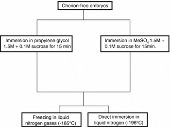
Three samples of 200 free-chorion embryos, from six females were used in the freezing experiment per treatment. The freezing equipment was composed of an insulated Styrofoam® box and a steel net tray. Two cryoprotectant solutions were used (Sol. A: MeSO4 1.5 M + sucrose 0.1 M; Sol. B: propylene glycol 1.5 M + sucrose 0.1 M). The embryos were stored into 0.5 ml straws (15 to 20 embryos/straw), in order to keep then dispersed within the straw and two freezing methods were performed: I: fast freezing using liquid nitrogen gases (−185 °C), where the straws were placed upon the tray, initially at 1 cm from the liquid nitrogen surface and, after 10 min, they were immersed in liquid nitrogen (Ninhaus-Silveira et al., Reference Ninhaus-Silveira, Foresti and Senhorini2006b); II: ultra-freezing, through direct immersion in liquid nitrogen (−196 °C).
Based on the parameters described above, four cryopreservation protocols were tested for P. lineatus embryos, as shown at Fig. 1.
Freezing protocols in Prochilodus lineatus embryos.

Thawing was carried out by immersing the straws in hot water bath for some seconds, using two distinct protocols: I: in a 36 °C water bath for 10s; II: in a 70 °C water bath for 5 s.
After thawing, four protocols to remove the cryoprotectant and reestablish embryo hydration were performed:
• I: they were placed directly under tap water (28 °C);
• II: they were placed into physiologic solution (NaCl 0.9%) for 5 min and then taken to the incubators (28 °C);
• III: progressive hydration with distilled water in three steps: A: adding water at a rate of 50% of the straw volume (5 min); B: completing up to 100% of the initial straw volume (5 min); C: putting the embryos in tap water incubators (28 °C);
• IV: progressive hydration with physiologic solution (NaCl 0.9%) in three steps: A: adding 50% of straw volume (5 min); B: completing up to 100% of the initial straw volume (5 min); C: putting the embryos in incubators under tap water (28 °C).
In order to detect ‘in toto’ morphological modifications within embryos, samples after performing freezing, thawing and hydration were analyzed under a stereomicroscope, using Petri dishes and photographed (Leica M212, coupled with an imaging system Leica MPS30).
For ultrastructural analysis of modifications in P. lineatus embryos, samples of thawed embryos were fixed in glutaraldehyde 2%, paraformaldehyde 8% in sodium phosphate buffer 0.1 M, pH 7.3, along several hydration periods. The samples were post-fixed in osmium tetroxide 1% for 2 h., counterstained in aqueous solution of uranyl acetate 0.5%, dehydrated in acetone and included into epoxy resin. Ultrafine cuts were counterstained in uranyl acetate (Watson, Reference Watson1958), washed in alcohol 50% and counterstained again in lead citrate (Reynolds, Reference Reynolds1963). The material was analyzed and photographed under transmission electron microscopy (Phillips-CM100).
Results
Chorion extraction
The utilization of enzymatic solution at 0.25 mg/ml for 30 s was satisfactory to remove completely the egg chorion. After exposure to the enzyme, the embryos were still enveloped by the chorion, but after washing and keeping them in incubator for some minutes, the chorion was totally removed by the water flow. The embryos remained viable, since most of them were able to reach the hatchery stage (Ninhaus-Silveira et al., Reference Ninhaus-Silveira, Foresti and Azevedo2006a).
Cryoprotectant toxicity
The toxicity test demonstrated the embryos at morula tolerated the chorion extraction, but not the exposure to cryoprotectants, presenting overall mortality in all treatments.
Embryos with four to six somites could not tolerate exposure to the following cryoprotectants (CPAs): ethylene glycol monomethyl ether (EGMME), propylene glycol monomethyl ether (PGMME) and methanol, with 100% of mortality in all samples.
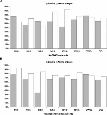
The embryos tolerated well all the procedures involving the cryoprotectants propylene glycol (PG) and dimethyl sulphoxide (MeSO4), being the highest percentage of survival (78.95%) and the lowest level of embryos with defections (6.67%) related to the treatment PG/1 M/1 min. However, the results from statistical analyses showed no significant differences between treatments (α = 0.05) (Fig. 2).
Graph representing toxicity experiments with the cryoprotectants dimethyl sulphoxide (MeSO4) (a) and propylene glycol (PG) (b), in relation to exposure time (1.5 and 15 min) and concentration (1 and 1.5 M) on the survival and normality of embryos. Time (min)/concentration (molarity).

Freezing, thawing and hydratation
None of the selected procedures (freezing, thawing and hydration) was effective on preserving the viability of P. lineatus embryos.
It was observed that in both thawing methods, even within the straws, the embryos presented a white coloration around the yolk region, while the liquid surrounding remained transparent, what would indicate the formation of ice crystals.
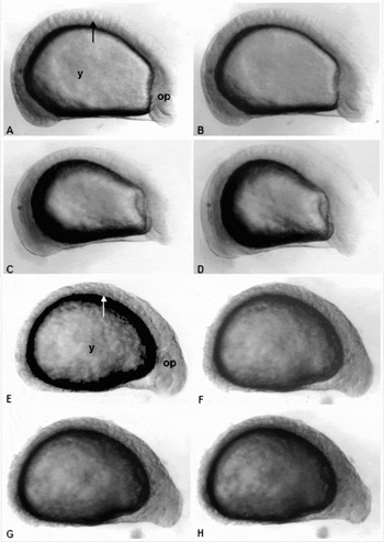
Microscope analysis revealed that the embryos frozen through ultra-fast process, when thawed, presented an external morphology apparently unchanged (Fig. 3a–d), while the embryos frozen by the fast procedure presented a folded surface (Fig. 3e–h). However, at the beginning of hydration process in all treatments, the embryos started swelling and were dissolved, and only the yolk sac remained intact.
Pictures of morphological modifications during the hydration process in frozen/thawing embryos with propylene glycol (PG) through ultra-fast (a–d) and fast methods (e–h). (a) 10 s; (b) 30 s; (c) 230 s; (d) 400 s; (d) 30 s; (f) 50 s; (g) 230 s; (h) 400 s. y, yolk; op, optical vesicle; s, somite.

Ultrastructural analysis
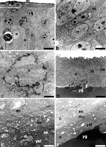
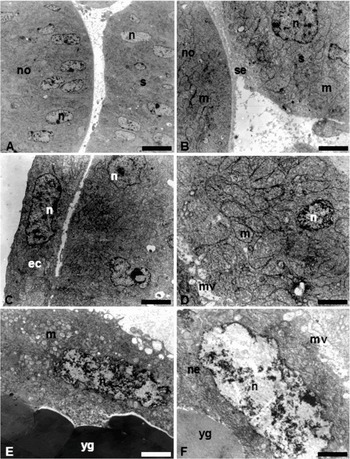
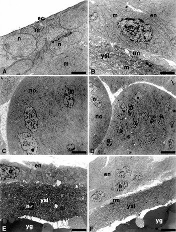
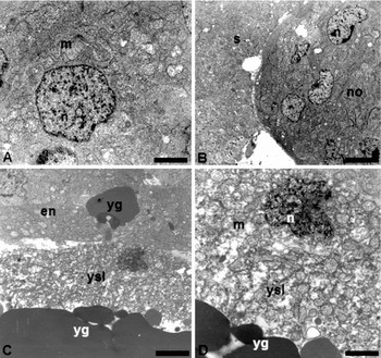
Ultrastructural modifications were observed when comparing embryos from control treatment (Fig. 4) with those submitted to cryopreservation (Figs. 5–7). Embryos exposed to freezing and thawing procedures presented ectoderm cells, somites, notochord and endoderm structurally unchanged, with well preserved nuclei and mitochondria (Figs. 5a–d, 6a–d, 7a, b).
Ultrastructure of embryos from control treatment. (a) Ectoderm, somite; (b) detail of notochord; (c) nucleus of endoderm cell; (d) yolk syncytial layer; (e) detail of the contact region between YSL and endoderm; (f) endoderm, yolk syncytial layer and yolk. n, nucleus; s, somite; no, notochord; m, mitochondria; yg, yolk globule; ysl, yolk syncytial layer; en, endoderm; mv, microvilli; ne, net of endomembranes; ec, ectoderm. Bars (a) 6.56 μm; (b) 3.07 μm; (c) 1.51 μm; (d) 2.79 μm; (d) 1.03 μm; (f) 4.33 μm.

Ultrastructure of embryos frozen with dimethyl sulphoxide through ultra-fast method. (a, b) somite and notochord; (c) ectoderm; (d) contact region between YSL and endoderm; (e, f) yolk syncytial layer. n, nucleus; s, somite; no, notochord; m, mitochondria; gv, yolk globule; ysl, yolk syncytial layer; en, endoderm; rm, ruptured microvilli; ne, net of endomembranes; es, extra-cellular substance; ec, ectoderm. Bars: (a) 11.11 μm; (b) 2.58 μm; (c) 3.28 μm; (d) 1.45 μm; (d) 2.58 μm; (f) 1.30 μm.

Ultrastructure of embryos frozen with propylene glycol through ultra-fast method. (a) ectoderm; (b) contact region between YSL and endoderm; (c) notochord; (d) somite; (e, f) yolk syncytial layer. n, nucleus; s, somite; no, notochord; m, mitochondria; yg, yolk globules; ysl, yolk syncytial layer; en, endoderm; rm, ruptured microvilli; ec, ectoderm. Bars: (a) 3.48 μm; (b) 2.65 μm; (c) 6.15 μm; (d) 11.11 μm; (d) 3.82 μm; (f) 6.15 μm.

Ultrastructure of embryos frozen with propylene glycol through fast method. (a) endoderm cell; (b) notochord and somite; (c, d) yolk syncytial layer. n, nucleus; s, somite; no, notochord; m, mitochondria; yg, yolk globules; ysl, yolk syncytial layer; en, endoderm. Bars: (a) 2.73 μm; (b) 7.47 μm; (c) 3.65 μm; (d) 1.51 μm.

Structural modifications in yolk globules were also absent in all treatments (Figs. 5e, 6e, 7c). However, the yolk syncytial layer from cryopreserved embryos presented an electron-dense cytoplasm, displaying a more compressed aspect, besides collapsed reticules, nuclei with unorganized chromatin and ruptures in the plasmatic membrane at the contact region between YSL and endoderm, leading to deformation of microvilli. The embryos indirectly submitted to freezing in nitrogen liquid, using propylene glycol as cryoprotectant, presented an YSL with unstructured cytoplasm, the net of endomembranes was destroyed and the nuclei presented altered chromatin while the mitochondria remained preserved (Fig. 7c, d).
Discussion
Dinnyés et al. (Reference Dinnyés, Urbányi, Baranyai and Magary1998), Hagedorn et al. (Reference Hagedorn, Kleinhans, Wildt and Rall1997), Urbanyi et al. (Reference Urbanyi, Baranyai, Magyary and Dinnyés1997, Reference Urbanyi, Baranyai and Dinnyés1998) and Zhang & Rawson (Reference Zhang and Rawson1995) suggested that the resistance to thermal shocks, to cryoprotectant toxicity and sensitivity to low temperatures in fish would be related to the embryo development stage, the more advanced the stage, the more propitious the freezing process. Results from the present work corroborate this suggestion, by revealing that P. lineatus embryos are also stage-dependent in relation to toxicity parameters and possibly sensible to low temperatures.
Prochilodus lineatus proved to be more sensitive to the chemical substances to remove the chorion and to cryopreservation than other species, such as Brachidanio rerio (Zhang & Rawsom, Reference Zhang and Rawson1996; Hagedorn et al., Reference Hagedorn, Kleinhans, Wildt and Rall1997) and Cyprinus carpio (Suzuki et al. Reference Suzuki, Komada, Takai, Keiji and Kozima1995; Dinnyés et al., Reference Dinnyés, Urbányi, Baranyai and Magary1998). In order to remove the chorion from zebrafish embryos, Westerfield (Reference Westerfield1993) used a pronase concentration eight times higher and for twice the time that we applied to P. lineatus embryos. Furthermore, a kind of forceps was used to remove the membrane completely. However, as with zebrafish, the P. lineatus embryos are not dependent on the presence of chorion to reach their development, as it acts as a barrier to mechanical shocks and, according to Hagedorn et al. (Reference Hagedorn, Kleinhans, Artemov and Pilatus1998), it would also act as an obstacle to the entrance of cryoprotectants.
Wowk et al. (Reference Wowk, Darwin, Harris, Russel and Rasch1999) have determined that EGMME and PGMME would be the best substances leading to formation of vitreous solution after freezing. However, they showed it to be highly toxic to P. lineatus embryos and inadequate for freezing embryos of this species in the present work conditions.
Hagedorn et al. (Reference Hagedorn, Hsu, Pilatus, Wildt, RalI and Blackband1996) demonstrated that methanol is more permeable to both MeSO4 and propylene glycol in zebrafish embryos. According to Dinnyés et al. (Reference Dinnyés, Urbányi, Baranyai and Magary1998), Zhang & Rawson (Reference Zhang and Rawson1998) and Zhang et al. (Reference Zhang, Rawson and Morris1993), methanol would be more efficient for the protection of zebrafish embryos against low temperatures, and 2 M would be the highest concentration to which a species could be exposed. The results from the present work showed that methanol, even in concentrations lower than 2 M, are toxic to P. lineatus embryos.
Dimethyl sulphoxide (MeSO4) is the most used substance in freezing fish semen (Maisse et al., Reference Maisse, Labbé, Ogier de Baulny, Leveroni Calvi and Haffray1998; Fogli da Silveira et al., Reference Fogli da Silveira, Kavamoto, Cestarolli, Godinho, Ramos and Silveira1990; Ribeiro & Godinho, Reference Ribeiro and Godinho2003; Ninhaus-Silveira et al., Reference Ninhaus-Silveira, Foresti, Tabata, Rigolino and Veríssimo-Silveira2002). However, according to Cabrita et al. (Reference Cabrita, Robles, Chereguini, de Paz, Anel and Herraéz2003), despite of its reduced utilization in freezing fish embryos, as its permeability is lower than that from methanol and propylene glycol (Hagedorn et al., Reference Hagedorn, Hsu, Pilatus, Wildt, RalI and Blackband1996; Cabrita et al. Reference Cabrita, Robles, Luna and Herraéz1999), it seems to be less toxic. In experiments involving marine invertebrates such as oysters and mussels (Chao et al., Reference Chao, Lin, Chen, Hsu and Liao1997), the cryopreservation process and the maintenance of post-freezing viability were accomplished successfully. For P. lineatus, the cryoprotectants MeSO4 and propylene glycol proved to be less toxic than the other cryoprotectants tested.
The effective survival of fish embryos after cryopreservation has not been accomplished yet, as, according to Robles et al. (Reference Robles, Cabrita, Real, Álvarez and Hérraez2003), there are many variables to consider. These authors, as well as Zhang & Rawson (Reference Zhang and Rawson1996), reported the formation of ice crystals within the embryos during the thawing process, which is indicated by embryo whitening. Zhang et al. (Reference Zhang, Rawson and Morris1993) suggested that the utilization of conventional freezing methods performed through exposure to liquid nitrogen vapour invariably results in the formation if intra-embryo ice crystals. In our experiment and treatments with P. lineatus, it was observed the opacity of yolk at some moment during thawing, characterizing, according to the authors cited above, the formation of intra-embryo crystals. Therefore, this feature can be considered an indicator of non-penetration of the cryoprotectant or that its concentration in tissues was not enough to protect the embryos against the negative effects of both the freezing and thawing processes.
Just after thawing, the embryos from P. lineatus presented intact external morphology. However, during hydration they started showing degradation features, similarly to what was observed by Robles et al. (Reference Robles, Cabrita, Real, Álvarez and Hérraez2003) in turbot embryos, excepting that they observed a folding at the yolk in the latter species, a feature that was absent in P. lineatus.
The hydration process goes on in thawed embryos up to the rupture of membranes, what may be related to structural features of their plasmatic membranes, as suggested by Hagedorn et al. (Reference Hagedorn, Lance, Fonseca, Kleinhans, Artemov, Fleischer, Hoque, Hamilton and Pukahenthi2002). These authors verified that B. rerio lacks aquaporin transmembrane proteins (AQPs), which would act in the transportation of water and ions in mammals. After inserting a type of aquaporin (AQP3) into the DNA of B. rerio specimens, they verified that the propylene glycol flow increased in those modified embryos.
Hagedorn et al. (Reference Hagedorn, Kleinhans, Artemov and Pilatus1998), analyzing the ultrastructure of B. rerio specimens before and after freezing and thawing processes, verified that the structures from blastoderm and yolk syncytial layer had been destroyed, suggesting that these alterations have occurred because of insufficient entrance of cryoprotectant into the cells. Ultrastructural analyses in P. lineatus indicated a better preservation of embryo cell structure (nucleus, endoplasmatic reticule and mitochondria), which might indicate that the cryoprotectant was able to penetrate and protect them. In relation to the YSL and yolk globules, the results from the present work seem to corroborate the report from Hagedorn et al. (Reference Hagedorn, Kleinhans, Artemov and Pilatus1998), as the YSL, at some degree, was unstructured after freezing and thawing treatments, indicating the lack or insufficient penetration of the cryoprotectant. As for the yolk, it seems to tolerate both freezing and thawing, remaining apparently unchanged.
It can be concluded that the protocols tested were unable to avoid the formation of intracellular ice crystals, which led to severe morphological alterations during the process of freezing and/or thawing, turning P. lineatus embryos non-viable. Thus, more research on the utilization of new cryoprotectants with a better and higher permeation and less toxicity, as well as the development of methods able to permeate these substances homogenously within the embryos is still necessary.
Acknowledgements
This work was supported by Aquaculture Section, at the Animal Production Department at Medicine, Veterinary e Zootecny College/UNESP/Botucatu/São Paulo/Brazil, which provided the fish and the facilities used in this study, and by CAPES (Coordenação de Aperfeiçoamento de Pessoal de Nível Superior) and CNPq (Conselho Nacional de Pesquisa).