Introduction
Sparganosis is a human infection caused by a plerocercoid tapeworm larva (sparganum) of the genus spirometra.Reference Norman and Kreutner1 The usual source of human sparganosis is related to eating habits, such as ingestion of the infected cyclops in contaminated water, or eating raw snake or frog meat. The majority of human infections involve the subcutaneous tissue or muscle of the chest abdominal wall or the brain.Reference Chang, Cho, Chi, Kim, Han and Kim2
In the head and neck region, the orbit is frequently affected.Reference Mukherjee, Biswas and Raman3–Reference Kubota and Itoh5 Rarely, sparganosis presents as a lateral neck mass, which is often initially misdiagnosed as cervical lymphadenopathy.Reference Chae, Choi, Lee and Lee6
There have been several previous reports of the radiological findings of cerebral sparganosis.Reference Chang, Cho, Chi, Kim, Han and Kim2, Reference Moon, Chang, Cho, Han, Cha and Chi7 In the head and neck region, however, reports of radiological findings are limited.
We present here the computed tomography (CT) and ultrasonography (US) findings of two sparganosis cases we have managed. We also present a review of the literature on sparganosis in the head and neck.
Case reports
Case one
A 72-year-old man presented with a one-month history of a right-sided, lateral neck mass at the level of the hyoid bone.
On physical examination, a movable, mildly tender, nodular mass was palpable in the right upper neck.
The first post-contrast CT scan of the neck revealed an ovoid mass (11 × 5 mm) with shaggy margins and mild contrast enhancement at the lateral aspect of the right sternocleidomastoid muscle (Figure 1a).
Investigations for case one. (a) The initial axial computed tomography (CT) scan, showing a small soft tissue mass with shaggy margins and mild contrast enhancement at the lateral aspect of the sternocleidomastoid muscle (arrow). (b) The initial ultrasound image reveals multiple coiled, nodular masses (arrows) with multi-layered, echogenic centres and a hypoechoic periphery with ill-defined margins. (c) Axial CT scan taken 8 months later, showing that the initial mass has disappeared; however, a new, similar lesion is seen at the lateral aspect of the right submandibular gland (arrow). The adjacent platysma is thickened. (d) to (f): Axial CT scans taken 13 months after presentation, showing that the second and third lesions at the lateral aspect of the right sternocleidomastoid muscle (thyroid cartilage level) have resolved (arrows). The new, cricoid-level lesion shows thin peripheral contrast enhancement and a tubular appearance on serial CT images. (g) Intra-operative photograph showing the excised sparganum larva, an ivory white, thread-like worm on yellow, nodular, fatty tissue. (h) Photomicrograph of the surgical specimen showing the characteristic segmental appearance of a sparganum. (H&E; 100)

On US, multiple conglomerated, nodular masses were noted at the lateral aspect of the right sternocleidomastoid muscle. The nodules had multi-layered, echogenic centres and a hypoechoic periphery with ill-defined margins (Figure 1b). Colour Doppler images did not reveal a significant vascular component. Ultrasound-guided fine needle aspiration cytology of the mass showed chronic granulomatous inflammation associated with a parasitic infection.
Excision of the mass was proposed, but the patient declined surgery.
Six months later, the patient revisited the clinic and presented with a new, right submandibular mass.
Repeated CT scanning at this time showed that the initial lateral neck mass was no longer present on the post-contrast scan; however, a small new mass was noted in the right submandibular space. A round, solid mass with shaggy margins was noted in the posterolateral aspect of the right submandibular gland (Figure 1c). The platysma muscle was thickened and the adjacent fatty tissue had increased linear densities.
Histological analysis of an US-guided needle biopsy showed normal fibrofatty tissue and a small piece of lymphoid tissue that was associated with a parasitic infection.
Again, the patient declined an excision; thus, a vermicide was prescribed.
Seven months after the second visit, the patient again revisited the clinic, with a new right lateral neck mass that was located at a lower position than the mass noted at the first visit.
A third CT scan was performed, 13 months after the first CT scan; post-contrast images revealed that the new mass was located in the subcutaneous space at the level of the right cricoid cartilage. The previously noted mass in the right submandibular space was seen to have resolved (Figure 1d to 1f). The new mass was enhanced peripherally, and was tubular on serial CT images.
Serological evaluation for human sparganosis, using an enzyme-linked immunosorbent assay, was positive.
The right neck mass was removed surgically.
Upon histopathological analysis, the mass contained an ivory white, thread-like worm within yellow, nodular, fatty tissue, measuring approximately 5 cm in length and 0.2 cm in width (Figure 1g). Histologically, the worm was confirmed as having the characteristic segmental structure of a sparganum (Figure 1h).
The patient underwent follow up for three years, with no recurrence.
Case two
A 60-year-old man presented with a one-month history of a left lateral neck mass. He had a history of eating raw snakes during his childhood.
The physical examination revealed a relatively firm, movable, painless mass within the left sternocleidomastoid muscle. There was no evidence of lymph node enlargement in the neck.
On US, multiple, ill-defined, nodular masses were noted within the left sternocleidomastoid muscle.
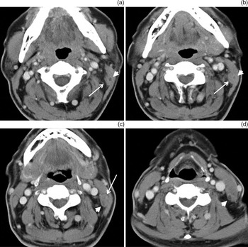
On post-contrast CT images, a minimally enhancing, soft tissue mass was noted at the posteromedial aspect of the left sternocleidomastoid muscle. This mass was craniocaudally oriented and showed a well-defined, central, hypodense, tubular structure on serial CT images (Figure 2).
Axial computed tomography scan for case two, showing a mildly enhancing, soft tissue mass at the posteromedial aspect of the left sternocleidomastoid muscle (arrows). The mass is craniocaudally oriented and shows a well-defined, central, hypodense, tubular structure on serial CT images (arrowheads).

Ultrasound-guided fine needle aspiration cytology revealed atypical cells and a granulomatous lesion.
The mass was removed surgically.
Upon histopathological analysis, the mass was seen to contain white, necrotic, nodular tissue measuring approximately 2 cm in length and 0.3 cm in width. Histologically, the typical thick, segmental structure of a sparganum was seen.
The patient underwent follow up for five years, with no recurrence.
Discussion
The most interesting findings in the present cases were the migratory nature and tubular appearance of the lesions on CT and US images. Migratory lesions have previously been reported as a CT finding in patients with cerebral sparganosis.Reference Chang, Cho, Chi, Kim, Han and Kim2, Reference Chang, Chi, Cho, Han, Han and Han8 However, migratory lesions in the head and neck have not previously been reported. When the sparganum is ingested by humans, the worm actively penetrates the intestinal wall to reach the peritoneal cavity, from which site it begins a slow systemic migration.Reference Chi, Chi and Lee9, Reference Mueller10 In our first case, short-term follow-up CT images demonstrated the migratory nature of the worm. Indeed, this migratory feature appears to be one of the significant findings of sparganosis.
The tubular appearance of sparganosis is another characteristic finding, and has been commonly described on US,Reference Park, Chai, Cho, Paek, Guk and Shin11–Reference Kim, Park and Paik13 CTReference Moon, Jung and Park14 and magnetic resonanceReference Cho, Lee, Yong, Kim, Park and Ryu15 images. This tubular pattern consists of empty tracts and the walls of a foreign body reaction.Reference Cho, Lee, Yong, Kim, Park and Ryu15 The tubular appearance needs to be distinguished from a small fistulous tract, abscess, or thyroglossal duct cyst in the neck.Reference Lin, Guo, Teng, Pan, Chen and Li16 In our cases, a smudged appearance of fatty tissue, shaggy margin, or platysmal thickening was noted around the tubular lesion. On histological evaluation, this peritubular change is seen to be due to a wide zone of chronic inflammatory cells.Reference Cho, Lee, Yong, Kim, Park and Ryu15
• Cervical sparganosis is a rare tapeworm larval infestation
• It can present as a lateral neck mass
• On serial follow-up scans, a migratory, tubular lesion should be considered significant for cervical sparganosis
The diagnosis of sparganosis can be established by serological analysis. An enzyme-linked immunosorbent assay test using sparganum-specific monoclonal antibodies is also useful.Reference Yeo, Yong and Im17 Immunodiagnosis for sparganosis is recommended to aid the pre-operative diagnosis, when a soft tissue tumour is suspected in an endemic area.Reference Chae, Choi, Lee and Lee6 Radiological evaluation is also helpful. Sparganosis is a form of granuloma, and CT scans may demonstrate non-specific findings. A pre-operative diagnosis of lipoma,Reference Chang, Lin and Yeh18 mucocele,Reference Iamaroon, Sukontason and Sukontason19 lymphadenopathy or malignant tumour should be considered.Reference Chuen-Fung and Alagaratnam20 Even though it is difficult to differentiate sparganosis from a soft tissue tumour based on pre-operative radiological data,Reference Chang, Cho, Chi, Kim, Han and Kim2, Reference Chae, Choi, Lee and Lee6 a migratory, tubular appearance in the head and neck is highly suggestive of sparganosis.
Conclusion
This is the first report in the world literature to describe the radiological findings of cervical sparganosis. A cervical mass in the head and neck area with a migratory, tubular appearance is suggestive of cervical sparganosis.