INTRODUCTION
The development of calcium indicators and imaging techniques has shed new light on the function of a subtype of glial cells widely distributed throughout the central nervous system (CNS): astrocytes. Indeed, although astrocytes have been known and described for more than a century, until recently very little was understood about their physiology. Their glycolytic metabolism, their tight coupling with blood vessels, their proximity with neurons and the variety of neurotransmitter transporters expressed at their membrane, naturally led researchers to describe them as passive cells, metabolic providers for neurons, devoted to homeostatic functions in the brain. In the mid-1990s, pioneering studies using permeant calcium indicators (reviewed in Nedergaard, Reference Nedergaard1994; Parpura et al., Reference Parpura, Basarsky, Liu, Jeftinija, Jeftinija and Haydon1994; Porter and McCarthy, Reference Porter and McCarthy1996) revealed that, in addition to their homeostatic functions, astrocytes are able to listen and talk back to neurons (Haydon, Reference Haydon2001). Since then, a growing number of studies have used calcium as an indicator of astrocytic activity. The resulting data indicate that calcium signaling in astrocytes is correlated with gliotransmitter release and modulation of neuronal activity (Angulo et al., Reference Angulo, Kozlov, Charpak and Audinat2004; Bowser and Khakh, Reference Bowser and Khakh2004; Fellin et al., Reference Fellin, Pascual, Gobbo, Pozzan, Haydon and Carmignoto2004; Fiacco and McCarthy, Reference Fiacco and McCarthy2004; Liu et al., Reference Liu, Xu, Arcuino, Kang and Nedergaard2004a; Perea and Araque, Reference Perea and Araque2005; Serrano et al., Reference Serrano, Haddjeri, Lacaille and Robitaille2006). This led researchers to consider calcium increases in astrocytes as the equivalent of the action potential in neurons.
This new-found communication between astrocytes and neurons raised the question of astrocyte involvement in synaptic plasticity, learning and memory. Using calcium to monitor and control astrocytic activity, evidence was gradually assembled establishing a role for astrocytes in the modulation of synaptic activity and synaptic networks. A conclusive answer was in sight with the demonstration that, through the elevation of intracellular calcium, astrocytic D-serine was a key player in the induction of long-term potentiation (LTP) (Henneberger et al., Reference Henneberger, Papouin, Oliet and Rusakov2010). However, shortly afterwards, work by McCarthy's group (Agulhon et al., Reference Agulhon, Fiacco and McCarthy2010) using transgenic mice in which calcium signaling was impaired cast doubt and confusion on this calcium doctrine. We will review and discuss the main facts that led researchers to believe that astrocytic calcium is the principal second messenger involved in astrocyte–neuron interactions and, in particular, synaptic plasticity.
CALCIUM REGULATION IN ASTROCYTES
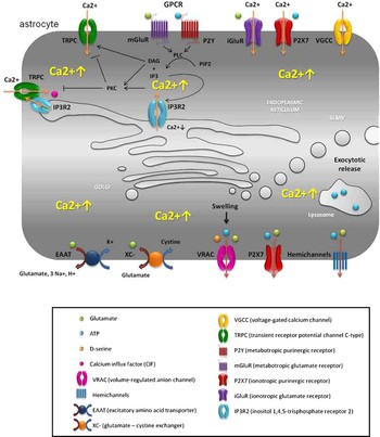
Astrocytes have been characterized using the glial fibriallary acidic protein (GFAP) expression as the main marker. Several studies have reported the coexistence of morphologically and functionally distinct populations of GFAP positive astrocytes with specific properties and distinct signaling pathways of [Ca2+]i regulation (Matthias et al., Reference Matthias, Kirchhoff, Seifert, Huttmann, Matyash and Kettenmann2003; Metea and Newman, Reference Metea and Newman2006). Within the hippocampus, two broad subtypes of GFAP-positive cells are observed: ‘complex’ glia are present in vivo at early developmental stages, while the protoplasmic, or ‘mature’ astrocytes, emerge at post-natal day 4, becoming the major population by three weeks of age. Interestingly, complex astrocytes are the major subtypes found in vitro, and so results obtained in cell culture have to be considered with care and need to be tested and validated in vivo or in slices (Zhou et al., Reference Zhou, Schools and Kimelberg2006). In addition, besides a heterogenous population of astrocytes, calcium signaling within individual astrocytes may be different in various cellular compartments. Below we detail some of the mechanisms of Ca2+ regulation described so far in astrocytes (Fig. 1).
Mechanisms of astrocytic calcium increase and gliotransmitter release. The most widely accepted mechanism for astrocytic Ca2+ increase is dependent on GPCR activation. GPCR activation leads to the depletion of intracellular Ca2+ stores via the canonical PLC/InsP3R pathway. In addition, the products of phosphatidylinositol 4,5-bisphosphate (PIP2) hydrolysis by PLC, InsP3 and DAG, gate Ca2+ enter through TRPCs. Alternatively, extracellular calcium can enter trough-activated ionotropic receptors (iGluR or P2X), or through VGCCs. Gliotransmitters can be released via Ca2+-dependent and -independent mechanisms. Ca2+-dependent vesicular exocytosis has been proposed as the main source of secreted astrocytic glutamate and D-serine. Glutamate, D-serine and ATP appear to be released from synaptic-like microvesicles (SLMVs), while ATP is also exocytosed from lysosomes. Ca2+ elevations can also lead to non-exocytotic gliotransmitter release through hemichannels (for ATP and glutamate) and the ATP-gated purinoreceptor channel P2X7. Ca2+-independent mechanisms include reversal of glutamate transporters (excitatory amino-acids transporters, EAATs) and transmitter efflux through volume-regulated anion channels (VRACs) (for ATP, glutamate and D-serine) or the glutamate–cystine exchanger XC.

Rises in astrocytic calcium come predominantly from the mobilization of Ca2+ stored in the endoplasmic reticulum (ER) (Yagodin et al., Reference Yagodin, Holtzclaw and Russell1995). Two types of calcium increase can be distinguished: spontaneous oscillations and increases due to neuronal activity both of which are believed to be inositol 1,4,5-trisphosphate (InsP3) dependent.
Astrocytes express a plethora of neurotransmitter receptors which largely correspond to G protein-coupled metabotropic receptors (GPCRs) (Verkhratsky et al., Reference Verkhratsky, Orkand and Kettenmann1998; Araque et al., Reference Araque, Parpura, Sanzgiri and Haydon1999), including purinergic receptors P2Y1,2,4,6,12,13,14 and glutamate receptors mGluR3 and mGluR5 (Verkhratsky, Reference Verkhratsky2010). Activation of these GPCRs coupled to phospholipase C (PLC) results in the breakdown of phosphatidylinositol 4,5-bisphosphate (PIP2) to produce InsP3 and diacylglycerol (DAG). InsP3 binding to its receptor (InsP3R) at the ER membrane leads to depletion of Ca2+ internal stores, and a subsequent rise in cytoplasmic [Ca2+]. Of the three InsP3R isoforms described, types 2 and 3 have been detected in astrocytes (Yamamoto-Hino et al., Reference Yamamoto-Hino, Miyawaki, Kawano, Sugiyama, Furuichi and Hasegawa1995; Sheppard et al., Reference Sheppard, Simpson, Sharp, Nucifora, Ross and Lange1997). Type two is likely the main functional receptor as in mice lacking InsP3R2 spontaneous calcium oscillations were absent and the stimulation of GPCR pathways did not trigger calcium changes in astrocyte cell bodies (Petravicz et al., Reference Petravicz, Fiacco and McCarthy2008). GPCR pathways are thought to be responsible for slow inward currents (SICs) in neurons (Angulo et al., Reference Angulo, Kozlov, Charpak and Audinat2004; Fellin et al., Reference Fellin, Pascual, Gobbo, Pozzan, Haydon and Carmignoto2004) and modulation of neuronal activity (Bowser and Khakh, Reference Bowser and Khakh2004).
Another potential site of calcium entry is through the Ca2+ permeable ionotropic glutamate channels (iGluRs) made up of N-methyl-D-aspartate receptors (NMDARs) and α-amino-3-hydroxy-5-methyl-4-isoxazole propionic acid receptors (AMPARs) that lack the GluR2 subunit (Steinhäuser and Gallo, Reference Steinhäuser and Gallo1996). Activation of these ligand-gated ion channels leads to an influx of cations, including Ca2+, into the cytoplasm. Both types of receptors have been detected by in situ hybridization and immunohistochemistry in various brain regions of the CNS. AMPARs are expressed by rat cortical (Conti et al., 2004) and hippocampal astrocytes (Petralia and Wenthold, Reference Petralia and Wenthold1992). However, functional AMPARs have only been detected in complex astrocytes (Matthias et al., Reference Matthias, Kirchhoff, Seifert, Huttmann, Matyash and Kettenmann2003); consequently, it is unlikely that AMPARs play a role in hippocampal protoplasmic astrocyte activation. In contrast, functional NMDARs have been found in cortical astrocytes (Schipke et al., Reference Schipke, Ohlemeyer, Matyash, Nolte, Kettenmann and Kirchhoff2001; Lalo et al., Reference Lalo, Pankratov, Kirchhoff, North and Verkhratsky2006), as well as in the hippocampus, where NMDA responses were evoked in both complex and passive astrocytes (Serrano et al., Reference Serrano, Robitaille and Lacaille2008). Therefore, it is likely that calcium entry through NMDARs could occur in mature hippocampal astrocytes.
Another ionotropic receptor, P2X7R, has been associated with glial calcium signaling (Alloisio et al., Reference Alloisio, Aiello, Ferroni and Nobile2006), and astrocytic release of excitatory amino acids and ATP in vitro (Duan et al., Reference Duan, Anderson, Keung, Chen, Chen and Swanson2003; Suadicani et al., Reference Suadicani, Brosnan and Scemes2006). Immunostaining in juvenile rat hippocampus showed expression of P2X1–4,6, and 7 proteins in astrocytes (Kukley et al., Reference Kukley, Barden, Steinhauser and Jabs2001). Yet, functional characterization of P2X receptors in acute slices found that the addition of ATP or P2X7R agonists did not induce P2X currents in hippocampal astrocytes (Jabs et al., Reference Jabs, Matthias, Grote, Grauer, Seifert and Steinhauser2007). These findings suggest that hippocampal glial cells in situ, under physiological conditions are devoid of functional P2X receptors, although they may have a role in the pathophysiology (Franke et al., Reference Franke, Gunther, Grosche, Schmidt, Rossner and Reinhardt2004).
Calcium elevation in astrocytes can also result from entry of extracellular Ca2+ through voltage-gated calcium channels (VGCCs). The presence of VGCCs in astrocytes has been reported in culture using both electrophysiology and molecular biology techniques (MacVicar, Reference MacVicar1984; Barres et al., Reference Barres, Chun and Corey1988; MacVicar and Tse, Reference MacVicar and Tse1988; MacVicar et al., Reference MacVicar, Hochman, Delay and Weiss1991; Duffy and MacVicar, Reference Duffy and MacVicar1994; D'Ascenzo et al., Reference D'Ascenzo, Vairano, Andreassi, Navarra, Azzena and Grassi2004). Expression of the diverse types of VGCCs was found to be dependent on the presence of neurons in culture (Corvalan et al., Reference Corvalan, Cole, de Vellis and Hagiwara1990) and the type of culture medium used (Latour et al., Reference Latour, Hamid, Beedle, Zamponi and Macvicar2003; D'Ascenzo et al., Reference D'Ascenzo, Vairano, Andreassi, Navarra, Azzena and Grassi2004). In protoplasmic astrocytes, gap junction coupling makes it difficult to isolate VGCCs currents. Moreover, these currents were also not detectable in freshly isolated astrocytes (Carmignoto et al., Reference Carmignoto, Pasti and Pozzan1998) either because the thin processes where VGCCs are expected to be found were missing, or because VGCCs are not expressed by mature astrocytes. Indeed, in situ expression studies show that VGCCs are likely expressed in immature astrocytes and that their expression declines with age (Akopian et al., Reference Akopian, Kressin, Derouiche and Steinhauser1996). The low density of these channels, the difficulties in isolating them in situ, and the lack of proof of their involvement in physiological responses to neurotransmitter probably account for a lack of interest by the scientific community.
Finally, the activation of transient receptor potential channels (TRPCs) C-type classical/canonical can be another entry point of extracellular Ca2+ into astrocytes. TRPCs are a subfamily of the TRP superfamily of non-selective, Ca2+ permeable cation channels first identified in Drosophila (reviewed in Clapham et al., Reference Clapham, Runnels and Strübing2001; Birnbaumer, Reference Birnbaumer2009). TRPCs have been found in cultured astrocytes (Pizzo et al. Reference Pizzo, Burgo, Pozzan and Fasolato2001; Grimaldi et al., Reference Grimaldi, Maratos and Verma2003; Goloniva, Reference Goloniva2005), and more recently in freshly isolated astrocytes from newborn and adult rat visual cortices (Malarkey et al., Reference Malarkey, Ni and Parpura2008). Furthermore, blocking the function of TPRC1 channel in cultured astrocytes resulted in a reduction of cytoplasmic Ca2+ elevations, which subsequently hampered glutamate release.
Mammalian TRPCs are commonly activated through the stimulation of PLC-coupled receptors: PIP2 hydrolysis leads to the generation of InsP3 and DAG, which open TRPCs through two different pathways (reviewed by Harteneck et al., Reference Harteneck, Plant and Schultz2000; Montell, Reference Montell2001; Birnbaumer, Reference Birnbaumer2009). These two mechanisms have been investigated in culture, presenting evidence that different TRPC proteins are gated through distinct mechanisms.
First, some TRPCs are activated by the production of InsP3 and consequent depletion of Ca2+ from internal stores (reviewed by Harteneck et al., Reference Harteneck, Plant and Schultz2000; Venkatachalam et al., Reference Venkatachalam, van Rossum, Patterson, Ma and Gill2002; Birnbaumer, Reference Birnbaumer2009). These channels have been implicated in store-operated Ca2+ entry (SOCE) in astrocytes (Goloniva, Reference Goloniva2005). Calcium influx through this pathway is thought to result from a diffusible messenger (Ca2+ influx factor) generated upon the release of Ca2+ from the internal stores (Randriamampita and Tsien, Reference Randriamampita and Tsien1993; Smani et al., Reference Smani, Zakharov, Csutora, Leno, Trepakova and Bolotina2004), or by conformational coupling between the activated-InsP3R at the ER membrane and TRPC at the plasma membrane (Kiselyov et al., Reference Kiselyov, Xu, Mozhayeva, Kuo, Pessah and Mignery1998; Boulay et al., Reference Boulay, Brown, Qin, Jiang, Dietrich and Zhu1999; Ma et al., Reference Ma, Patterson, van Rossum, Birnbaumer, Mikoshiba and Gill2000; Rosado et al., Reference Rosado, Brownlow and Sage2002). In favor of the latter, TRP and InsP3R were found to co-immunoprecipitate (Boulay et al., Reference Boulay, Brown, Qin, Jiang, Dietrich and Zhu1999), and high-resolution microscopy revealed the presence of TPRC1 protein in close proximity to the ER membrane (Goloniva, Reference Goloniva2005).
Second, TRPC proteins can also be activated in vitro by an increase in DAG (Hofmann et al., Reference Hofmann, Obukhov, Schaefer, Harteneck, Gudermann and Schultz1999) reviewed by Trebak et al. (Reference Trebak, Vazquez, Bird and Putney2003), through an InsP3R-independent pathway. DAG acts as a dual regulator of the TRPC through PKC-independent activation, probably by direct binding and indirect PKC-dependent inhibition (Venkatachalam et al., Reference Venkatachalam, Zheng and Gill2003). In addition, PLC activation can alter the concentration of TRPC at the plasma membrane through the regulation of vesicular cycling of channels localized to intracellular compartments (Yao et al., Reference Yao, Ferrer-Montiel, Montal and Tsien1999; Cayouette et al., Reference Cayouette, Lussier, Mathieu, Bousquet and Boulay2004; Singh et al., Reference Singh, Lockwich, Bandyopadhyay, Liu, Bollimuntha and Brazer2004).
Lastly, it is thought that TRPCs may also be implicated in the elevation of intracellular calcium via SOCE, independently of PLC and receptor activation (Birnbaumer, Reference Birnbaumer2009).
Several in vitro studies in cell lines from different mammalian species have provided evidence supporting the mechanisms of mammalian TRPC activation described above. However, only in a few cases have they been validated in vivo. The small amount of data obtained for cultured astrocytes suggests a contribution of TRPCs to Ca2+ signaling and the ensuing glutamate release (Malarkey et al., Reference Malarkey, Ni and Parpura2008), but reveals no information relative to their gating. Additional work both in vitro and in vivo is critical to confirm a functional role of TRPCs in astrocyte Ca2+ signaling and characterize their regulatory mechanisms.
Since the majority of studies have focused on the measurement of calcium transients within the somatic region, very little is known about calcium transients within astrocytic microdomains. Indeed, it is unclear whether the calcium transients of distal astrocytic processes are synchronous with those of the soma, or compartmentalized and independent of the soma. In acute slices, work in Bergmann glial cells supports the existence of microdomain fluctuations as a result of calcium release in response to electrical stimulation of Purkinje cell parallel fibers (Grosche et al., Reference Grosche, Matyash, Moller, Verkhratsky, Reichenbach and Kettenmann1999). Consistent with these observations, local in vivo monitoring of calcium in the cortex of mice indicates that calcium fluctuations rarely spread to the cell body under non-pathological conditions in anesthetized mice, while calcium increases in the cell body were observed after status epilepticus (Takano et al., Reference Takano, Tian, Peng, Lou, Libionka and Han2006; Ding et al., Reference Ding, Fellin, Zhu, Lee, Auberson and Meaney2007, Reference Ding, Wang, Cui and Haydon2009). More recently, Shigetomi's group attempted to measure differences in calcium signals between the soma and processes of rat astrocytes co-cultured with neurons. Calcium signals were evoked either pharmacologically or by single action potential, and were detected using a genetically encoded calcium sensor GcaMP2 with a membrane tethering domain Lck (Shigetomi et al., Reference Shigetomi, Kracun, Sofroniew and Khakh2010). They found that local, compartmentalized calcium transients exist in astrocyte processes, and that they are probably similar to those observed in previous studies with Bergmann glia. In conclusion, it is likely that several mechanisms of calcium regulation coexist in astrocytes and that different calcium pools can be mobilized independently, notably in the tiny astrocytic processes that are found in close apposition with synapses.
ROLE OF CALCIUM ELEVATION IN GLIOTRANSMITTER RELEASE
In order to link calcium elevation in astrocytes to the regulation of neuronal activity and synaptic plasticity, it is important to identify the mechanism(s) by which calcium could influence the release of gliotransmitters. Several studies have already established a correlation between an increase in astrocytic [Ca2+]i and the release of gliotransmitters (Fig. 1) such as glutamate, ATP, D-serine, γ-amino butyric acid-A (GABA) and prostaglandins (Parpura et al., Reference Parpura, Basarsky, Liu, Jeftinija, Jeftinija and Haydon1994; Bezzi et al., Reference Bezzi, Carmignoto, Pasti, Vesce, Rossi and Rizzini1998; Newman, Reference Newman2001; Mothet et al., Reference Mothet, Pollegioni, Ouanounou, Martineau, Fossier and Baux2005). Mechanisms for Ca2+-dependent, as well as Ca2+-independent glutamate release from astrocytes, and to a lesser extent D-serine, have been thoroughly studied. In what follows, we summarize the ones described so far for these two gliotransmitters.
Calcium-dependent exocytosis is probably the main source of secreted astrocytic glutamate and D-serine, although this method of release is still controversial. Several reviews have already detailed the evidence in favor of exocytotic release (Volterra and Meldolesi, Reference Volterra and Meldolesi2005; Malarkey and Parpura, Reference Malarkey and Parpura2008; Milanese et al., Reference Milanese, Bonifacino, Zappettini, Usai, Tacchetti, Nobile, Begley and Crews2009) and the recent review by Hamilton and Attwell thoroughly addresses the question by combining morphofunctional evidence to verify the likelihood of this theory (Hamilton and Attwell, Reference Hamilton and Attwell2010). Briefly, astrocytes in vitro and in vivo were shown to contain synaptic-like microvesicles (SLMVs) (Bezzi et al., Reference Bezzi, Gundersen, Galbete, Seifert, Steinhauser and Pilati2004) and express the protein machinery required for Ca2+-dependent exocytosis (Parpura et al., Reference Parpura, Fang, Basarsky, Jahn and Haydon1995; Hepp et al., Reference Hepp, Perraut, Chasserot-Golaz, Galli, Aunis and Langley1999; Maienschein et al., Reference Maienschein, Marxen, Volknandt and Zimmermann1999; Araque et al., Reference Araque, Li, Doyle and Haydon2000; Bezzi et al., Reference Bezzi, Gundersen, Galbete, Seifert, Steinhauser and Pilati2004; Zhang et al., Reference Zhang, Fukuda, Van Bockstaele, Pascual and Haydon2004) although in at least one study the corresponding mRNAs could not be detected by GeneChip arrays (Cahoy et al., Reference Cahoy, Emery, Kaushal, Foo, Zamanian and Christopherson2008). Inhibition of the (H+) ATPase, which creates the voltage and pH gradient required for transmitter accumulation, blocks the release of glutamate (Araque et al., Reference Araque, Li, Doyle and Haydon2000; Pasti et al., Reference Pasti, Zonta, Pozzan, Vicini and Carmignoto2001) and D-serine (Mothet et al., Reference Mothet, Pollegioni, Ouanounou, Martineau, Fossier and Baux2005) from cultured astrocytes. Furthermore, monitoring of SLMVs in vitro and in slices revealed that they exocytose glutamate and D-serine in response to stimuli that increased intracellular calcium (Bezzi et al., Reference Bezzi, Carmignoto, Pasti, Vesce, Rossi and Rizzini1998, Reference Bezzi, Domercq, Brambilla, Galli, Schols and De Clercq2001, Reference Bezzi, Gundersen, Galbete, Seifert, Steinhauser and Pilati2004; Crippa et al., Reference Crippa, Schenk, Francolini, Rosa, Verderio and Zonta2006; Domercq et al., Reference Domercq, Brambilla, Pilati, Marchaland, Volterra and Bezzi2006; Martineau et al., Reference Martineau, Galli, Baux and Mothet2008). Conversely, it was shown in vivo that blocking astrocytic Ca2+ prevents glutamate and D-serine release (Bezzi et al., Reference Bezzi, Domercq, Brambilla, Galli, Schols and De Clercq2001; Kang et al., Reference Kang, Kang, Lovatt, Torres, Zhao and Lin2008; Henneberger et al., Reference Henneberger, Papouin, Oliet and Rusakov2010; Reyes-Haro et al., Reference Reyes-Haro, Muller, Boresch, Pivneva, Benedetti and Scheller2010).
Are glutamate and D-serine stored and released from the same SLMVs? As yet there is no clear evidence for this, although some data suggest they may be. Indeed, D-serine accumulates in vesicles that contain VGlut (Mothet et al., Reference Mothet, Pollegioni, Ouanounou, Martineau, Fossier and Baux2005) and are morphologically similar to those storing glutamate (Williams et al., Reference Williams, Diaz, Macnab, Sullivan and Pow2006). Independently of whether they are co-stored or not, astrocytic glutamate and D-serine are likely to be released synchronously, as degradation of D-serine by the application of D-amino acid oxydase was observed to reduce NMDA-evoked currents (Reyes-Haro et al., Reference Reyes-Haro, Muller, Boresch, Pivneva, Benedetti and Scheller2010). The concerted secretion of NMDAR activator and co-activator could have a potent effect on synaptic plasticity. Thus, further research is needed to determine not only if glutamate and D-serine are stored in the same vesicles but also how their co-release might be regulated.
It has been shown that addition of tetanus neurotoxin (TeNT) inhibits all solube N-ethylmaleimide-sensitive factor attachment protein receptor (SNARE)-dependent mechanisms and blocks glutamate release (Jourdain et al., Reference Jourdain, Bergersen, Bhaukaurally, Bezzi, Santello and Domercq2007; Perea and Araque, Reference Perea and Araque2007). This was taken as evidence in support of regulated vesicular release. However, as pointed out by Hamilton and Attwell (Reference Hamilton and Attwell2010), addition of TeNT also affects the lysosomal and constitutive secretory pathways (Stern et al., Reference Stern, Edwards and Sakmann1992; Proux-Gillardeaux et al., Reference Proux-Gillardeaux, Gavard, Irinopoulou, Mege and Galli2005). Indeed, lysosomes are membrane-bound compartments whose fusion with the plasma membrane appears to be Ca2+-dependent (Jaiswal et al., Reference Jaiswal, Fix, Takano, Nedergaard and Simon2007; Li et al., Reference Li, Ropert, Koulakoff, Giaume and Oheim2008). Lysosomes are a potential source of secreted ATP in primary astrocyte cultures (Zhang et al., Reference Zhang, Chen, Zhou, Song, Xu and Luo2007), but as yet have not been shown to contain glutamate (Zhang et al., Reference Zhang, Chen, Zhou, Song, Xu and Luo2007) or D-serine (Martineau et al., Reference Martineau, Galli, Baux and Mothet2008). The secretory pathway is similarly regulated by changes in [Ca2+]I and thus could control gliotransmitter release by adjusting the targeting and concentration of membrane proteins responsible for their release (including AMPARs (Lu et al., Reference Lu, Man, Ju, Trimble, MacDonald and Wang2001), TRPC3 (Singh et al., Reference Singh, Lockwich, Bandyopadhyay, Liu, Bollimuntha and Brazer2004) and glutamate transporters (Stenovec et al., Reference Stenovec, Kreft, Grilc, Pangrsic and Zorec2008)) through the regulation of the constitutive secretory and recycling pathways (Hay, Reference Hay2007). Still, as noted by Hamilton and Attwell, the timescale of insertion/removal of membrane proteins is likely to be too slow for the kinetics of release observed for these gliotransmitters (Hamilton and Attwell, Reference Hamilton and Attwell2010).
In addition to vesicular release, changes in astrocytic [Ca2+]i could regulate glutamate efflux through P2X7 purinergic receptors (Duan et al., Reference Duan, Anderson, Keung, Chen, Chen and Swanson2003; Fellin et al., Reference Fellin, Sul, D'Ascenzo, Takano, Pascual and Haydon2006) and hemichannels (Ye et al., Reference Ye, Wyeth, Baltan-Tekkok and Ransom2003). Indeed, the permeability of these channels is increased under conditions of low [Ca2+]EC (Stout et al., Reference Stout, Costantin, Naus and Charles2002; Duan et al., Reference Duan, Anderson, Keung, Chen, Chen and Swanson2003).
Alternatively, several Ca2+-independent mechanisms have also been described. These include volume-regulated anion channels (VRACs) (Kimelberg et al., Reference Kimelberg, Goderie, Higman, Pang and Waniewski1990; Takano et al., Reference Takano, Kang, Jaiswal, Simon, Lin and Yu2005; Rosenberg et al., Reference Rosenberg, Kartvelishvily, Shleper, Klinker, Bowser and Wolosker2010), cystine-glutamate exchangers (Bender et al., Reference Bender, Reichelt and Norenberg2000) and reversal of glutamate transporters (Szatkowski et al., Reference Szatkowski, Barbour and Attwell1990) reviewed in Volterra and Meldolesi (Reference Volterra and Meldolesi2005), Malarkey and Parpura (Reference Malarkey and Parpura2008) and Hamilton and Attwell (Reference Hamilton and Attwell2010). To date, transport of D-serine via these pathways has only been described in the case of VRACs (Rosenberg et al., Reference Rosenberg, Kartvelishvily, Shleper, Klinker, Bowser and Wolosker2010).
Although there is some evidence that VRACs could operate under physiological conditions, it is thought that glutamate release through these non-exocytotic mechanisms mostly occurs under pathological conditions of, for example, high [ATP] and low [Ca2+]EC (Fellin et al., Reference Fellin, Sul, D'Ascenzo, Takano, Pascual and Haydon2006; Thompson et al., Reference Thompson, Zhou and MacVicar2006), hypo-osmolarity (Phillis et al., Reference Phillis, Song and O'Regan1997; Seki et al., Reference Seki, Feustel, Keller, Tranmer and Kimelberg1999), high [Cys]EC (Cavelier and Attwell, Reference Cavelier and Attwell2005) or high [K+]EC (Longuemare and Swanson, Reference Longuemare and Swanson1997; Rossi et al., Reference Rossi, Oshima and Attwell2000).
So far the majority of research strongly favors Ca2+-dependent regulated vesicular release of glutamate and D-serine. Nevertheless, we cannot exclude the participation of other systems. In fact, it is probable that all these mechanisms coexist, and that their relative contribution depends on the brain region and the specific physio- or pathological conditions.
Regardless of the mechanism for gliotransmitter release, in slices, increasing calcium by uncaging of InsP3 or o-nitrophenyl-ethylene glycol-bis(β-aminoethylether)-N,N,N′,N′-tetraacetic acid (NP-EGTA) was found to increase excitatory post-synaptic current (EPSC) amplitude (Gordon et al., Reference Gordon, Iremonger, Kantevari, Ellis-Davies, MacVicar and Bains2009), and modulate the frequency of both inhibitory and excitatory neuronal activity by means of neuronal mGluR activation (Fiacco and McCarthy, Reference Fiacco and McCarthy2004; Liu et al., Reference Liu, Xu, Arcuino, Kang and Nedergaard2004a, Reference Liu, Xu, Kang and Nedergaardb). Calcium elevation in astrocytes also triggers NMDA slow inward currents; although their physiological relevance is disputed as they are thought to be non-physiological (Fiacco et al., Reference Fiacco, Agulhon and McCarthy2009). SICs have been triggered by calcium photolysis, electrical stimulation and activation of GPCRs (Angulo et al., Reference Angulo, Kozlov, Charpak and Audinat2004; Fellin et al., Reference Fellin, Pascual, Gobbo, Pozzan, Haydon and Carmignoto2004; Perea and Araque, Reference Perea and Araque2005; D'Ascenzo et al., Reference D'Ascenzo, Fellin, Terunuma, Revilla-Sanchez, Meaney and Auberson2007; Reyes-Haro et al., Reference Reyes-Haro, Muller, Boresch, Pivneva, Benedetti and Scheller2010). Buffering of astrocytic calcium is another widely used method that alters synaptic transmission (Serrano et al., Reference Serrano, Haddjeri, Lacaille and Robitaille2006) and SIC generation (Zhang et al., Reference Zhang, Wang, Ye, Ge, Chen and Jiang2003; Shigetomi et al., Reference Shigetomi, Bowser, Sofroniew and Khakh2008; Reyes-Haro et al., Reference Reyes-Haro, Muller, Boresch, Pivneva, Benedetti and Scheller2010). The above-mentioned studies were seen as undeniable proof that calcium was a key second messenger in astrocyte-neuron communication. Recently, however, these results were challenged by McCarthy's group using transgenic tools (Agulhon et al., Reference Agulhon, Fiacco and McCarthy2010).
CALCIUM AND PLASTICITY
Although many studies have implicated astrocytes in neuronal plasticity (Ben Achour and Pascual, Reference Ben Achour and Pascual2010) very few looked at the inherent astrocytic mechanisms involved and the effect of astrocytes on synapses. In this review, we will focus on the few papers that directly addressed the question of astrocytic calcium involvement in the modulation of short-term potentiation and LTP in the hippocampus. Perea and Araque (Reference Perea and Araque2007) were the first to demonstrate that astrocyte stimulation concomitant with pre-synaptic stimulation increased the probability of release and synaptic efficacy (Perea and Araque, Reference Perea and Araque2007). Stimulation of astrocytes was achieved using ATP or by uncaging calcium NP-EGTA in individual astrocytes. Interestingly, mild pre-synaptic stimulation paired with post-synaptic depolarization triggered LTP only when astrocytes were stimulated. As this synaptic potentiation was blocked by infusion of tetanus toxin into astrocytes, as well as by group 1 mGluR antagonist, the authors favored a Ca2+ and SNARE-dependent release of glutamate that would bind to pre-synaptic group 1 mGluRs.
In contrast to the previous study in which NMDA receptors were found not to be involved, Henneberger et al. looked at the role of astrocytes in NMDA-dependent LTP (Henneberger et al., Reference Henneberger, Papouin, Oliet and Rusakov2010). In this study, rather than buffering astrocytic calcium with chelators, they used a calcium-clamp, arguing that this technique, in addition to blocking rapid calcium transients, allows inhibition of slow calcium oscillations. Under these conditions, tetanic stimulation failed to induce NMDA-dependent LTP. Dissection of the mechanism indicated that lack of D-serine release from astrocytes under calcium-clamp conditions was responsible for the impaired potentiation. This was further confirmed when potentiation was prevented by infusion of a serine racemase inhibitor and depletion of D-serine pools. Conversely, exogenous application of D-serine restored potentiation. Finally, infusion of light-chain tetanus toxin into astrocytes mimicked the calcium clamp, indicating that D-serine is probably released through SNARE-dependent mechanism.
The results of these two studies present calcium as an important second messenger required for the modulation of synaptic plasticity. However, the Agulhon et al. study cast doubt on this scheme. Indeed, they found that increasing astrocytic calcium via an exogenous Gq-coupled receptor expressed specifically in astrocytes, did not reproduce the studies that had used InsP3/Ca2+ uncaging, or other pharmacological approaches (cf previous section). Their results were supported by InsP3R2 knockout mice where stimulation by well-known stimuli such as ATP, DHPG and carbachol failed to increase calcium in astrocyte cell bodies (Petravicz et al., Reference Petravicz, Fiacco and McCarthy2008), and the responses to tetanic stimulation, paired pulse facilitation and post titanic stimulation were indistinguishable from those elicited in wild-type mice. These results led the authors to conclude that G protein-coupled receptors and their downstream signaling cascades were not involved in synaptic plasticity.
Altogether the results reviewed above appear overwhelmingly conflicting. Yet, looking more closely the first two studies probe astrocyte contribution to neuronal plasticity, whereas the last one challenges the signaling pathway thought to be involved. Although each approach has its caveats, we would like to push the debate and look for alternative interpretations which could validate the two approaches. First, both the Henneberger and Agulhon studies apparently challenge a potential involvement of calcium and Ins3P signaling pathways. Indeed, the concentration of EGTA (10 mM) in the intracellular pipette solution used in the Henneberger study should be enough to prevent calcium increases induced by Ins3P activation; however, the induction of LTP is unaffected. This suggests that D-serine release from astrocytes does not rely on a large increase in calcium but rather on slow calcium oscillations, referred to as ‘active homeostatic mechanisms’, which are inhibited by the calcium clamp. The recent work of Shigetomi et al. detailed in the previous section revealed that calcium fluctuations at the membrane can differ from the ones observed in the cytoplasm. Importantly, in this report, calcium membrane microdomains were insensitive to GPCR activation suggesting that other mechanisms of intracellular calcium regulation exist. One interesting possibility is that calcium permeable channels, such as TRPs or VGCCs, are present at the proximity of the SNARE complex as it is supposedly the case for neurons.
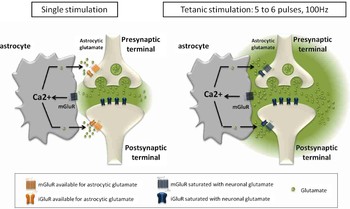
It would seem that the three papers discussed here also present discrepancies concerning an eventual role of astrocytic glutamate. Indeed, in the Henneberger study, impaired potentiation due to calcium clamp was fully rescued by the addition of D-serine, implying that glutamate is either not released by astrocytes or does not affect synaptic plasticity. Meanwhile, the Perea study indicates that astrocytic glutamate acts pre-synaptically to potentiate synapses. A more detailed observation of the experimental protocols used by the three groups reveals that all apply stimuli of radically different strengths, ranging from single minimal and mild stimulations (Perea), to 0.33 Hz theta-burst stimulation repeated five times or 100 Hz 1 s tetanic stimuli repeated twice (Agulhon), or 100 Hz 1 s tetanic stimuli repeated thrice (Henneberger). The differences in the stimulation protocols directly affect not only the amount of extrasynaptic glutamate but also the saturation levels of NMDA and mGlu receptors, which have an EC50 of 2 and 10 µM, respectively (Clements and Westbrook, Reference Clements and Westbrook1991; Hayashi et al., Reference Hayashi, Momiyama, Takahashi, Ohishi, Ogawa-Meguro and Shigemoto1993). Analytical and modeling studies have shown that, after a single action potential, glutamate barely escapes the synaptic cleft due to dilution effects and transporter activity (Barbour, Reference Barbour2001). This was experimentally confirmed using an extracellular glutamate sensor: a single stimulation in the cerebellum barely raised the extracellular concentration of glutamate to sub-micromolar levels (Okubo et al., Reference Okubo, Sekiya, Namiki, Sakamoto, Iinuma and Yamasaki2010). This concentration, lower than the EC50 of NMDA and mGluR receptors, leaves room for astrocytic glutamate to act on extrasynaptic glutamate receptors. However, with repeated stimulations at 100 Hz, after five to six pulses, glutamate concentrations quickly reach a concentration of 8 µM in the extracellular space (Okubo et al., Reference Okubo, Sekiya, Namiki, Sakamoto, Iinuma and Yamasaki2010). Due to differences in synaptic organization and astrocytic coverage in the cerebellum as compared with the hippocampus, the buildup of glutamate is even higher in the hippocampus: after two pulses, concentrations close to half the saturation of NMDA receptors are reached. Consequently, hundreds of pulses, such as those used in tetanic stimulation, would rapidly generate a buildup of glutamate that saturates NMDA and mGluR receptors (Fig. 2). Furthermore, the accumulation of glutamate is all the more important when experiments are run at room temperature due to decreased transporter activity (Diamond and Jahr, Reference Diamond and Jahr2000). Thus, excessive amounts of neuronal glutamate would not only compete out astrocytic glutamate but also saturate NMDARs and mGluRs, thus preventing any possible effect of astrocytes on neuronal plasticity. Although the above interpretations are purely speculative, they could account for the apparently conflicting results obtained by the various groups.
Competition between neuronal and astrocytic glutamate as a result of different stimulation protocols. After a single action potential, due to restricted spill over, the extracellular glutamate concentration is below the EC50 of NMDA and mGluR receptors, allowing astrocytic glutamate to act on extrasynaptic glutamate receptors (left panel). In contrast, following a tetanic stimulation (100 Hz, 5 to 6 pulses), extracellular glutamate concentrations of neuronal origin rapidly saturate NMDA and mGluR receptors and prevent any potential effect of astrocytic glutamate on neuronal plasticity (right panel).

CONCLUSION
As yet, it is too early to definitively rule out calcium as the key second messenger in astrocyte–neuron communication and neuronal plasticity. The questions raised by the papers discussed in this review indicate that a better understanding of calcium dynamics, signaling and gliotransmitter release is necessary, and that the variety of approaches being used are effective tools to tackle the issue. In the future, it will be important to study calcium signaling not only at the whole astrocyte level but also within astrocytic microdomains in fine processes close to synapses, at the sites of glutamate release.
Statement of interest
None.