Case details
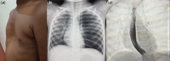
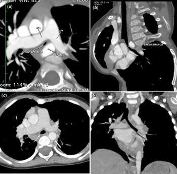
A 3-year-old boy presented with history of recurrent hospitalisations due to respiratory distress and wheeze since 6 months of age. There was no history of stridor and dysphagia. He had pectus excavatum (Fig 1a), which was also present in a milder form in the asymptomatic mother and maternal uncle, suggestive of familial pectus.Reference Creswick, Stacey and Kelly1 Breath sounds were diminished on left side of the thorax. There was no cyanosis. He was developmentally normal with no dysmorphic features. Chest X ray revealed obstructive emphysema of the left lung (Fig 1b). Echocardiogram revealed structurally normal heart, normal pulmonary arterial pressure, and left aortic arch. Pulsatile extrinsic compression of the left main bronchus was noted on flexible fiberoptic bronchoscopy. CT pulmonary angiogram confirmed the presence of left lung emphysema and rightward shift of mediastinum. Haller index was 3.4, suggestive of moderate degree of pectus excavatum. The aortic arch was left sided with normal branching pattern. However, the course of the descending thoracic aorta was abnormal. The descending thoracic aorta traversed from left to right of the midline and descended along the right anterior para-spinal region. At the level of bronchus, the descending thoracic aorta was pre-spinal as it coursed towards the right. Severe compression of the left main bronchus was noted between the right pulmonary artery anteriorly and abnormally coursing descending thoracic aorta posteriorly (Fig 2a and b). The course of descending aorta was not retro-esophageal which differentiated this condition from circumflex aorta. On barium swallow, the entire oesophagus was pushed to right side of spine (Fig 1C). There was no posterior compression of the oesophagus, thus ruling out retro-oesophageal course of aortic arch.
a ) Image of the chest wall showing pectus excavatum. b ) Left lung emphysema and mediastinal shift to the right on chest X ray. c ) Barium swallow image showing oesophageal shift to the right side of the spine.

Pre-operative CT angiogram: a ) Axial image showing left bronchial compression between right pulmonary artery and descending aorta. Left bronchus is slit like. Note the pre-vertebral position of the descending aorta. b ) Oblique view showing significant compression of the left main bronchus between right pulmonary artery and descending thoracic aorta. Post-operative CT angiogram: c ) Axial image showing normal caliber of left bronchus (arrow). Note the normal left para-spinal position of the descending aorta (bold arrow) following aortopexy. d ) Coronal view showing normal left main bronchus. LPA=left pulmonary artery; RPA=right pulmonary artery

After complete evaluation, a diagnosis of vascular compression of left main bronchus between the right pulmonary artery and abnormally coursing descending thoracic aorta was made. Aortopexy was performed through left postero-lateral thoracotomy, under general anaesthesia. The upper descending thoracic aorta was mobilised away from left main bronchus to the left of the spine and fixed to the periosteum of thoracic vertebrae using 2-0 Ethibond sutures (Ethicon Inc. Johnson and Johnson, Belgium) with Teflon felt pledgets. The ductal ligament was also ligated and divided. The child recovered well with an uneventful post-operative period. Surgical correction of pectus excavatum was not considered as there was no significant compression of the heart by the sternum and as the child was very young. Two years follow-up revealed good symptomatic improvement with no further hospitalisation. Repeat CT pulmonary angiogram showed good relief of left main bronchus compression; the dimension having improved from 2.1 mm pre-operatively to 5.56 mm at one-year follow-up (Fig 2C,D). Repeat bronchoscopy revealed normal caliber of left main bronchus, with no evidence of residual bronchomalacia.
Discussion
Left main bronchus narrowing can occur due to intrinsic structural wall abnormalities resulting in bronchial collapse, or extrinsic compression of the bronchus, most often by adjacent vascular structures. Compared to right bronchus, left bronchus is more vulnerable for compression due to the longer course and smaller caliber compared to right. Any abnormality in the surrounding structures like pulmonary artery, descending aorta, vertebral body, and oesophagus can result in bronchial compression. They can manifest with stridor, wheeze, recurrent pneumonia, and apneic spells.
Abnormal pre-vertebral course of descending thoracic aorta resulting in left main bronchial compression is a rare entity, first described by Donnelly and his colleagues.Reference Donnelly, Bisset and McDermott2 They described the magnetic resonance images of two infants where the descending aorta, instead of coursing in the left paraspinal space, had an abnormal pre-vertebral course resulting in bronchial compression, caused by the pincer effect of aorta and right pulmonary artery. In our patient too, the descending thoracic aorta coursed abnormally, crossing from left to right and subsequently descended in the anterior right paraspinal space. At the level of bronchus, the aorta crossed the midline and was pre-vertebral, slightly onto the right. Goldman et al had reported associated CHDs in 50% of their patients.Reference Goldman, Rimell and Meza3 In their series of 18 patients, Arcieri et al reported associated malformations in 16 children, which included CHDs, oesophageal atresia, and trachea-oesophageal fistula.Reference Arcieri, Serio and Nenna4 Our patient had a structurally normal heart, but had associated pectus excavatum. This association was earlier reported by Speggiorin et al.Reference Speggiorin, Atamanyuk and Wallis5 Whether associated chest wall abnormality contributes to the abnormal course of the aorta is speculative.
A left aortic arch with right descent, most often has a retro-oesophageal course as it crosses the midline, and is described as circumflex aorta. This often forms a complete vascular ring and causes airway compression. In our patient, though the arch was left sided with right descent, the aortic arch was not retro-oesophageal, as evidenced on CT angiogram and barium swallow. It is important to differentiate abnormal pre-vertebral course of descending aorta from circumflex aorta, as the surgical approach is different. Posterior aortopexy gives good symptom relief in the former, while a more extensive aortic uncrossing procedure is required for the latter.
Our patient showed a dramatic and sustained symptom relief following posterior aortopexy. This has been described as an effective mode of management by most previous authors too.Reference Donnelly, Bisset and McDermott2,Reference Arcieri, Serio and Nenna4–Reference Hungate, Newman and Meza6 Speggiorin and his colleagues adopted a combined approach of Nuss procedure and aortopexy in two cases, where pectus excavatum was associated. However, we decided to do aortopexy alone initially, as there was no significant compression of the trachea and heart and had planned to consider Nuss procedure only if child continued to remain symptomatic. Symptoms may sometimes persist after aortopexy, due to secondary bronchomalacia, and may require bronchial stenting for symptom relief.Reference Arcieri, Serio and Nenna4 Arcieri et al reported residual bronchomalacia in 44% of their cases.Reference Arcieri, Serio and Nenna4 Repeat bronchoscopy at one-year follow-up revealed normal caliber of bronchus with no residual malacia in our patient.
Conclusion
Abnormal pre-vertebral course of descending aorta is a rare cause of airway obstruction and has to be differentiated from vascular ring. Recognition of this entity is important as the surgery (aortopexy) is not complex and offers good symptomatic relief in these children.
Acknowledgements
None.
Financial Support
This research received no specific grant from any funding agency, commercial, or not-for-profit sectors.
Conflicts of Interest
None.
Ethical Standards
The authors assert that all procedures contributing to this work comply with the ethical standards of the relevant national guidelines on human experimentation and with the Helsinki declaration of 1975, as revised in 2008, and has been approved by the institutional committees.