INTRODUCTION
Expansion of human activities usualy results in habitat fragmentation, a phenomenon widely recognized as a major threat to the Earth's biodiversity (Fahrig, Reference Fahrig2003; Castro and Fernandez, Reference Castro and Fernandez2004). The transformation of a continuous forest into isolated fragments results in a hyper-dynamic landscape with unpredictable consequences to biodiversity and, consequently, overall ecosystem health (Ruedas et al. Reference Ruedas, Salazar-Bravo, Tinnin, Armién, Cáceres, Garcia, Ávila Diaz, Gracia, Suzán, Peters, Yates and Mills2004; Pardini, Reference Pardini2004; Gillespie et al. Reference Gillespie, Chapman and Greiner2005; Hudson et al. Reference Hudson, Dobson and Lafferty2006). A decrease in community richness and changes in community composition, as well as in species interaction, is apparent as a major consequence of this process (Fahrig, Reference Fahrig2003; Castro and Fernandez, Reference Castro and Fernandez2004; Pardini, Reference Pardini2004; Redeker et al. Reference Redeker, Andersen, Pertoldi, Madsen, Jensen and Jorgensen2006).
However, studies concerning the effects of habitat fragmentation upon infection by parasites and consequentely on the emergence or re-emergence of human infectious diseases are still scarse. Studies with gastrointestinal helminths and protozoans in primates (Gillespie et al. Reference Gillespie, Chapman and Greiner2005; Gillespie and Chapman, Reference Gillespie and Chapman2006), and Lyme disease in New York, USA (Allan et al. Reference Allan, Keesing and Ostfeld2003) concluded that the habitat fragmentation process modulates parasite infection dynamics, increasing the potential risk of infection. These studies indicate that infections by parasites in fragmented habitats may increase, due to host species diversity loss and a probable increase in the population density of a more suitable reservoir (Allan et al. Reference Allan, Keesing and Ostfeld2003; Gillespie et al. Reference Gillespie, Chapman and Greiner2005; Gillespie and Chapman, Reference Gillespie and Chapman2006). It is important to emphasize that the last factor changes according to the parasite, the study area and time-scale.
Despite this, little is known about the consequences of habitat fragmentation on Trypanosoma cruzi infection dynamics in the wild. Studies showed that mammals and vectors in the same region, and even in the same forest fragment, may be involved in distinct transmission cycles with distinct host competence (Pinho et al. Reference Pinho, Cupollito, Mangia, Fernandes and Jansen2000; Lisboa et al. Reference Lisboa, Mangia, De Lima, Martins, Dietz, Baker, Ramon-Miranda, Ferreira, Fernandes and Jansen2004). This shows that the dispersal of T. cruzi in the wild occurs in complex transmission cycles ruled by factors still unknown (Jansen et al. Reference Jansen, Pinho, Lisboa, Cupollito, Mangia and Fernandes1999). In addition, habitat fragmentation or deforestation processes, by decreasing the biodiversity and/or favouring the maintenance of more generalist and tolerant species (Castro and Fernandez, Reference Castro and Fernandez2004; Gillespie et al. Reference Gillespie, Chapman and Greiner2005), can explain the re-emergence of Chagas disease in Brazil. In the localities where new cases of this disease were recorded in the last 2 years the epidemiological conditions observed were: a high human pressure on environmental and consequently low mammal diversity with the maintenance of the most common and competent reservoir of T. cruzi in the environment, the common opossum (Didelphis aurita), a species known as a habitat generalist that tends to increase its abundance in areas affected by human activities (Jansen et al. Reference Jansen, Pinho, Lisboa, Cupollito, Mangia and Fernandes1999; LoGiudice et al. Reference LoGiudice, Ostfeld, Schimidt and Keesing2003; Olifiers et al. Reference Olifiers, Gentile and Fiszon2005).
The aim of this study was to understand the effects of habitat fragmentation on infection by T. cruzi among small wild mammals. For this, we analysed the direct effect of this process on small mammal communities, comparing a fragmented habitat with a continuous forest. We associated the community results with the prevalence of infection by T. cruzi observed, analysing also the time-scale of this pattern.
MATERIALS AND METHODS
Study area
Our study was carried out, over 2 years (2004–2005), in 2 areas of the Teresópolis municipality (22°24′ S; 42°58′ W), state of Rio de Janeiro, Brazil. Area (i) is a fragmented landscape in the Venda Nova district, located in the surroundings of the municipality. In this area a total of 12 isolated fragments (sites) were randomly selected and arranged into 3 size classes (4 per class): small (<10 ha), medium (10–40 ha) and large (>40 ha). The matrix among fragments was composed of agricultural and cattle-raising fields. Area (ii) Serra dos Órgãos National Park (PARNASO), a biological reserve, founded in 1984 with a total area of 10 527 ha, is a continuous forest site selected as the preserved control area. It is worth mentioning that no human dwellings are present in the any of the studied areas.
These areas was selected for 2 reasons: firstly because a previous study conducted 10 years ago in Caleme village, a district on the surroundings of Teresópolis, demonstrated that 50% of marsupials were infected (patent parasitaemia) by T. cruzi (Pinho et al. Reference Pinho, Cupollito, Mangia, Fernandes and Jansen2000); and secondly both are under the same natural conditions: Cwb–height tropical climate according to Köppen classification, presented a short dry season between June and August. The altitude varies from 800 to 1100 meters above sea level, the mean maximum and minimum annual temperatures are, respectively, 23·9°C and 13·3°C and rainfall is around 1600 mm/year. The vegetation has been classified as Lower Mountain Atlantic Rain Forest (Oliveira-Filho and Fontes, Reference Oliveira-Filho and Fontes2000).
We returned to Caleme to check the actual epidemiological profile and compare it with the areas of the present study.
Capture of small mammals
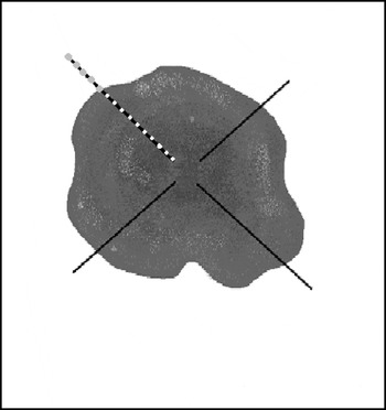
Small mammals were collected with live traps (Tomahawk® and Sherman® models) baited with a mixture of peanut butter, banana, oat and bacon. The traps were placed along linear transects, at 20 m intervals among capture stations in all study sites. Each site was composed of 4 transects with a distinct number of capture stations according to its class size: 7 per transect in the sites of the small class size (5 on the ground – with 2 live traps; and 2 on the understory – with 1 trap); 14 in the sites of the medium class size (10; and 4 respectively); and 21 in the sites of the large class size and PARNASO (15; and 6 respectively). In the sites of area (i) (fragmented habitat), transects received 5 further capture stations per transect in their surroundings (Fig. 1). The trapping period was 5 nights per site in each survey. Two surveys per site were carried out (one in wet and other in dry season in the same year). The total sampling efforts were: 1920 traps-nights in the small set; 3840 in the medium set; 5760 in the large set; 1440 in the PARNASO site (total effort=12 960 traps-nights).
Site design of fragments of the medium set. The 4 linear transects (black lines) with 10 capture stations per transect inside the fragment (white points) and the further 5 capture stations per transect in its surroundings (grey points).

Two kinds of study were conducted with the small wild mammals. (i) A study of capture-recapture (twice) with the marsupials D. aurita and Philander frenatus. All marsupials captured were ear-tag marked, with individual numbers, and released at the same point of capture. These procedures were done at the end of the trapping period. These species were chosen due to their longevity and their importance in the maintenance of T. cruzi in the wild. (ii) A removal study with the other small mammals. In Caleme, the trapping design was the same as in PARNASO, but only 1 survey was carried out during the dry season. The total sampling effort was 720 trap-nights. At this site, the marsupials were released and all the other small mammals were sacrificed.
Parasitological data
Blood samples (a maximum of 1·5 ml per specimen) were collected by cardiac puncture under anaesthesia (50 mg/kg body weight of Ketamin® by intramuscular injection). These samples were used to perform fresh blood smears, haemoculture and serology. If insufficient material was collected, priority was given to the haemoculture.
A total of 466 haemocultures, 435 serum samples and 376 fresh blood smears of individual animals were analysed.
Parasitological follow-up
One fresh blood smear from each individual was examined for verification of patent parasitaemia. Two tubes containing NNN medium with a liver infusion triptose (LIT) overlay were inoculated with 0·2 ml of blood from each specimen. The tubes were examined in the laboratory twice a month up to a maximum of 5 months for the serologically positive specimens and 2 months for seronegative ones. These procedures were performed using a light microscope at 100× magnification.
Serological follow up
The immunofluorescense assay (IFA) was performed as proposed in the literature (Camargo, Reference Camargo1966) and done with an adapted IFI-Chagas/Bio- Manguinhos Kit®. Briefly, rodent sera, maintained at −20°C, were tested with commercial fluorescein isothiocyanate-labelled anti-rat IgG (FITC, Sigma®), while marsupial sera, conserved under the same conditions, were tested with a specific intermediary anti-opossum serum raised in rabbits and the reaction was visualized using commercial FITC-labelled anti-rabbit serum (Sigma®). The cut-off values adopted for the serological titres of rodents and marsupials were ⩾1:10 and ⩾1:40, respectively, since these were the lowest serum dilutions of individual animals with positive haemocultures (Herrera et al. Reference Herrera, D'Andrea, Xavier, Mangia, Fernades and Jansen2005). As controls, sera from laboratory reared, experimentally infected and uninfected animals were used throughout.
Data analyses
The species richness (S′) was calculated as the number of species captured, and the Alpha diversity (Shannon Index, H′), as a local index of diversity for each site. Only those species with more than 10 individuals captured had their abundance considered (N/T; where N=total number of individuals captured per species and T=total sampling effort). Beta w diversity, a broad sense metric which incorporates differences in composition attributable to species richness gradient, was calculated for pairs of sites, considering each site related to all others from the same fragments class size, as described by Pardini et al. (Reference Pardini, Marques de Souza, Braga-Neto and Metzger2005). To obtain 1 value for each class size, the mean of the paired Beta w values was calculated. These values were used to calculate Beta w diversity among fragment classes and PARNASO. The Alpha diversity for the wet and dry seasons was calculated for each site. The differences between the Alpha and Beta diversities were tested by one-way analysis of variance. To construct the diversity diagram, over the H′, it was necessary to calculate the maximum (H′max) and minimum (H′min) diversity as follow: H′max=LnS′; and H′min=[LnN−(N-S′+1/N)]∗(N-S′+1); where N=1000 (standardized).
The independence among the fragment classes and PARNASO and between the seasons was tested by the Chi-Square test. The relationship between site classes and prevalence was tested with SPSS.10 ‘curve estimation’ utility, for this analysis we considered the proportion among sampling effort displayed area in each site.
The Maintenance Host Range (MHR), i.e the number of infected species/total number of species of a given area, was evaluated by the index proposed in the literature as host specificity index (Std*) (Poulin and Mouillot, Reference Poulin and Mouillot2005). The minimum and maximum values for MHR evaluation varied from 1 to 6. This analysis was run in the program TaxoBiodiv2 (available at http://www.otago.ac.nz/zoology/downloads/poulin/ TaxoBiodiv2). For these analyses the Caleme site was not considerated.
RESULTS
Small mammal diversity
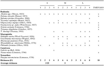
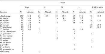
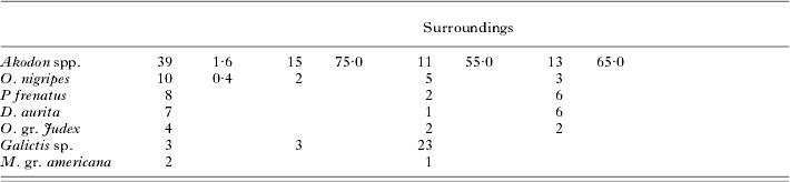
A total of 494 specimens belonging to 16 genera (2 carnivores, 5 marsupials, 8 rodents and 1 xenarthra) were captured (Tables 1 and 2). The rodents Akodon spp. and Oligoryzomys nigripes, and the marsupials D. aurita and P. frenatus were the most abundant in all sites where human activity was observed (fragmented habitat), while in PARNASO, unaffected by human activity, marsupials were less abundant than rodents. Indeed, the rodents Delomys dorsalis, Trinomys dimidiatus, Akodon spp. and O. nigripes were the most abundant in this site (Table 2). The rodents D. dorsalis, Sciurus aestuans and T. dimidiatus; and the carnivore Nasua nasua seemed to be less able to bear habitat fragmentation, and were captured only in the PARNASO (Table 1). A lower recapture rate, 10% for D. aurita and 13% for P. frenatus, was observed.
Species richness in the fragments (S, small; M, medium; L, large sets) and continous forest (PARNASO) in Teresópolis (Rio de Janeiro, Brazil)

(x, Presence.)
Total number (N) and total abundance (Abund.) of small mammals, only for the species that had more than 10 individuals captured, in the fragments (S, small; M, medium; L, large sets), PARNASO (continous forest), and surroudings of the fragments in Teresópolis (Rio de Janeiro, Brazil)

Fragmentation and small mammal communities
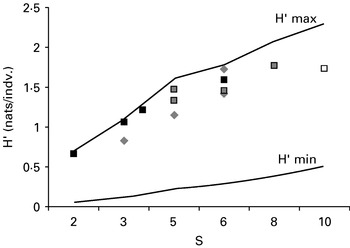
The richness and Alpha diversity tended to decrease with the site size (Fig. 2). The Alpha diversity indices among the sites were not statistically different (F=1·971; P=0·189). Nevertheless, when both Alpha diversity and the species richness were analysed together, it was possible to point out some differences according to the site size: (i) small wild mammal diversity in smaller sites and (ii) higher diversity in larger sites and PARNASO (Fig. 2). The Alpha diversity did not differ significantly between the seasons (F=4·975; P=0·067).
Diversity diagram with the Richness (S′) and the Shannon Index (H′) in the 12 fragments (black square, small set; grey rhombus, medium set; grey square, large set) and the PARNASO (white square) associated with the hypothetical maximun (H′max) and minimum (H′min) Shannon Index.

Contrary to the Alpha diversity, the Beta diversity (Beta w) was significantly different among the fragments (F=4·818; P=0·024). In this context, the mean Beta diversity was higher among sites of the smaller class size (1·2465) and reduced in the medium and large sites (1·1983 and 1·1589 respectively). When we considered all studied sites (12 sites in the fragmented habitat and 1 site in the continous forest), the mean Beta diversity was the highest (1·5283), and the difference between control area and fragment classes was statistically significant (F=45·09; P=0·001).
Only 1 individual of P. frenatus dispersed between different fragments in the dry season.
Parasitological data
An extremely mild controlled infection by T. cruzi was detected in all seroprevalent animals since no patent parasitaemia could be detected in fresh blood samples, and no parasites were isolated by haemoculture, inclusively in the Caleme area.
Serological data
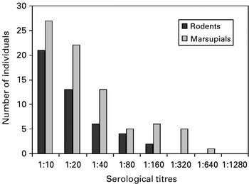
A total seroprevalence of 18% was detected by IFA in all small mammal assemblages. The serological titres varied from 1:10 to 1:160 in rodents and 1:40 to 1:640 in marsupials (Fig. 3).
Serological titre variation determined by immunofluorescence assay (IFA) among rodents and marsupials in the study area. The cut-off values adopted for the serological titres of rodents and marsupials were ⩾1:10 and ⩾1:40 respectively.

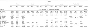
Infection by T. cruzi was detected in the rodents Akodon spp., O. nigripes, Oxymicterus gr. judex and D. dorsalis; and in the marsupials D. aurita and P. frenatus. Akodon spp. was the only rodent found to be seropositive by T. cruzi in all studied sites. Marsupials were found seropositive only in the fragments and in Caleme sites (Table 3).
Total seroprevalence of Trypanosoma cruzi infection by immunofluorescence assay inside the fragments, PARNASO and Caleme and surrounding the fragments; and prevalence of infection inside the fragments and PARNASO during the wet and dry seasons in Teresópolis (Rio de Janeiro, Brazil)


The recapture index was 15% (14/91), of these 2 specimens (1 D. aurita and 1 P. frenatus) seroconverted. This result showed a very low transmission rate.
T. cruzi and habitat fragmentation
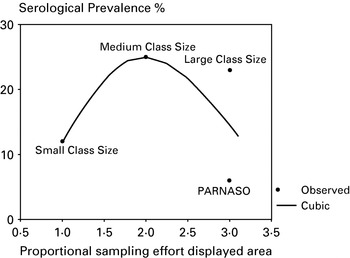
The observed seroprevalence varied among the fragments and the control area (χ2=15·78, P<0·005; d.f.=3), and among the surroundings of the fragments (χ 2=11·16, P<0·005; d.f.=2) (Table 3). The seroprevalence was lowest in the highest richness area (PARNASO). The fragmented habitat showed the higher seroprevalence. Among them, the seroprevalence was lower in the small class size, where the human pressures and the small mammal diversity loss were highest (Figs 2 and 4).
Model of the habitat fragmentation effect on the Trypanosoma cruzi transmission cycle among small mammals in fragmented areas (small, medium and large sets) in comparison to PARNASO (continuous forest) in Teresópolis (Rio de Janeiro, Brazil).

Despite this, no statitiscal difference was observed between class sizes and serological prevalence (F=0·34; P=0·76). The class sizes and serological prevalence relationship was described by a cubic equation: serological prevalence=1·8+(14·56∗site set2)−(4·38∗site set3).
Significant anti-T. cruzi seroprevalence was observed in the medium (χ2=17·87, P<0·0001; d.f.=1) and large classes (χ2=21·96, P<0·0001; d.f.=1) in the wet season (Table 3). This difference between the seasons was associated with the increased abundance of D. aurita, since the Alfa diversity between the seasons was not different. No statistical difference of the infection prevalence between the seasons was observed in the small class or in the PARNASO control area.
In these study areas, T. cruzi apparently increased its host range according to the increase in the fragmentation process and decrease host diversity, exploring more taxonomically distant hosts in the small class (Sdt* (MHR)=5·00) than in the medium (Sdt* (MHR)=4·06) and large (Sdt* (MHR)=4·01) classes. This aspect is confirmed by the lower MHR values in PARNASO (Sdt* (MHR)=2·00), the preserved and higher richness site.
DISCUSSION
The results showed that at least 3 main factors are modulating the transmission cycle of T. cruzi among small mammals in this fragmented Atlantic Rain Forest landscape: (i) biodiversity loss; (ii) increase of marsupial abundance and (iii) seasonal variation.
Lower small mammal diversity (Alpha diversity) in the smaller set was expected (McArthur and Wilson, Reference McArthur and Wilson1967). However, since the fragmentation process in the area is already interrupted the current small mammal community seems to be under other natural pressures apart from fragment size: (i) age of fragment; (ii) matrix quality on the surroundings; (iii) habitat structure and (iv) the stochastic effect (Lomolino and Weiser, Reference Lomolino and Weiser2001; Castro and Fernandez, Reference Castro and Fernandez2004; Pardini, Reference Pardini2004; Redeker et al. Reference Redeker, Andersen, Pertoldi, Madsen, Jensen and Jorgensen2006).
Considering the studied fragmented sets as an assemblage, no overall diversity loss (Beta w diversity) was observed in comparison with the continuous forest (PARNASO). However, distinct community composition in the fragments was observed. This feature shows the environmental heterogeneity that may be displayed by fragments even in the same region, pointing to a probable consequent epidemiological mosaic.
The data allow two interpretations to explain the observed higher prevalence of infection by T. cruzi in the fragmented habitat in comparison to PARNASO: (i) the loss of local small mammal diversity that consequently decreases the dilution effect, by decreasing the proportion of inefficient potential hosts; and/or (ii) the increase of vector population as the consequence of the overall biodiversity maintenance and feeding opportunity (high Beta w values) (Schmidt and Ostfeld, Reference Schmidt and Ostfeld2001).
The first hypothesis seems to be more probable, considering that the dispersal among non-connected fragments in this study is not expected (Pardini et al. Reference Pardini, Marques de Souza, Braga-Neto and Metzger2005) and that the study area with greatest diversity richness (PARNASO) presented the lowest prevalence of infections by T. cruzi.
The Maintenance Host Range index (the capacity of the parasite to infect a high number of taxonomically distant hosts) observed in this study confirms that, also in the case of T. cruzi, the decrease of abundance and richness of potential host species in the fragmented habitat, causes parasites to become more eclectic, infecting a higher number of available host species (Poulin and Mouillot, Reference Poulin and Mouillot2005).
Thus, the fragmentation process may have favoured T. cruzi transmission since it resulted in a high MHR index and increased competent host species population (mainly D. aurita and P. frenatus). Nevertheless, it did not alter the profile of infection of the animals, since all displayed subpatent parasitaemia throughout the study.
The higher prevalence of infection observed in the wet season may be explained by the lower small mammal diversity and higher abundance of marsupials observed during this period, as a result of the marsupials dispersed by the lack of food and by the recruitment population period. A possible outcome is that T. cruzi may display 2 different strategies of transmission for the distinct seasons in the study area: (i) an increase of the area distribution of infection occurrence in the dry season and (ii) maintenance of the transmission cycle in the wet season (Pavlovsky, Reference Pavlovsky1966).
Besides being dependent on spatial variation, the T. cruzi transmission cycle in the wild environment was demonstrated to be a temporal phenomenon, since a previous study in the Caleme area (in 1996) showed that 29% of D. aurita and 48% of P. frenatus were positive by haemoculture (Pinho et al. Reference Pinho, Cupollito, Mangia, Fernandes and Jansen2000). The change in the infection profile of the animals in Caleme that displayed an impressive parasitaemia in the 1996 survey (detectable by haemocultures to a very low parasitaemia detectable only by an indirect diagnostic method in the present study) showed that the competence as reservoir of the different host species of T. cruzi displays multi-year fluctuations. In spite of the overall mild character of the infection, transmission of T. cruzi was maintained, as observed by the two individual seroconversions. The factors that define the profile of infection in a given wild environment are still unknown. In fragmented areas annual changes in landscape use are observed, new interactions among host species, larger and diverse parasitic fauna interchange, alterations of host community composition and hosts health status (Kunz et al. Reference Kunz, Matz-Resing, Stolte, Hamilton and Kaup2002; McKay, Reference McKay2006). Consequently, in the current scenario, two conditions may emerge: the increase of transmissibility of the parasite due to an increase of parasitaemias or the extinction of the enzootic transmission cycle.
Regarding parasitism phenomena, the presented data showed that habitat fragmentation leads to unpredictable results for wildlife or human disease emergence. Furthermore, this study also showed the importance of micro-regional peculiarities to understand the factors that modulate the focal transmission cycles of parasites in health monitoring.
Ethical clearance and Licenses
The present work has the endorsement of the Comissão de Ética e Uso de Animais-CEUA/FIOCRUZ, RJ, Brazil, registration number: P0083-01. This study was licensed by Instituto Brasileiro do Meio Ambiente e dos Recursos Naturais Renováveis-IBAMA, license number: 02001.003618/03-06 and 02001.006721/2004.
The authors thank Dr Vera Bongertz and Dr Caludio Bidau for insightful comments and revision of the English, and the staff of the Laboratorio de Biologia e Parasitologia de Maniferos Silvestres Reservatórios and Laboratorio de Biologia de Tripanosomatideos for technical support. This study was supported by: CNPq/ITT/FH Köln-FMER N°. 690145/2001-2, CAPES, IOC/FIOCRUZ.