Natural nanoclays have gained significant attention due to their environmentally friendly and cost-effective properties as nanofillers (Nazir et al., Reference Nazir, Mohamad Kassim, Mohapatra, Gilani, Raza, Majeed, Jawaid and Oualid2016; Guo et al., Reference Guo, Aryana, Han and Jiao2018; Elmi, Reference Elmi2023). Halloysite nanotubes (HNTs), a subtype of kaolinite, stand out due to their high aspect ratio, abundance, tuneable surface chemistry and porosity (Biddeci et al., Reference Biddeci, Spinelli, Colomba and Di Blasi2022). These unique properties position HNTs as an attractive green alternative to synthetic nanomaterials such as carbon nanotubes (Liu et al., Reference Liu, Jia, Jia and Zhou2014; Ma et al., Reference Ma, Wu, Higaki and Takahara2018; Çankaya et al., Reference Çankaya, Ünal and Korcan2024), particularly for applications in biomedicine, packaging and catalysis, as has been discussed in recent reviews (Kouser et al., Reference Kouser, Sheik, Nagaraja, Prabhu, Prashantha and D’Souza2020; Boccalon et al., Reference Boccalon, Sassi, Pioppi, Ricci, Marinozzi, Gorrasi and Nocchetti2022; Massaro et al., Reference Massaro, Noto and Riela2022b; Butun Sengel et al., Reference Butun Sengel, Tunca, Deveci, Bas and Butun2023; Fahimizadeh et al., Reference Fahimizadeh, Wong, Baifa, Sadjadi, Auckloo and Palaniandy2024).
Structurally, HNTs form through the weathering of rocks in tropical and subtropical climates. They typically exhibit a tubular morphology, although they can also appear as spheroids or plates (Wong et al., Reference Wong, Pasbakhsh, Arabi, Keeling and Tan2021). These nanotubes follow the chemical formula Al2Si2O5(OH)4·nH2O, consisting of a dioctahedral 1:1 two-layered hollow structure made up of aluminosilicate layers rolled together to form their walls, held together by van der Waals forces and hydrogen bonds. The dimensions of HNTs vary, with lengths ranging from 100 to 2000 nm and inner diameters of 10–30 nm, and the outer diameters range from 30 to 50 nm (Joussein et al., Reference Joussein, Petit and Delvaux2007; Rawtani & Agrawal, Reference Rawtani and Agrawal2012; Prishchenko et al., Reference Prishchenko, Zenkov, Mazurenko, Fakhrullin, Lvov and Mazurenko2018; Ouyang et al., Reference Ouyang, Liu, Yang and Zhang2019; Kassa et al., Reference Kassa, Benhamida, Kaci and Bruzaud2020).
The surface complexity of HNTs includes distinct hydrophilic and hydrophobic regions, as reported by Biddeci et al. (Reference Biddeci, Spinelli, Colomba and Di Blasi2023) and Ganapathy et al. (Reference Ganapathy, Shanmugam, Pitchiah, Murugan, Chinnathambi and Alharbi2022): a hydrophilic internal lumen with aluminol (Al–OH) groups, a less hydrophilic external surface with siloxane (Si–O–Si) groups, an interlayer surface chemically similar to the internal lumen and edge surfaces terminated by both aluminol and silanol (Si–OH) groups. This structural complexity contributes to their exceptional thermal stability, mechanical strength and functional properties, as explained by Gaaz et al. (Reference Gaaz, Sulong, Kadhum, Al-Amiery, Nassir and Jaaz2017), making them ideal for reinforcing polymers such as polyethylene, epoxy resins and rubbers (Cheng et al., Reference Cheng, Chang, Liu, Qin and Zhu2017; Lisuzzo et al., Reference Lisuzzo, Cavallaro, Milioto and Lazzara2020). Even at low concentrations (3–7 wt.%), HNTs significantly enhance thermal resistance, mechanical properties and flame retardancy (Bugatti et al., Reference Bugatti, Sorrentino and Gorrasi2017; Petková et al., Reference Petková, Ryba, Hrabovská, Ujhelyiová and Hricová2019), enabling their application in coatings, tissue engineering and active packaging (Kouser et al., Reference Kouser, Prabhu, Prashantha, Nagaraja, D’Souza and Meghana2022; Qin et al., Reference Qin, Dong, Nie, Fakhrullin, Zhang and Zhang2024; Sun et al., Reference Sun, Yu, Liu, Yan, Xu and Cheng2024; Kim et al., Reference Kim, Kim, Lee, Yang, Yu and Kim2025; Zhang et al., Reference Zhang, Huang, Yang, Dong, Zhou and Yan2025).
In the light of these features, the inherent hydrophilicity of HNTs remains a significant challenge, driven by abundant hydroxyl groups on their inner and outer surfaces, along with their strong surface polarity, leading to poor interfacial adhesion and aggregation when integrated into various organic and inorganic matrices (Meng et al., Reference Meng, Wang, Tang, Hong, Gao and Chen2017). To overcome these limitations, researchers have explored various surface modification strategies for both the outer and inner surfaces of HNTs, including acid etching, alkali treatment, polymers, surfactants and nanomaterials. These approaches have proven effective for improving the dispersion and compatibility of HNTs in nanocomposites (Yuan et al., Reference Yuan, Tan and Annabi-Bergaya2015; Tharmavaram et al., Reference Tharmavaram, Pandey and Rawtani2018; Tarasova et al., Reference Tarasova, Naumenko, Rozhina, Akhatova and Fakhrullin2019).
Previous research has focused primarily on modifying the external surfaces of HNTs, exploiting their elongated, nanorod-like shape (Abdullayev et al., Reference Abdullayev, Joshi, Wei, Zhao and Lvov2012; Sargazi et al., Reference Sargazi, Laraib, Barani, Rahdar, Fatima and Bilal2022), which facilitates interactions through van der Waals forces, hydrogen bonding and electrostatic and covalent reactions (Kokulnathan et al., Reference Kokulnathan, Wang, Thangapandian and Alaswad2020; Zhao et al., Reference Zhao, Zhou and Liu2020). These interactions make the surface amenable to functionalization with various molecules through, for example, the grafting of different metallic nanoparticles (Ni, ZnO, Pt) or functional organic molecules such as organophosphonic acid and organosilanes (Sun et al., Reference Sun, Liu, Lv, Dong, Wu and Wang2015; Zhang et al., Reference Zhang, Tang, Yang and Ouyang2016; Jawwad Saif et al., Reference Jawwad Saif, Muhammad Asif and Naveed2018; Taroni et al., Reference Taroni, Meroni, Fidecka, Maggioni, Longhi and Ardizzone2019; Sadjadi, Reference Sadjadi2020; Lisuzzo et al., Reference Lisuzzo, Bertini, Lazzara, Ferlito, Ferrante and Duca2023).
Among these, organofunctional alkoxysilanes have been widely adopted as a strategy for modifying HNTs. This approach requires a good understanding of silanization chemistry, which explains how alkoxysilane hydrolysis forms silanol groups, as they can attach various functional groups on HNT surfaces through covalent bonding. The covalently attached silane molecules form a monolayer or multilayer depending on the reaction conditions, thereby functionalizing the HNT surface with the desired groups, such as vinyl, epoxy, methacrylate, phenyl, thiol and amine groups, which have been extensively studied for their effectiveness at improving HNT compatibility and functionality with various matrices (Zhang et al., Reference Zhang, Tang, Yang and Ouyang2016; Yang et al., Reference Yang, Chen, Leng, Huang, Wang and Tian2017; Jafazadeh & Haddadi-Asl, Reference Jafazadeh and Haddadi-Asl2023; Bao et al., Reference Bao, Yan and Han2024; Senyel & Dike, Reference Senyel and Dike2024; Wieczorek et al., Reference Wieczorek, Tatarchuk, Skórczewska, Szulc and Tomaszewska2024).
Although surface modifications of the external walls of HNTs are well-documented, strategies targeting the inner lumen, defined by aluminol-rich groups and typically air-filled cavities that serve as a host for guest molecules, remain less explored, primarily due to the challenges related to limited access and confined internal spaces. This inner lumen presents a significant challenge for the efficient loading of functional molecules (Biddeci et al., Reference Biddeci, Cavallaro, Di, Lazzara, Massaro and Milioto2016, Reference Biddeci, Spinelli, Colomba and Di Blasi2022; Sahnoune et al., Reference Sahnoune, Taguet, Otazaghine, Kaci and Lopez-Cuesta2017; Kaur et al., Reference Kaur, Gupta, Prakash, Rodriguez, Sheremet, Mehta and Sharma2024). Researchers have proposed methods such as acid etching to widen the lumen, enabling the encapsulation and release of active agents such as drugs, enzymes, dyes, nucleic acids or nanomaterials. These modifications can facilitate applications such as controlled drug release, catalysis and nanoreactor functionalities, as demonstrated in recent reviews and research papers (Rawtani et al., Reference Rawtani, Pandey, Tharmavaram, Pathak, Akkireddy and Agrawal2017; Zhang et al., Reference Zhang2017; Lazzara et al., Reference Lazzara, Cavallaro, Panchal, Fakhrullin, Stavitskaya, Vinokurov and Lvov2018; Santos et al., Reference Santos, Ferreira, Veiga, Ribeiro, Panchal, Lvov and Agarwal2018; Massaro et al., Reference Massaro, Licandro, Cauteruccio, Lazzara, Liotta and Notarbartolo2022a; Lei et al., Reference Lei, Zhou, Liu, Kong, Liao and Li2023; Zhang et al., Reference Zhang, Huang, Yang, Dong, Zhou and Yan2025).
In this study, distinct modification approaches are explored to prepare organic–inorganic compounds. These approaches involve silanization with triethoxy(octyl)silane (OTES) and caffeic acid (CA) loading, aiming to address the inherent limitations of the aggregation and poor dispersibility of HNTs, particularly Algerian HNTs, which remain largely underexplored despite their local abundance. This paper also provides a comparative evaluation of the effects of these two treatments, which have been minimally explored in surface modification studies.
As a chemical agent, OTES has proven effective at converting HNT surfaces to a hydrophobic state by covalently binding to the external silanol groups, forming hydrophobic alkyl layers that maintain their dispersion in polymer matrices. This medium-length alkyl chain can be expected to improve compatibility without causing excessive steric hindrance, making it suitable for applications in flexible packaging films, thermally resistant composites and hydrophobic coatings (Sánchez-Fernández et al., Reference Sánchez-Fernández, Peña-Parás, Vidaltamayo, Cué-Sampedro, Mendoza-Martínez and Zomosa-Signoret2014; Siy et al., Reference Siy, Tan, Viron, Sajor, Santos and Penaloza2020).
However, effective lumen modification requires overcoming the challenge of limited accessibility. To address this, acid etching with weak acids such as acetic acid is applied prior to loading. This treatment selectively removes impurities and increases the lumen diameter for effective loading of guest molecules such as CA, which stands out as a bioactive phenolic compound with strong antioxidant and antimicrobial properties (Maurya & Devasagayam, Reference Maurya and Devasagayam2010; Katuwavila et al., Reference Katuwavila, Perera, Karunaratne, Amaratunga and Karunaratne2016).
The impact of both treatments on the structural, chemical, textural and thermal properties of HNTs will be assessed using advanced techniques, including Fourier-transform infrared (FTIR) spectroscopy, X-ray fluorescence (XRF), nitrogen adsorption–desorption isotherms (Brunauer–Emmett–Teller; BET), scanning electron microscopy (SEM), thermogravimetric analysis (TGA) and X-ray diffraction (XRD).
The modifications are expected to significantly enhance the dispersion, thermal stability and compatibility of Algerian HNTs, promoting the development of an eco-friendly, high-performance polymer nanocomposite based on HNTs, making them viable for applications such active packaging and thermal insulation coatings.
Materials and methods
Materials
The Algerian HNTs were collected from the Djebel Debbagh deposit in Guelma region, located in north-eastern Algeria, distinguished by their greyish colour due to their 1.22 wt.% MnO2 content. The material was supplied by SOALKA (Société Algérienne des Kaolins), an Algerian kaolin production company. The HNTs exhibited an average internal diameter range of 10–30 nm and an external diameter of 30–50 nm, a density of 2.1 g cm–3 and a specific surface area of 54 m2 g–1 (Kennouche et al., Reference Kennouche, Le, Kaci, Quantin, Caro-Bretelle and Delaite2016).
For prior processing, raw HNT stones were first manually ground into a coarse powder using a hand mortar to facilitate the ball mill processing. The coarse powder was then refined using a laboratory ball mill for 1 h and sieved through a 38 µm mesh to yield microparticles with an average diameter of 25 µm, containing a fraction of nanoparticles.
For surface modification, CA and OTES were procured from Sigma Aldrich (France) for surface treatment of the HNTs. Acetic acid (99.7% CH3COOH) was employed as a mild acid to enhance the lumen size of the HNTs.
HNT silanization
Silane modification followed the procedure outlined by Carli et al. (Reference Carli, Daitx, Soares, Crespo and Mauler2014). Approximately 20 g of HNTs was dispersed in 120 mL of 96% ethanol solution. OTES was added at a 1.0:0.2 (w:v) ratio. The mixture was stirred for 2 h, then collected, filtered and washed with ethanol before being dried at 70°C for 8 h.
HNT acid treatment and CA loading
CA loading into the HNTs was performed following a basic protocol adapted from several research groups (Garcia-Garcia et al., Reference Garcia-Garcia, Garcia-Sanoguera, Fombuena, Lopez-Martinez and Balart2018; Satish et al., Reference Satish, Tharmavaram and Rawtani2019; Prinz Setter & Segal, Reference Prinz Setter2020), which involved mixing the nanotubes in a concentrated CA solution, enabling efficient interaction between the nanotubes and the acid solution. To facilitate the infiltration of the CA into the HNT lumen, multiple vacuum cycles were applied, ensuring thorough penetration of the guest molecules. After infiltration, the mixture was subjected to centrifugation to separate unbound CA and remove excess solution. The nanotubes were then washed to eliminate any remaining impurities, followed by drying to obtain the CA-loaded HNTs. The critical step involved selective etching of the lumen diameter using a mild acid treatment. In this case, acetic acid was chosen for its etching properties without impacting structural integrity, which effectively increased the inner surface area of the nanotubes, thereby improving the loading capacity (Garcia-Garcia et al., Reference Garcia-Garcia, Ferri, Ripoll, Hidalgo, Lopez-Martinez and Balart2017).
Characterization techniques
The crystal structures of both modified and unmodified HNTs were determined using XRD with a Panalytical Empyrean powder diffractometer at room temperature in reflection mode with incident Cu-Kα radiation (K = 1.5405 Å) at 40 kV and 40 mA. The data were collected over a Bragg angle range of 5–70°2θ with a scanning speed of 2° min–1.
The crystalline domain size of the HNTs was estimated using the Scherrer equation (Equation 1; Boucenna et al., Reference Boucenna, Layachi, Cherfia, Laoutid and Satha2023), analysing the full width at half-maximum (FWHM) of the major diffraction peak:
 \begin{equation}L = {\text{ }}0.9\lambda {\text{ }}/{\text{ }}\left({2\Delta \unicode{x03B8} \mathrm{cos}\unicode{x03B8} } \right)\end{equation}
where L represents the crystallite size, λ is the X-ray wavelength (0.154 nm for Cu-Kα radiation), Δθ is the FWHM of the diffraction peak and θ is the angle of the peak.
The chemical compositions of HNTs before and after the corresponding treatments were obtained using a Bruker S8 Tiger wavelength-dispersive XRF spectrometer, with a rhodium target X-ray tube operated at 50 kV.
The surface area and porosity of the HNTs were measured using a Quantachrome NOVA Touch 2LX analyser. Samples were degassed at 90°C for 10 h under vacuum prior to N2 adsorption at 77 K. BET surface area and Barrett–Joyner–Halenda (BJH) pore volume were calculated using a Nova Touch 2LX analyser.
Chemical structure changes were investigated using a Bruker Alpha spectrometer equipped with an attenuated total reflectance module. Each sample was scanned over the range of 400–4000 cm–1 with 4 cm–1 resolution and 32 accumulated scans.
Surface morphology changes were examined using a JOEL 840 A LOGS scanning electron microscope (Tokyo, Japan) operated at 1–5 kV on metal-coated samples broken in liquid nitrogen.
Thermal stability was assessed using TGA with a PerkinElmer STA 6000 thermal analyser. For this assessment, ∼12 ± 0.5 mg of each sample was heated from 30°C to 800°C at 10°C min–1 under a nitrogen atmosphere.
Results and discussion
Treatment effects on HNT crystal structure
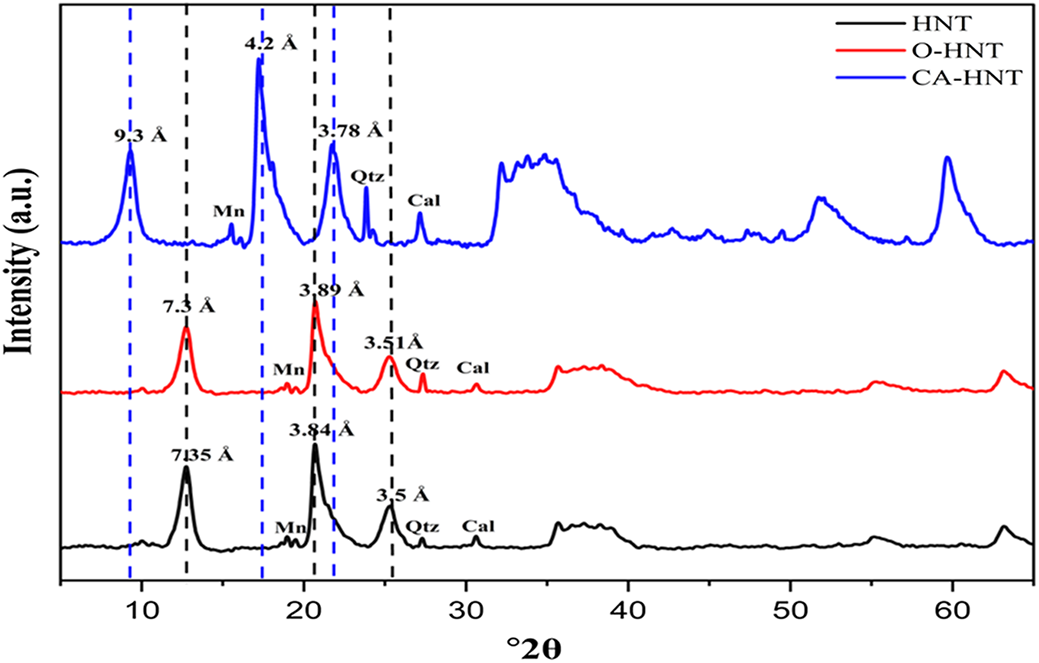
The impact of HNT functionalization on the crystal structure of both untreated and chemically treated HNTs was examined. The XRD traces of untreated HNTs and those subjected to silane and CA treatment were analysed (Fig. 1).
XRD traces of untreated HNTs, silanized HNTs treated with OTES (O-HNTs) and CA-loaded HNTs (CA-HNTs). Cal = calcite; Mn = manganese; Qtz = quartz.

The characteristic diffraction peaks of HNTs at 12.74°2θ and 25.40°2θ, corresponding to (001) and (002) diffraction planes with basal spacings of 7.35 Å and 3.44 Å, respectively, signify the typical peaks of untreated HNTs indicative of a dehydrated halloysite structure of 7 Å (Chen et al., Reference Chen, Yang and Wang2018). The absence of a significant peak at ∼10 Å or another one migrating towards 8.5 Å further confirmed the absence of the hydrated 10 Å form or the intermediate dehydrated state, as was found by Liu et al. (Reference Liu, Chen, Zhang, Liu and Zhang2021) and Siranidi et al. (Reference Siranidi, Hillier and Chryssikos2024). Additionally, the intense peak observed at 20.70°2θ, attributed to the (020) diffraction plane with a basal spacing of 3.89 Å, strongly supports the presence of a tubular structure to the halloysite (Joussein et al., Reference Joussein, Petit and Delvaux2007). The presence of manganese, responsible for the grey colouration of the Algerian HNTs, is evident from the peak at ∼4 Å, with additional peaks indicating quartz and calcite impurities, which are common in natural HNT samples.
In the case of silane treatment, nearly identical peaks were observed in the XRD traces of OTES-modified HNTs (O-HNTs), the crystalline form of the HNTs following OTES treatment remained intact. This preservation of the crystalline structure is attributed to the robust hydrogen bonds between the layers, which resist structural modification and provide stability, resisting significant alteration even after chemical treatment. Furthermore, the intensity of these diffraction peaks diminished progressively due to the delamination process induced by organic silane coating on the HNT surface that reduced the coherence of the crystalline lattice, lowering the intensities of the peaks without shifting their positions. This is typical of silane-treated clays presented in the literature (Yuan et al., Reference Yuan, Southon, Liu, Green, Hook, Antill and Kepert2008; He et al., Reference He, Xu, Tang, Zhang and Yang2015).
Unlike the silane treatment, CA treatment resulted in a shift towards lower angles and an increase in the intensity of the HNT diffraction peaks, indicating the insertion of CA molecules between the HNT interlayers and the disruption of the orderly tubular staking arrangement due to the acid etching and internal loading (Wong et al., Reference Wong, Pasbakhsh, Arabi, Keeling and Tan2021). This observation confirmed the successful loading of CA into the HNT lumen and the chemical bonding with HNT functional groups.
The crystallite size of the untreated HNTs, calculated using the Scherrer equation, was found to be 111.08 Å. Significant changes in crystallite size were observed following silane and CA treatments. Specifically, silane treatment reduced the crystallite size to 103 Å. This decrease can be attributed to the formation of a surface coating on the nanotubes, probably due silane organic alkyl chain grafting, which acts as a physical barrier that restricts the nucleation and growth of crystallites by limiting interlayer interactions and nucleation sites. In contrast, CA increased the crystallite size to 113.54 Å, which is thought to have resulted from acid etching removing amorphous regions, permitting CA to interact with the HNT functional groups and enabling the formation of larger, more defined crystalline structures within the nanotubes through chemical bonding within the lumen (Abdullayev et al., Reference Abdullayev, Joshi, Wei, Zhao and Lvov2012; Machowska et al., Reference Machowska, Klara, Ledwójcik, Wójcik, Dulińska-Litewka and Karewicz2022).
The observed changes are similar to those seen in the XRD traces (Fig. 1), with diminished peak intensities after silane treatment, indicating smaller crystallites and delamination, while the shift to lower angles and increased intensities with CA loading reflect larger crystallites and greater structural order. These results suggest that the diffraction behaviour is directly correlated with the crystal-size variations, confirming the chemical modification impacts on HNTs. The silane inhibits growth by forming a surface barrier, whereas CA increases structural order through acid etching and interlayer interactions.
Treatment effects on HNT chemical composition
XRF analysis was conducted to measure the concentrations of the major chemical elements in both the untreated and chemically modified HNTs (Table 1). The analysis showed that the raw HNTs consist of SiO2 (47.9 wt.%) and Al2O3 (37.6 wt.%) as the predominant components, confirming the typical aluminosilicate composition of the kaolinite-type halloysite structure (Stor et al., Reference Stor, Czelej, Krasiński and Gradoń2023). Minor oxides including Fe2O3, SO3 and ZnO were also detected.
Chemical composition of the raw and treated HNTs as obtained using XRF analysis.

Following silanization with OTES, the silica content increased to 49.1 wt.%, suggesting successful silane grafting, where the silane, which is rich in silicon, effectively coats the outer siloxane surface of the HNTs. However, alumina decreased to 36.2 wt.%, which is attributed to the hydroxylation process, which led to partial alumina dissolution (Fu et al., Reference Fu, Wang, Li and Chen2016).
Moreover, CA loading onto HNTs decreased both Al2O3 and SiO2 levels to 45.2 and 33.8 wt.%, respectively, suggesting that a hydrophobic layer formed, as has been demonstrated previously by Garcia-Garcia et al. (Reference Garcia-Garcia, Garcia-Sanoguera, Fombuena, Lopez-Martinez and Balart2018). The reduction in alumina, along with other trace elements, can also be attributed to the partial leaching of alumina cations from the octahedral layer due to the acidic hydrolysis step with acetic acid, which primarily affects the inner surface (Sidorenko et al., Reference Sidorenko, Kurban, Aho, Ihnatovich, Kuznetsova and Heinmaa2021).
These compositional changes align with the XRD results, supporting the conclusion that both surface treatments effectively induced selective modifications to the HNTs.
Treatment effect on HNT porous texture
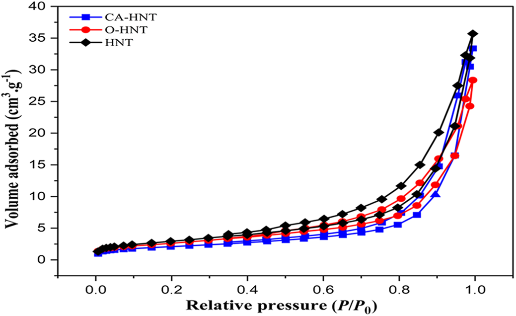
The nitrogen adsorption–desorption isotherms of raw HNTs, O-HNTs and CA-HNTs are shown in Fig. 2. According to the International Union of Pure and Applied Chemistry (IUPAC) classification, the three samples display type-IV isotherms that correspond to mesoporous materials (Sangwichien & Aranovich, Reference Sangwichien and Aranovich2002). The hysteresis loops observed in the isotherms are typical of type H3, suggesting that the pores are slit-shaped and associated with aggregates of plate-like particles. The hysteresis loops extend over the relative pressure (P/P 0) range of 0.4–1.0, indicative of capillary condensation within mesopores (pore width: 2–50 nm; Panda et al., Reference Panda, Mishra, Mishra and Singh2010; Lim et al., Reference Lim, Park and Sohn2020).
Nitrogen adsorption isotherms of untreated HNTs, silanized HNTs treated with OTES (O-HNTs) and CA-loaded HNTs (CA-HNTs).

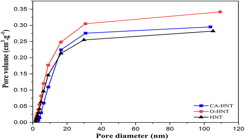
The BET surface areas and total pore volumes of the untreated and modified HNTs are shown in Fig. 3 & Table 2. The raw HNTs exhibited a BET surface area of 54 m2 g–1 and a pore volume of 0.28 cm3 g–1, which align with their natural tubular morphology and mesoporosity as presented in the literature (Zhang et al., Reference Zhang, Pan, Zhang, Liu, Ye, Xia and Chen2012). After functionalization with the OTES silane agent, the surface area and pore volume increased significantly to 74 m2 g–1 and 0.341 cm3 g–1, respectively. This enhancement can be attributed to the improved dispersion of the nanotubes and the exposure of previously inaccessible pores due to the silane moieties.
Total pore volume of untreated HNTs, silanized HNTs treated with OTES (O-HNTs) and CA-loaded HNTs (CA-HNTs).

BET surface areas and total pore volumes of HNTs, HNTs treated with OTES (O-HNTs) and CA-loaded HNTs (CA-HNTs).

However, modification with CA resulted in a decrease in surface area measured at 42 m2 g–1 and an increase in pore volume to 0.295 cm3 g–1. The decrease in surface area is possibly due to pore blocking caused by the deposition of CA molecules on the surface edges and within the pores of the nanotubes. However, the increase in pore volume is probably due to the acid etching widening the existing pores, which increases the volume capacity to hold guest molecules (Yu et al., Reference Yu, Xu, Hao, Yuan, Zhang and Wang2024).
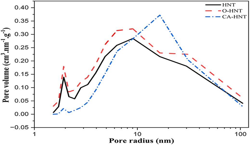
Across all samples, the BJH pore-size distribution curves (Fig. 4) revealed distinct alterations in textural properties, reflecting the effects of the chemical modifications on the porous texture of the HNTs.
Pore-size distribution of untreated HNTs, silanized HNTs treated with OTES (O-HNTs) and CA-loaded HNTs (CA-HNTs).

The untreated HNT sample exhibited a mesoporous nature with two distinct pore-size distributions: a peak pore radius centred at ∼2 nm attributed to small internal and external pores, alongside a broader peak at 8–10 nm, corresponding to the lumen and consistent with the porosity that arises predominantly from the lumen and surface defects (Pasbakhsh et al., Reference Pasbakhsh, Churchman and Keeling2013).
The O-HNT sample, modified with OTES, displayed a shift in the peak pore radius towards 10–12 nm, suggesting minor surface reorganization improvement due to the organosilane chain grafting. This probably removed surface impurities and reduced aggregation without significantly altering the pore structure, leading to enhanced compatibility with organic matrices.
The CA-HNT sample, initially subjected to acid etching in the acetic acid step followed by CA loading, showed the most pronounced change, with a notable peak shift to 16 nm. The acid etching step with a weak acid such as acetic acid effectively removes structural impurities and widens the already-existing pores, promoting macropore formation through proton penetration-induced dehydroxylation and selective leaching of Al3+ cations from octahedral layers, creating larger voids in the HNT matrix without introducing significant heterogeneity, thereby facilitating enhanced CA loading (Jauković et al., Reference Jauković, Krajišnik, Daković, Damjanović, Krstić, Stojanović and Čalija2021). This is different from when using strong acids such as sulfuric acid, as Garcia-Garcia et al. (Reference Garcia-Garcia, Ferri, Ripoll, Hidalgo, Lopez-Martinez and Balart2017) reported, as these acids introduce heterogeneous porosity with multiple pore-size peaks.
Treatment effects on HNT chemical structure
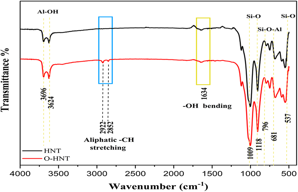
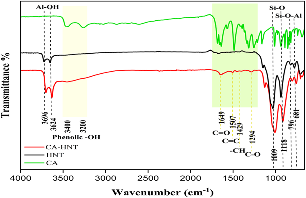
The FTIR spectrum of unmodified HNTs exhibited characteristic peaks at 3696, 3624 and 914 cm–1, corresponding to the stretching of inner-surface Al-OH, the stretching of inner surface Al-OH and the bending vibration of inner surface Al-OH, respectively (Sabahi et al., Reference Sabahi, Khorami, Rezayan, Jafari and Karami2018). The peaks at 3456 and 1637 cm–1 relate to –OH stretching and the bending vibration of adsorbed water, respectively. The appearance of the 1637 cm–1 peak, often referred to as the zeolitic peak in palygorskite and sepiolite clays (Bukas et al., Reference Bukas, Tsampodimou, Gionis and Chryssikos2013), is suggested to result from incomplete removal of residual bending water. This water may remain trapped in defect sites or within the slit-like crevices that form during the water loss process, as noted previously by Siranidi et al. (Reference Siranidi, Hillier and Chryssikos2024). The peaks at 1110, 1039 and 536 cm–1 can be attributed to apical Si–O stretching vibrations, in-plane Si–O stretching vibrations and Si–O bending vibrations, respectively. Additionally, the peaks at 759 and 686 cm–1 correspond to the symmetric stretching of Si–O–Si and the perpendicular stretching of Si–O–Al, respectively (Yuan et al., Reference Yuan, Southon, Liu, Green, Hook, Antill and Kepert2008; Hillier et al., Reference Hillier, Brydson, Delbos, Fraser, Gray and Pendlowski2016).
The FTIR spectrum of O-HNTs (Fig. 5) showed a decrease in the peak of zeolitic water. This can be understood by considering that silanol groups, formed when the alkoxy groups in OTES are hydrolyzed, interact with hydroxyl groups in the inner layers of the HNTs, as well as with the edges and surface defects on the HNTs. These interactions lead to a reduction in the generation of water that is subsequently removed during the drying process, possibly due to increased hydrophobicity of the HNTs after silanization, limiting water adsorption and so reducing the overall amount of adsorbed water between the layers (Senyel & Dike, Reference Senyel and Dike2024). Furthermore, new absorption bands appeared in the O-HNT FTIR spectrum, notably at 2922 and 2852 cm–1, which can be attributed to the asymmetric and symmetric stretching vibrations of aliphatic –CH2 groups, respectively. Shifts in peak positions and changes in peak intensities further confirm the success of the silane treatment (Li et al., Reference Li, Zhu, Zhou and Zhong2015; Pal et al., Reference Pal, Kundu, Malas and Das2015).
FTIR spectrum of untreated HNTs and silanized HNTs treated with OTES (O-HNTs).

However, upon treatment with CA, and through a comparative analysis of the spectrum curves (Fig. 6), the emergence of new peaks becomes evident, notably at ∼1644, 1507, 1429 and 1294 cm–1. These peaks can be attributed to the C=O stretching, C=C stretching of the aromatic ring group, –CH stretching of the aromatic hydroxyl group and C–O phenolic stretching from CA (Arat & Uyanık, Reference Arat and Uyanık2017), respectively. Additionally, a notably broader peak corresponding to the presence of phenolic –OH from CA at ∼3400–3200 cm–1 can be observed (Liu et al., Reference Liu, Chen, Zhang, Liu and Zhang2021). This serves as compelling evidence of the successful CA loading on the HNTs.
FTIR spectrum of untreated HNTs and CA-loaded HNTs (CA-HNTs).

Treatment effects on HNT morphology
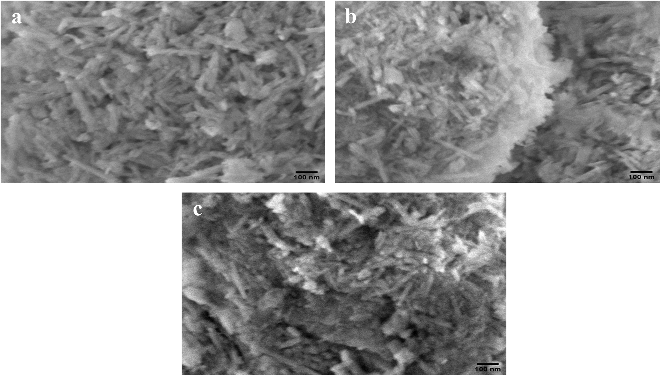
SEM morphological analysis indicated that pristine HNTs (Fig. 7a) exhibit well-defined tubular structures with smooth, uniform and consistent surfaces that tend to aggregate due to their hydrophilic nature, which is typical of unmodified clays. This aggregation arises from the presence of hydroxyl groups.
SEM images of (a) unmodified HNTs, (b) HNTs modified with OTES (O-HNTs) and (c) HNTs loaded with CA (CA-HNTs).

Following treatment, the observed morphology noticeably changed in the O-HNT and CA-HNT samples (Fig. 7b,c), and this could be ascribed to the interaction between the hydroxyl moieties on the HNT surface and the added OTES and CA compounds. The O-HNTs appear to be aggregates with slightly roughened surfaces. These changes, as Garcia-Garcia et al. (Reference Garcia-Garcia, Garcia-Sanoguera, Fombuena, Lopez-Martinez and Balart2018) described, can be attributed to silane organic moieties being coated onto HNT external surfaces, thereby masking the HNT walls and edges, promoting surface roughness and decreasing hydrophilicity.
Furthermore, the analysis reveals that CA-HNTs exhibited a rougher and more compact surface with significant clustering of nanotubes. This observation confirms the greater packing density of HNT aggregates, suggesting that the HNTs are stacked or bundled more tightly (Zhang et al., Reference Zhang, Meng, Zhou, Liu and Guo2022). The loss of tube visibility with increased density indicates a significant alteration in the morphological characteristics of the CA-HNTs compared to the unmodified HNTs, which can be attributed to the successful loading of CA.
Such modifications are expected to improve the compatibility and homogeneity of these HNTs as their morphological shape remained unaffected by the two modification processes; however, their size and degree oaded with Czange (Arat & Uyanık, Reference Arat and Uyanık2017; Abu El-Soad et al., Reference Abu El-Soad, Pestov, Tambasova, Osipova, Martemyanov and Cavallaro2020, Reference Abu El-Soad, Lazzara, Pestov, Tambasova, Antonov, Cavallaro and Kovaleva2021).
Treatment effects on HNT thermal stability
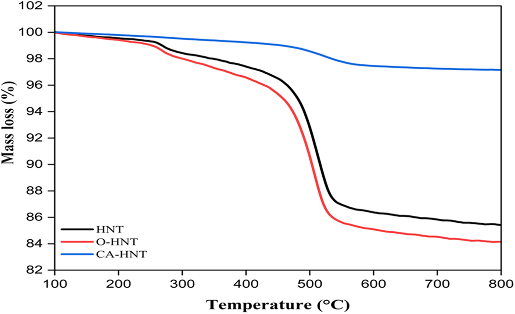
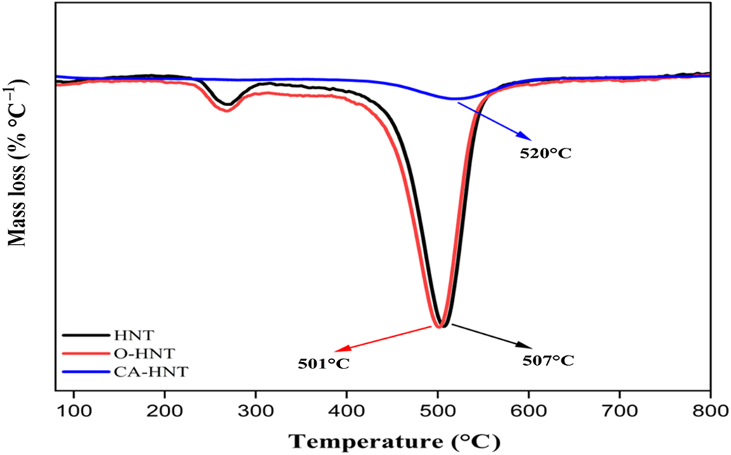
The TGA and derivative thermogravimetry (DTG) curves for both untreated and treated HNTs (Figs 8 & 9) provide crucial insights into their thermal degradation behaviour, the effects of modification with OTES and the loading efficiency of CA into the HNT lumen.
TGA of unmodified HNTs, HNTs modified with OTES (O-HNTs) and HNTs loaded with CA (CA-HNTs).

TGA of unmodified HNTs, HNTs modified with OTES (O-HNTs) and HNTs loaded with CA (CA-HNTs).

The untreated HNTs showed two distinct mass loss stages. A minor mass loss of 1.33% occurred between 200°C and 300°C, which can be attributed to the desorption of crystallization water and adsorbed water from the surface and internal channels of the nanotubes. The second, more substantial mass loss of 12.76% occurred between 400°C and 600°C, with a DTG peak at 507°C, corresponding to the dihydroxylation of the structural Al-OH groups (Yu et al., Reference Yu, Wang, Hu and Guo2017; Abu El-Soad et al., Reference Abu El-Soad, Pestov, Tambasova, Osipova, Martemyanov and Cavallaro2020).
Similar thermal behaviour was observed in the HNTs organically modified with OTES (O-HNTs), but with slight yet significant differences. A reduction in crystallization and adsorbed water mass to 1.22% can be noted, which can be attributed to the hydrophobic nature of the silane treatment due to the long octyl alkyl chain extending from its triethoxy groups (Açışlı et al., Reference Açışlı, Karaca and Gürses2017; Rozhina et al., Reference Rozhina, Panchal, Akhatova, Lvov and Fakhrullin2020). Additionally, an increased mass loss estimated at 13.89% during dihydroxylation can be observed, with a slightly broadened DTG peak estimated at 501°C, probably due to gradual degradation of the organic compounds grafted onto the silane during the surface modification, which serves as a determining factor in achieving less abrupt mass losses compared to the raw HNTs. The obtained results align with those reported in the literature (Belkassa et al., Reference Belkassa, Bessaha, Marouf-Khelifa, Batonneau-Gener, Comparot and Khelifa2013; Bischoff et al., Reference Bischoff, Daitx, Simon, Schrekker, Liberman and Mauler2015). This implies that the grafting of the organic moieties of the silane played a crucial role in modifying the surface chemistry, reducing the inherently hydrophilic nature of the HNTs by promoting the dehydration process.
For the CA-HNTs, notable variations in behaviour were identified compared to both HNTs and O-HNTs. Crystallization and adsorbed water mass losses were no longer observed. Additionally, the secondary mass loss at 2.75% in CA-HNTs was significantly lower than the overall mass loss observed in HNTs. This reduction in mass loss, leading to improved thermal stability, can be attributed to HNT etching with acetic acid, which removes the hydroxyl groups typically associated with thermal decomposition recorded between 400°C and 600°C (Prinz Setter et al., Reference Prinz Setter, Dahan, Abu Hamad and Segal2022). This process, coupled with the loading of CA, results in a shift of the decomposition peak to a higher temperature of ∼520°C, confirming the improved stability (Liu et al., Reference Liu, Wang, Bai, Zhang, Kan and Jin2018; Lim et al., Reference Lim, Chow and Pung2019). The CA-HNT characterization was repeated three times to ensure the accuracy of these findings.
These results suggest that the absence of CA decomposition, combined with reduced mass loss at the decomposition stage, elimination of both crystallization and adsorbed water and increase in degradation temperature of 13°C compared to raw HNTs, indicated that loading of CA facilitated the elimination of water molecules as well effectively shielded the HNTs from heat-induced alterations, improving their thermal stability. However, this improvement was not substantial, which can be explained by the small amount of CA loaded, as reported previously by Zahidah et al. (Reference Zahidah, Raja, Kakooei, Kermanioryani, Mohebbi, Ismail and Bothi Raja2017). These findings align with the results from the XRF, XRD, BET and FTIR spectroscopy analyses, which confirm the presence of CA either chemically bonded to the HNT surface or encapsulated within the lumen. The lack of CA decomposition may be explained by the formation of strong covalent bonds between the HNTs and CA.
Conclusion
This research successfully demonstrated the functionalization of Algerian HNTs through two distinct methods: silanization with OTES and CA loading.
These surface modifications were confirmed through comprehensive characterization. XRD analysis showed that for O-HNTs the core crystalline structure remained intact, with a slight reduction in crystallite size and decreased peak intensities, revealing the formation of a surface coating with the OTES moieties. Conversely, CA treatment led to an increase in crystallite size alongside noticeable shifts in the diffraction peaks, probably due to molecular interactions or subtle acid-etching effects. XRF analysis revealed a greater silica content after silanization, whereas the CA treatment reduced both alumina and silica contents due to the acid etching, indicating successful surface modification and the formation of a hydrophobic layer. FTIR spectroscopy analysis revealed new absorption bands unique to each grafted agent, further confirming the success of the chemical modifications. SEM analysis revealed that OTES treatment resulted in surface roughening, whereas CA treatment led to a rougher, more compact and tightly packed appearance to the HNT aggregates, supporting successful grafting. TGA analysis revealed that O-HNTs exhibited a reduction in initial adsorbed water losses (from 1.33% to 1.22%) and a slightly altered dehydroxylation profile (peak at 501°C). By contrast, CA-HNTs demonstrated notably improved thermal stability by eliminating initial water loss and shifting the primary decomposition temperature to 520°C (13°C increase over raw HNTs), demonstrating improved resistance to degradation. BET analysis revealed that OTES treatment significantly increased the surface area from 54 m2 g–1 for raw HNTs to 74 m2 g–1, whereas CA treatment reduced it to 42 m2 g–1, suggesting pore blocking by the loaded CA molecules.
Overall, these modifications improved the properties of Algerian HNTs by effectively preventing HNT aggregation and reducing their inherent hydrophilicity, which could potentially lead to improved particle dispersion, compatibility with non-polar matrices and greater thermal stability. This paves the way for modified HNT applications in advanced polymer nanocomposites, particularly in active packaging and thermal insulating materials, making them promising high-performance green nanofillers.
Author contributions
Conceptualization: Zeyneb Abir Mekhania; methodology: Zeyneb Abir Mekhania; investigation: Zeyneb Abir Mekhania, Nora Benkacher; visualization: Zeyneb Abir Mekhania; writing – original draft: Zeyneb Abir Mekhania; writing – review and editing: Kahina Iggui, Aida Benhamida; supervision: Kahina Iggui, Aida Benhamida, Nadjette Bengourna; formal analysis: Kahina Iggui; data curation: Nora Benkacher, Abdelheq Layachi; project administration: Aida Benhamida, Nadjette Bengourna; data analysis: Atmane Djermoune, Farid Ait Merzeg, Abdelheq Layachi; validation: Atmane Djermoune, Farid Ait Merzeg; resources: Hamid Satha.
Acknowledgements
None.
Financial support
None.
Competing interests
The authors declare none.