Isolated subclavian artery refers to loss of its aortic continuity along with its ductal connection to the pulmonary artery.Reference Chen, Cheng and Lin1 It is often associated with other intra-cardiac defects.Reference Smith, Hirschklau and Reitz2–Reference McMahon, Thompson, Kearney and Nihill4 In this anomaly, vertebral steal during arm exercise leads to vertebrobasilar insufficiency and neurological symptoms.Reference Carano, Piazza, Agnetti and Squarcia3,Reference Potter and Pinto5 If duct is patent, there is a second steal from the subclavian artery to the lungs.Reference Koneti, Qureshi and Sivakumar6 Surgical re-implantation to aorta resumes arm perfusion. A young child with DiGeorge syndrome, large ventricular septal defect, and severe hyperkinetic pulmonary hypertension is described to discuss the anatomy, haemodynamics, and echocardiographic clues to diagnosis.
Case report
A 15-month-old child with a birth weight of 1.75 kg had growth failure weighing 6.8 kg(<3rd centile) with lung infections and heart failure. She had broad forehead, low-set ears, and broad nasal bridge and 22q11 deletion on genetic analysis. Clinical exam showed increased respiratory efforts, equal pulses and saturations in all extremities, hyperkinetic precordium, loud pulmonary component of second sound, and a pansystolic murmur. Chest X-ray showed cardiomegaly, left aortic arch, and plethoric lungs. Biventricular hypertrophy and normal QRS axis were seen in the electrocardiogram.
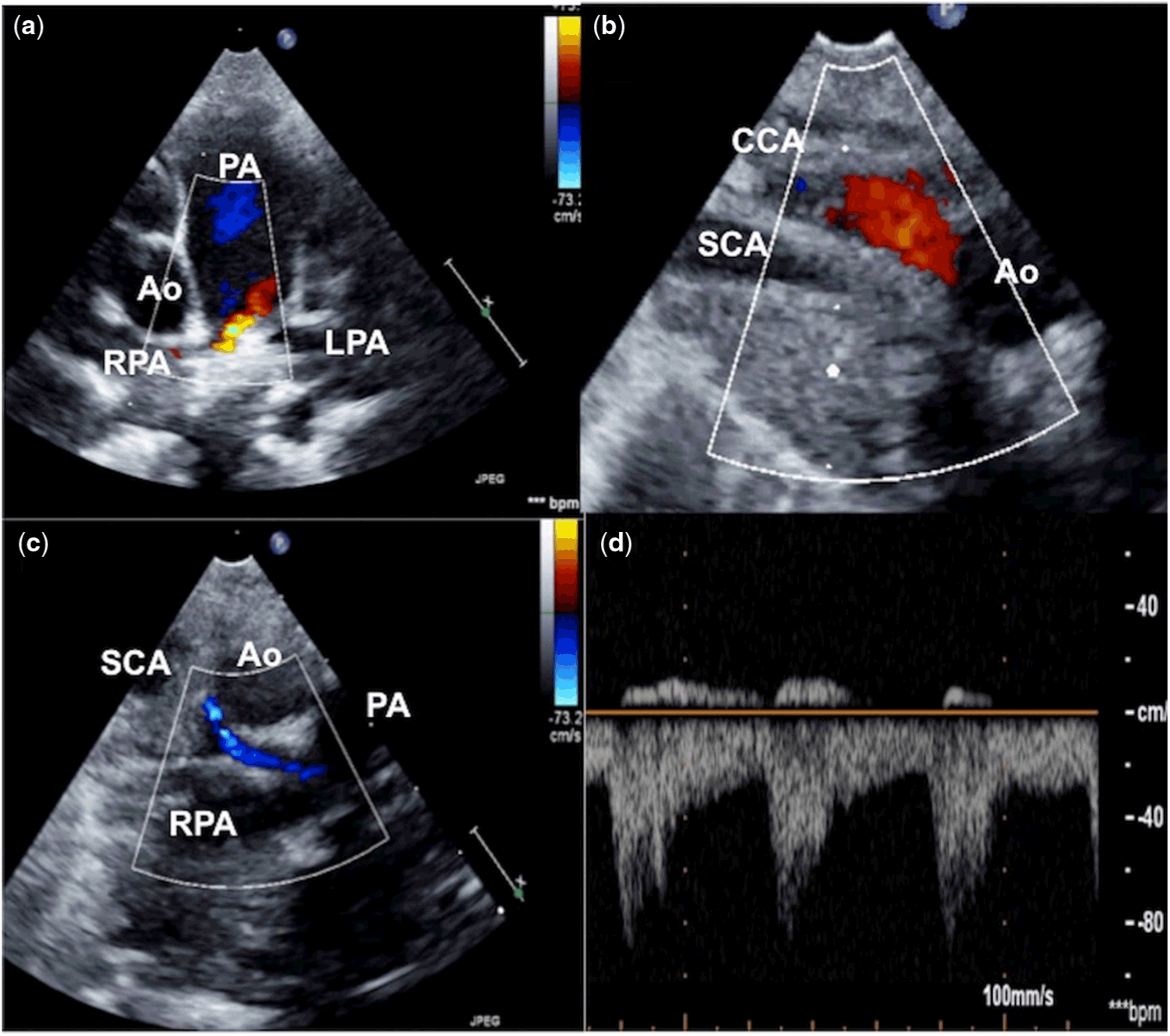
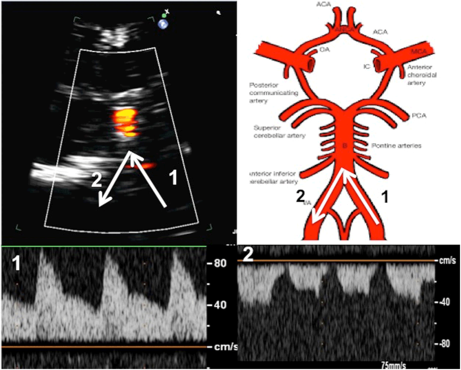
A 6-mm perimembranous ventricular septal defect shunted left to right with an inter-ventricular gradient of 10 mmHg with left ventricular dilation. A continuous colour flow into the mediastinal right pulmonary artery suggested an unusual duct insertion (Fig 1, Supplementary Video S1). The first branch from the left aortic arch was right carotid artery as it was unbranched. A cranial ultrasound from the anterior fontanelle showed forward flows towards the basilar artery in the left vertebral artery and reversal of flow in the right vertebral artery (Fig 2). This suggested a diagnosis of isolation of the right subclavian artery with subclavian steal from the posterior circulation.
High parasternal short axis view (a) on echocardiogram shows a continuous flow in unusual location from the right pulmonary artery. Suprasternal view (b) shows the first vessel from the left arch to the right without any branching indicating that the vessel is right carotid artery. A vessel parallel to the right carotid artery (c) that shows flow reversal in blue colour is the isolated right subclavian artery (d) that connects through the duct to the right pulmonary artery.

Transcranial ultrasound in coronal plane from anterior fontanelle (a) shows the red colour flows towards the transducer in the basilar artery through the left vertebral artery (marked as 1). There was no similar colour flow from the right vertebral artery in the right side (marked as 2). Spectral Doppler with a small sample volume in both the left (1) and right (2) vertebral artery shows characteristic flow reversal in the right vertebral artery which clinches the diagnosis.

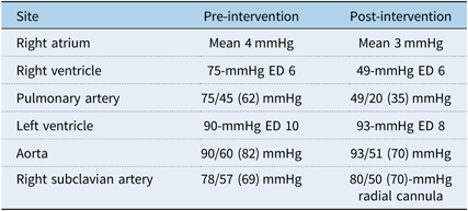
Catheter haemodynamics (Table 1) showed near-systemic pulmonary artery pressures, a shunt ratio of 1.8, and an indexed pulmonary vascular resistance of 6.6 Wood units. Aortogram showed late-filling right subclavian artery through vertebral steal which subsequently filled the right pulmonary artery through the arterial duct, indicating double steal (Supplementary Video S2). A catheter advanced from the pulmonary artery into the right subclavian artery showed retrograde flows into the right pulmonary artery through a 4-mm arterial duct. As the perimembranous ventricular septal defect had an adequate aortic margin, it was closed with a 6–4 Amplatzer duct occluder II device (Abbott, St Paul, Minnesota, United States of America). Duct closure with a 5–4 Piccolo Occluder (Abbott) reduced the pulmonary pressures and equalised the pressures in right radial artery cannula to near systemic levels (Supplementary Video S3).
Haemodynamics.

ED = end diastolic
After 2 years, her weight was above 50th centile, right arm pulses were well felt with no significant systolic blood pressure difference between the upper limbs, though with radio-radial delay. Echocardiogram showed absence of residual shunts and normal pulmonary artery pressures.
Discussion
In the embryo, the right subclavian artery gets isolated from the aorta when the seventh inter-segmental artery separates from the right fourth arch and connects to the right sixth arch through the arterial duct.Reference Lee, Park, Ko and Seo7 Surgical re-implantation of the isolated vessel to aorta or ipsilateral carotid artery is a therapeutic option, which needs long mobilisation, division of ductal remnants, and may stretch the vessel leading to stenosis.Reference Ghasemi, Serati, Emami and Movahed8 Transcatheter duct closure prevents the second steal, thereby improving the arm perfusion.Reference Koneti, Qureshi and Sivakumar6,Reference Jones, Garabedian and Grifka9 The combined shunt from ventricular septal defect and duct in our patient caused severe pulmonary hypertension. The margins of the ventricular septal defect permitted a safe transcatheter closure with the low-profile occluder, which advances through a small guide catheter and lacks radial force to injure conduction tissues. Even though surgical correction of both the defects would be considered the ideal option, the lack of clinically perceivable pulse difference between the arms on 2-year follow-up indicated the adequacy of the arm perfusion by the compensated vertebral steal. While other complex arch anomalies associated with tracheo-oesophageal compression will need a pre-interventional tomographic imaging, ducts in isolated subclavian artery are not known to compress airways.Reference Lee, Park, Ko and Seo7
The challenging echocardiographic identification of this anomaly is guided by three findings: 1) the absence of branching of the first aortic branch suggesting abnormal subclavian origin; 2) colour Doppler flows at an unusual location into the branch pulmonary artery due to ductal flows; and 3) reversed blood flows on transcranial Doppler in vertebral artery. Precise echocardiographic recognition of these three findings serves in diagnosis of this rare anomaly. The diagnosis is often confirmed by advanced computed tomographic or magnetic resonance imaging techniques.Reference Ghasemi, Serati, Emami and Movahed8,Reference Crystal, Rivenes and Ing10
Conclusions
Isolated subclavian artery is diagnosed by the following findings: first arch branch is non-branching, ductal flows in the pulmonary artery from an unusual location, and reversal of blood flow in the vertebral artery. Transcatheter duct closure preventing the second steal is an alternative to surgical reanastamosis. This report is unique due to simultaneous non-surgical correction of the associated large ventricular septal defect.
Acknowledgements
None.
Financial Support
This research received no specific grant from any funding agency, commercial or not-for-profit sectors.
Conflicts of Interest
None.
Ethical Standards
The authors assert that all procedures contributing to this work comply with the ethical standards of the Indian Council of Medical Research and with the Helsinki Declaration of 1975, as revised in 2008, and has been approved by the institutional committees of Madras Medical Mission, Chennai, India.
Supplementary Material
To view supplementary material for this article, please visit https://doi.org/10.1017/S1047951119001963