INTRODUCTION
Abnormalities in the elasmobranch group have been reported in several species around the world (Devadoss, Reference Devadoss1983). In the Mexican north-west have been reported different kinds of abnormalities in the elasmobranchs including albinism in Narcine entemedor and Galeocerdo cuvier (Sandoval-Castillo et al., Reference Sandoval-Castillo, Mariano-Meléndez and Villavicencio-Garayzar2006), functional bicephaly in Rhinoptera steindachneri (Castro-Aguirre & Torres-Villegas, Reference Castro-Aguirre and Torres-Villegas1979) and Prionace glauca (Galván-Magaña et al., Reference Galván-Magaña, Escobar-Sánchez and Carrera-Fernández2011), foetal abnormalities in P. glauca (Bejarano-Álvarez et al., Reference Bejarano-Álvarez, Galván-Magaña and Ochoa-Báez2011), morphological abnormality in Dasyatis longa (Escobar-Sánchez et al., Reference Escobar-Sánchez, Galván-Magaña, Downton-Hoffmann, Carrera-Fernández and Alatorre-Ramírez2009) and ocular malformations in Urobatis halleri (Rubio-Rodríguez et al., Reference Rubio-Rodríguez, Navarro-González and Vergara-Solana2011).
Abnormalities in Myliobatis spp. have not been commonly reported, only one record of albinism was reported in the California bat ray Myliobatis californica Gill, 1865 from the west coast of Baja California (B.C.), Mexico (De Jesús-Roldan, Reference De Jesús-Roldan1990).
The California bat ray, occurs in temperate and tropical seas (Ebert, Reference Ebert2003), and is commonly found in coastal waters from central Oregon, USA to Gulf of California, Mexico (Miller & Lea, Reference Miller and Lea1972). It has been reported on the west coast of Baja California Peninsula (Beltrán-Félix et al., Reference Beltrán-Félix, Hammann, Chagoya-Guzman and Álvarez-Borrego1986; Villavicencio-Garayzar, Reference Villavicencio-Garayzar1996). It is common in shallow inshore water, estuaries and on beds of seaweed (Miller & Lea, Reference Miller and Lea1972; McEachran & Notarbartolo-di-Sciara, Reference McEachran, Notarbartolo-di-Sciara, Fischer, Krupp, Schneider, Sommer, Carpenter and Niem1995).
The California bat rays have histotrophic viviparity, their gestation period is estimated between 9 and 12 months and embryo number in pregnant females ranges between two and five pups (Martin & Cailliet, Reference Martin and Cailliet1988). They feed on worms, molluscs and crustaceans (Gray et al., Reference Gray, Mulligan and Hannah1997).
This species has commercial importance, because it is the second species in the batoids catches in the west coast of Baja California Sur (B.C.S.) (Ramírez-Amaro, Reference Ramírez-Amaro2011). Also, this ray was assessed as at least concern on the IUCN Red List of Endangered Species (Cailliet & Smith, Reference Cailliet and Smith2006). The aim of this paper is to report the first description of abnormal horns in the cephalic area in M. californica.
MATERIALS AND METHODS
The specimen was caught in a gillnet (25.4 cm mesh size) in April 2010 by artisanal elasmobranch fishermen at San Pablo (27°21′N 114°77′W) located on the west coast of B.C.S. and was caught 10 miles offshore of San Pablo. Unfortunately the specimen was not donated by the fishermen; however morphometric measurements (according to McEachran & Notarbartolo-di-Sciara, Reference McEachran, Notarbartolo-di-Sciara, Fischer, Krupp, Schneider, Sommer, Carpenter and Niem1995) and digital photographs were taken. For batoids identification we used the McEachran & Notarbartolo-di-Sciara (Reference McEachran, Notarbartolo-di-Sciara, Fischer, Krupp, Schneider, Sommer, Carpenter and Niem1995) keys.
RESULTS
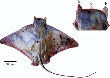
The abnormal specimen found in San Pablo was a male measuring 385 mm of disc width (DW), the specimen captured was a juvenile, because it presented small claspers (clasper length of 15 mm), uncalcified clasper that did not rotate easily, and also its DW was smaller than the size at maturity for males reported (AD: 400 mm) by Villavicencio-Garayzar (Reference Villavicencio-Garayzar1996). The specimen had three cephalic horns, two in the cephalic lobes zone (which we call ‘lateral horns', Figure 1a, b) and one more at the rostral middle zone (which we call ‘medial horns', Figure 1c). The malformed California bat ray was alive at the time of capture.
The malformed specimen in dorsal side with three cephalic horns, two in the cephalic lobes (a, b) and one in the rostral middle zone (c).

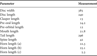
The horns showed as flashy extensions, apparently fused to the rostral cartilages, which were projected in front in the head, similar to the cephalic fins in the Mobulidae subfamily. The lateral horns had similar lengths (a: 22.3 mm; b: 25.1 mm), while the medial horn presented a smaller length (c: 5.3 mm). The morphological measurements are shown in Table 1.
Morphometric measurements of an abnormal Myliobatis californica expressed in millimetres (mm).

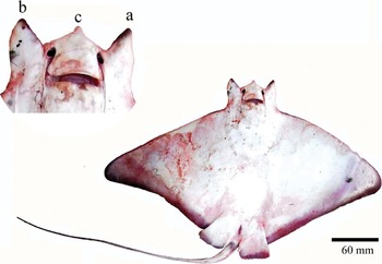
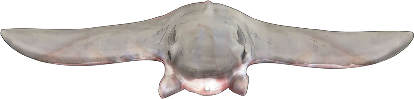
The specimen presented a normal mouth, which is connected to the two nostrils by oro-nasal channels and covered with a nose lobe (Figures 2 & 3). Dental plates showed a normal arrangement. Also during the dissection of the specimen it was observed that internal organs showed a normal development.
The specimen with ventral side with three cephalic horns, two in the cephalic lobes zone (a, b) and one in the rostral middle zone (c).

Drawing of malformed ray with front side.

DISCUSSION
There are several explanations that would explain these deformities in the California bat ray: external factors such as environmental degradation or pollution could be related to embryonic abnormalities (Casarini et al., Reference Casarini, Gomes and Tomas1997). However, the fishing area close to the west cost of B.C.S. is clear of pollution and we cannot consider it as a cause for abnormalities, as considered in other shark studies (e.g. Heupel et al., Reference Heupel, Simpfendorfer and Bennet1999; Galván-Magaña et al., Reference Galván-Magaña, Escobar-Sánchez and Carrera-Fernández2011). Other causal factors of malformations could be parasitic infection, arthritis, injury, tumours, bad nutrition, congenital abnormality (Heupel et al., Reference Heupel, Simpfendorfer and Bennet1999) or a natural mutation during embryonic development (Mancini et al., Reference Mancini, Casas and Amorim2006).
There are reports of morphogenetic movement of the anterior cranial area, accompanied by anterior extension of the pectoral fin, which would drastically change the size and shape of embryonic configuration in the anterior cranial areas (Miyake et al., Reference Miyake, McEachran, Walton and Hall1992). In some cases, the structural organization of anterior cranial areas, as seen in the development of Myliobatis and Rhinoptera, suggest that morphogenetic movements accompanied by differentiation of the underlying structures would affect the pattern of anterior cranial areas, including the nasal capsules and rostral cartilage (Miyake et al., Reference Miyake, McEachran, Walton and Hall1992).
Two subfamilies (Rhinopteridea and Mobulidae) belonging to the Myliobatidae family have a pair of cephalic fins, which are projected from the anterior limit of the head (Garman, Reference Garman1913). These cephalic fins are composed of a separated propterygium of the pectoral fin ornamented with fin rays and are supported dorsally by a pair of the presumptive rostral projections (Miyake et al., Reference Miyake, McEachran, Walton and Hall1992).
In the mobulids such as Mobula spp. and Manta spp., the cephalic fins are externally separated from a major portion of the pectoral fins and form a pair of paddles-shaped projection in front; however the horns found in M. californica do not show paddles-shape (Miyake et al., Reference Miyake, McEachran, Walton and Hall1992). The lateral horns could have a similar role to the cephalic lobes in myliobatids, which are used to detect prey via electrosensory pores, pin prey to the substrate to prevent their escape, and manipulate prey toward their mouth as do cownose rays (Rhinoptera bonasus) (Sasko et al., Reference Sasko, Dean, Motta and Hueter2006; Wilga et al., Reference Wilga, Motta and Sanford2007). It could be that development of horns could not affect the survival of the M. californica specimen, because it presented a size near the size at maturity reported by Villavicencio-Garayzar (Reference Villavicencio-Garayzar1996).
Based on the embryonic stage, the formation of the lateral horns found in the ray, would be indicative of a morphogenetic plasticity in these taxa that may have facilitated the evolution of rhinopterid and mobulid species from a common myliobatid ancestor.
We conducted an exhaustive literature search to find out if there is some background about the formation of cephalic horns in the batoid group; however we did not find any corroborating information. Some studies report abnormalities of anterior lobes of the pectoral fins, which were not fused with the head and were tapering forward and inward in the form of a pair of what appeared to be horns; however these are not strictly horns (e.g. Templeman, Reference Templeman1965; Somasekhara-Nair & Chellam, Reference Somasekharan-Nair and Chellam1971; Escobar-Sánchez et al., Reference Escobar-Sánchez, Galván-Magaña, Downton-Hoffmann, Carrera-Fernández and Alatorre-Ramírez2009; Mejía-Falla et al., Reference Mejía-Falla, Navia and Muñoz2011). Therefore this is the first record of cephalic horns found in batoids.
ACKNOWLEDGEMENTS
The present study was supported by CA Sea Grant, the Moore Family Foundation, the Tinker Foundation, the Save Our Seas Foundation, Sharks and Rays Project of CICIMAR-IPN and the Scripps Institution of Oceanography. We thank Carmina Salinas, for field data collection and for providing the photograph and Diana Alejandra Saavedra Cervantes for drawing one of the pictures. The first author thanks CONACYT and PIFI for providing fellowships. F.G.M thanks Instituto Politecnico Nacional (COFFA and EDI) for providing fellowships.