




No CrossRef data available.
Article contents
Impact of parental onchocerciasis and intensity of transmission on development and persistence of Onchocerca volvulus infection in offspring: an 18 year follow-up study
Published online by Cambridge University Press: 17 October 2003
Abstract
This study analysed the impact and the extent by which parental Onchocerca volvulus infection, intensity of transmission of O. volvulus infective 3rd-stage larvae (L3) and anthropometric factors may influence the acquisition, development and persistence of O. volvulus infection in offspring. A total of 15 290 individuals in 3939 families with 9640 children were surveyed for microfilariae of O. volvulus, and prevalence and level of O. volvulus infection in children aged 0 to 20 years from infected and non-infected parents were followed longitudinally for 18 years. Children from O. volvulus-infected mothers had not only a substantially higher risk to become infected; they also acquired infection earlier in life and developed higher infection levels. Multiple logistic regression analysis showed that maternal O. volvulus infection and children's age are the predominant predictors for patent O. volvulus infection, while the intensity of transmission, measured by the annual transmission potential (ATP) of O. volvulus L3, was less decisive. Longitudinal follow up of children showed that during vector control activities by the Onchocerciasis Control Programme (OCP) and in low-level transmission areas, infection persisted at higher levels in children from O. volvulus-positive mothers. In summary, the dominant risk factor for children to become infected is maternal onchocerciasis, and also age-associated factors will strongly impact on the development of patent O. volvulus infection in offspring.
Keywords
Information
- Type
- Research Article
- Information
- Copyright
- 2003 Cambridge University Press
INTRODUCTION
Maternal infection with parasites during pregnancy is considered as a risk factor which may increase the susceptibility to homologous infection in their offspring after birth. For several protozoa and helminth parasite species it has been shown that parasite-derived antigens and even entire parasites may pass the placenta and reach the foetus (Loke, 1982). In lymphatic filariasis, higher levels of circulating microfilariae but a diminished cellular responsiveness to parasite-specific antigens and an impaired TH1- and TH2-type cytokine production were observed in children from infected than from non-infected mothers (Elson et al. 1996; Steel et al. 1994). In onchocerciasis, familial predisposition to infection and infection-associated pathology has been suggested (Meyer et al. 1994; Murdoch et al. 1997) and in utero sensitization of children from O. volvulus-infected mothers has been observed (Soboslay et al. 1999). Exposure to parasite antigens during prenatal and neonatal life, together with the immaturity of the foetal immune system and its susceptibility to tolerogenic signals, may induce parasite-specific immune tolerance and thus facilitate parasite persistence when infected in later life (Hightower, Lammie & Eberhard, 1993; Carlier & Truyens, 1995). Prenatal exposure to filarial antigens is known to activate specific immune responses in newborns (Weil et al. 1983; Steel et al. 1994), early life sensitization and tolerance induction will create immune responses with a preferential TH2-type pattern (Ridge, Fuchs & Matzinger, 1996), and such neonatally induced antigen-specific responses will persist upon secondary antigen contact later in life (Barrios et al. 1996; Adkins & Du, 1998).
Immune competence in neonates and young children, as compared to adults, has been considered as immature and intrinsically deficient (Wilson, 1986). However, recent studies have shown that neonatal T cells can be appropriately activated, i.e. in utero exposure to helminth and mycobacterial antigens will generate cytokine responses similar to those observed in adults (Ridge et al. 1996; Forsthuber, Yip & Lehmann, 1996; Sarzotti, Robbins & Hoffman, 1996; Malhotra et al. 1997; Soboslay et al. 1999). Furthermore, balanced TH1/TH2 primary effector responses to antigens will develop in newborns, but following secondary antigen challenge in later life cellular immune responses were biased to TH2 which persisted into adulthood, both in mice (Barrios et al. 1996; Kovarik et al. 1999) and humans exposed to infection (Malhotra et al. 1999). However, it remains unclear to what extent maternal infection will increase the risk of children becoming infected, or predispose them to higher infection levels. Therefore, this study analysed the impact and the consequences of parental onchocerciasis on development, intensity and persistence of O. volvulus infection in their children.
MATERIALS AND METHODS
The analyses were based on demographic, parasitological and entomological data collected by the Onchocerciasis Control Programme (OCP) in Ouagadougou, Burkina Faso. The study populations were located in Burkina Faso, Ghana and Togo. The data consisted of a complete census of sentinel village populations and parasitological examination for microfilariae (mf) of O. volvulus. Skin snips were taken from each iliac crest of the villagers, incubated in distilled water for 30 min and subsequently examined microscopically for the presence of mf of O. volvulus. Negative skin snips were incubated for another 24 h and re-examined (Prost & Prod'Hon, 1978; Boatin et al. 1997). For every individual, the sex, age, country of origin, village, family number and family affiliation were registered. The parasitological data considered for analyses were prevalence and the geometric mean of the number of mf in both skin snips. For each of the parents, the infection status and infection levels were determined before the start of vector control measures as initiated by the OCP. Parents and children were re-examined repeatedly for the presence of O. volvulus mf for a period of 18 years by taking skin biopsies and reconfirming the individual familial affiliation. The intensity of transmission of O. volvulus L3 was determined by the OCP in the vicinity of each sentinel village by regular fly catches, microscopic dissection of caught black flies and the calculation of the number of transmitted L3 per year, i.e. the annual transmission potential (ATP).
The data were analysed using the JMP 5 statistical package. Differences in microfilarial levels were analysed by multiple Wilcoxon rank sum tests, adjusting the level of significance by a Bonferroni correction. The probability of being patently infected (Pinf) with O. volvulus and Odds Ratios (OR) were calculated by multiple logistic regression with predictors country, age, parental infection status, ATP and sex. Predictors were grouped as follows: country by [BF=Burkina Faso], [TG=Togo] and [GH=Ghana], age by [0–5], [6–10], [11–15] and [16–20] years, parental infection status by [M+F+] (both mother and father O. volvulus (O.v.) microfilariae (mf)-positive), [M+F−] (mother O.v. mf-positive and father O.v. mf-negative), [M−F+] (mother O.v. mf-negative and father O.v. mf-positive) and [M−F−] (both mother and father O.v. mf-negative), ATP by [low] (0–10 L3/year), [medium] (11–100 L3/year) and [high] (>100 L3/year) and sex by [boy] and [girl]. Multiple logistic regression yielded a fit with r2=0·264 and 76·2% of children being correctly predicted by the model (sensitivity: 63%, specificity: 84%, area under the ROC-curve: 0·83). Interactions did not improve the fit considerably and were omitted for purposes of clear interpretation. A maximum saturated fit for which interactions were selected from all possible interactions of degree two by forward stepwise selection of variables yielded only r2=0·281 with 77·4% of children being correctly predicted.
RESULTS
Probability of children being patently infected with O. volvulus
The probability (Pinf) of patent O. volvulus infection in children aged 0–20 years from infected and non-infected parents is shown in Fig. 1. The sample size for logistic regression analysis, for which all predictors were available, was n=4502 children, i.e. about half of all the children in the data set (Table 1), this reduction of sample size was due to unavailability of ATP values. Multiple logistic regression showed that the predictor variables parental infection status, age, ATP, country (for each P<10−4) and sex (P<0·005) contributed significantly to the probability (Pinf) of a child being patently infected with O. volvulus. Although statistically significant due to the large sample size, ATP, country and sex determined Pinf quantitatively with a minor effect size, whereas age and parental infection status were the predominant predictors. Pinf continuously increased with age (Fig. 1A,J,N,R) and clearly depended on the parental infection status contributing in the order M+F+>M+F−>M−F+>M−F− (Fig. 1A). In these sigmoid-like functions of Pinf, we noted that the increase of Pinf occurred earlier in the children's life the higher the ‘infection rank’ of the parents. This ordinal influence of the parental infection status was seen as well in combination with the other predictors (Fig. 1A–D). Also age clearly separated Pinf in combination with the other predictors (Fig. 1E–H). The impact of ATP was rather weak and we noted that Pinf tended to be lower in regions with medium transmission (ATP level of 10–100 L3/year) as compared to regions with low transmission (ATP level of 0–10 L3/year) (effect statistically not significant, compare Odds Ratios in Fig. 3).

Fig. 1. Multiple logistic regression analysing the probability (Pinf) for patent Onchocerca volvulus infection in children born to O. volvulus-infected or non-infected parents from Burkina Faso (BF), Ghana (GH) and Togo (TG) before start of vector control by the Onchocerciasis Control Programme for West Africa. Children were grouped according to the parents' infection status, the country of origin (BF, GH, TG), by age and sex, and the local intensity of transmission (ATP level) of infective 3rd-stage larvae (L3) of O. volvulus. The parents' infection status was: M+F+, both mother and father O. volvulus (O.v.) microfilariae (mf)-positive; M+F−, mother O.v. mf-positive and father O.v. mf-negative; M−F+, mother O.v. mf-negative and father O.v. mf-positive; M−F−, both mother and father O.v. mf-negative. Children were grouped according to age (0–5 years, 6–10 years, 11–15 years, and 16–20 years) and sex (boy, girl). For the intensity of transmission of L3 of O. volvulus, the annual transmission potential (ATP) in the respective sentinel villages where parents and their children were living was determined, and ATP levels were grouped as low (0–10 L3/year), medium (11–100 L3/year) or high (>100 L3/year).
Table 1. Prevalence of onchocerciasis in parents and their children and intensity of transmission of infective third-stage larvae (L3) of Onchocerca volvulus before the start of vector control measures as conducted by the Onchocerciasis Control Programme (OCP) for West Africa (Participants originating from Burkina Faso, Ghana and Togo were examined for the presence of microfilariae of O. volvulus by skin biopsy.)

Microfilarial densities in children born to O. volvulus-infected parents
A total of 15290 individuals in 3939 families with 9640 children were surveyed for microfilariae of O. volvulus (Table 1) and levels of infection in children are shown in Fig. 2. Disparate levels of infection were observed between children from mf-positive and mf-negative mothers (M+ versus M−, P<0·0001) with fathers being mf-positive in both groups (Fig. 2). Mf-densities in children (age groups 6–10 years and 11–15 years) born to O. volvulus-positive mothers and fathers were clearly higher than in those children born to mf-positive mothers but mf-negative fathers (group M+F+ versus M+F−, P=0·001), and also higher than in offspring from mf-negative mothers and mf-positive fathers (M+F+ versus M−F+, P<0·0001), but in [M−F−] when compared with [M+F−] significant differences (α<0·017, after Bonferroni correction) were present only in the age group of 6–10 years (Fig. 2). In 16 to 20-year-old children, infection levels were higher in children born to mf-positive parents (M+F+ versus M+F−, P=0·005) (Fig. 2). In the oldest age group, infection levels were higher only in those children from mf-positive parents (P<0·017, M+F+ versus M−F+ or M+F−, Bonferroni adjusted for 3 tests) (Fig. 2).

Fig. 2. Levels of Onchocerca volvulus microfilariae per skin snip in children from infected and non-infected parents from Burkina Faso, Ghana and Togo before start of vector control by the Onchocerciasis Control Programme for West Africa. Children were grouped according to the parents' infection status: M+F+, both mother and father O. volvulus (O.v.) microfilariae (mf)-positive; M+F−, mother O.v. mf-positive and father O.v. mf-negative; M−F+, mother O.v. mf-negative and father O.v. mf-positive; M−F−, both mother and father O.v. mf-negative. Data are given as geometric means of both skin snips (ss). For purposes of illustration, geometric means were transformed by logarithm to base ten. Box plots show median with the 25% and 75% quartiles and 1·5× of the interquartile range. Data were analysed by the Wilcoxon Rank Sum Test. After Bonferroni correction for multiple testing, the level of significance is α=0·017 (3 tests per age group).
Odds ratios for children to become infected
Odds ratios (OR) for children (age 0–20 years) were calculated referring parental infection status to [M−F−], children's age to age group [0–5 years], the intensity of transmission to [low] (ATP 0–10 L3/year) and country to [BF] (Fig. 3). Children from mf-positive mothers and fathers had the highest risk to become infected with O. volvulus with an OR of 10·5 (95% CL [6·1, 19·1]). The OR was 4·0 (95% CL [2·1, 7·8]) for children from mf-positive mothers and mf-negative fathers and the OR reduced further to 1·9 (95% CL [1·0, 3·6]) when mothers were mf-negative despite mf-positive fathers. When children were grouped according to age, the ORs were 6·3 (6–10 years, 95% CL [5·3, 7·6]), 23·7 (11–15 years, 95% CL [19·1, 29·6]) and 43·9 (16–20 years, 95% CL [30·5, 64·3]). According to ATP-levels, OR increased to 1·8 (95% CL [1·5, 2·3]) when the ATP exceeded 100 L3/year, while the OR decreased to 0·8 (95% CL [0·7, 1·0]) when the ATP level was 11–100 L3/year. Therefore, the predominant risk factors for the development of patent infection are the infection status of the mother and the children's age.

Fig. 3. Odds ratios (OR) with 95% confidence intervals for Onchocerca volvulus infection in 0 to 20-year-old children in Burkina Faso (BF), Ghana (GH) and Togo (TG) before start of vector control by the Onchocerciasis Control Programme for West Africa. Children were grouped according to their parents' infection status: M+F+, both mother and father O. volvulus (O.v.) microfilariae (mf)-positive; M+F−, mother O.v. mf-positive and father O.v. mf-negative; M−F+, mother O.v. mf-negative and father O.v. mf-positive; M−F−, both mother and father O.v. mf-negative [reference group]. Children were grouped according to age (Group 1: 0–5 years [reference group], Group 2: 6–10 years, Group 3: 11–15 years, and Group 4: 16–20 years) and sex. For the intensity of transmission of O. volvulus L3, the annual transmission potential (ATP) in the respective sentinel communities where parents and their children were living was determined, and ATP levels were grouped as low (0–10 L3/year [reference group]), medium (11–100 L3/year) and high (>100 L3/year).
Prevalence of O. volvulus infection in children before and during vector control measures
Development and persistence of O. volvulus infection in children and parents were followed longitudinally for 18 years, and prevalence of infection is shown in Fig. 4. Before the start of vector control through the OCP, infection prevalence in the age groups I to III (0–5 years, 6–10 years and 11–15 years) was highest in those children from mf-positive parents (M+F+) as compared to the other parent groups. In 16 to 20-year-old children, prevalence of O. volvulus infection was similar between M+F+ and M+F− groups but clearly differed in children from mf-positive (M+F+ and M+F−) and mf-negative mothers (M−F+ and M−F−) (data not shown). During the first years of vector control by the OCP, prevalence of O. volvulus mf remained the highest in children born to mf-positive parents (M+F+). After 18 years of continuous vector control (time group 216 months), prevalence of infection was strongly reduced and similar in parents and all offspring groups. Noteworthy, during the first 10 years of control (time group 85 months), overall prevalence of infection in Burkina Faso, Ghana and Togo remained unchanged, and thereafter prevalence of O. volvulus infection in children decreased gradually.

Fig. 4. Prevalence (%) of Onchocerca volvulus microfilariae and its exact 95% confidence intervals in children and their parents from Burkina Faso, Ghana and Togo during 18 years of vector control by the Onchocerciasis Control Programme for West Africa (OCP). Children were grouped according to the parents' infection status. M+F+, both mother and father O. volvulus (O.v.) microfilariae (mf)-positive; M+F−, mother O.v. mf-positive and father O.v. mf-negative; M−F+, mother O.v. mf-negative and father O.v. mf-positive; M−F−, both mother and father O.v. mf-negative. Time groups are given in months after the beginning of vector control.
DISCUSSION
In onchocerciasis, the spectrum of disease ranges from asymptomatic to severe skin lesions and blindness (Partono, 1987; Murdoch, 1992), and indigenous populations often develop less pathology even though chronically infected, while newly exposed or those migrating into an endemic area experience the acute form of infection and severe disease, both in lymphatic filariasis (Beaver, 1970) and onchocerciasis (Kron et al. 1993). Different factors such as genetic predisposition, the pattern of exposure and acquired immunity have been attributed for those differences (Ottesen, Weller & Heck, 1977; Meyer et al. 1994; Yazdanbakhsh et al. 1995). Parasite antigen exposure during prenatal and neonatal life has also been considered as a factor which may induce tolerance and facilitate parasite persistence upon infection in later life (Weil, 1983; Steel et al. 1994; Loke, 1982). The present study shows that maternal infection with O. volvulus is strongly associated with an increased risk for their children becoming infected earlier in life; they develop higher infection levels and our longitudinal follow up showed as well that O. volvulus infection persisted at higher levels in children from infected parents. The intra-uterine exposure to filarial antigens has been attributed to increased risk of filarial infection in offspring (Lammie et al. 1991; Hightower et al. 1993) and, furthermore, in offspring from infected mothers cellular responses to parasite antigens were reduced (Steel et al. 1994). This supports the hypothesis that acquisition of filarial infection in offspring is strongly related to maternal infection status and prenatal antigen exposure will contribute to depressed parasite-specific cellular immune responses in children from infected mothers. In his large-scale epidemiological study Das et al. (1991) could show that parental, and not only maternal infection is an important risk factor for offspring to become infected, suggesting that development of infection in children was only minimally influenced by factors other than household exposure. In the present analysis, fathers' infection status did also impact on odds ratios (OR=1·9) and mf-levels in their offspring, but most prominently the probability and risk for infection increased with age and this again supports the suggestion that life-style and family exposure will influence parasite acquisition and levels of infection in children.
In children from mf-positive parents infection prevalence remained the highest during vector control by the OCP. Previous epidemiological evaluations have demonstrated a partial failure of vector control in eastern areas of the OCP (de Sole & Remme, 1991). At this time, some children from Togo became mf-positive due to incomplete vector control and recurred transmission of O. volvulus L3 and, interestingly, these were the offspring of mf-positive as well as mf-negative mothers, irrespective of the fathers' infection status (data not shown). This finding may indicate that under low-level exposure, acquisition of infection will not preferentially occur in children born to mf-positive mothers. Therefore, environmental factors, age-dependent life-style and family exposure may be equally important parameters for children becoming mf-positive in early years.
In the present study, an intense L3 transmission (ATP>100 L3/year) did not substantially increase the risk for infection in children (OR=1·8). It is known that microfilarial prevalence will increase with transmission intensity in a strongly non-linear relationship implying that disease control measures have to decrease ATP levels very substantially before changes of microfilarial prevalence can be expected (Remme et al. 1986; Basanez et al. 2002). Recent findings suggested that the intensity of parasite transmission will also determine the expression of specific immunity in filariasis, but this may be significant only in areas of extremely high transmission. An increasing ‘inoculum size’ may facilitate tolerance induction (Borghans & de Boer, 1998) and intense transmission of infective larvae (ATP>2000 L3/person/year) may favour immune hypo-responsiveness (King, 2001; Akue & Devaney, 2002) and parasite persistence, while low-intensity exposure to infection may rather promote appropriate immune reactivity (King et al. 2001; Soboslay et al. 1997). It is noteworthy that newborns and young children exposed to parasite antigens in utero will generate in vitro cellular immune responses similar to those observed in adults (Malhotra et al. 1997, 1999; Soboslay et al. 1999; Pit et al. 2000).
From our investigation we may therefore conclude that maternal infection with O. volvulus will impose an increased risk for children of becoming infected post-natally; these children will develop higher infection levels and may then become a parasite reservoir in areas with ongoing low level or recurred O. volvulus L3 transmission. Even though the control activities of the OCP have largely eliminated the parasite from the human population, children from infected parents may remain the most susceptible host for recrudescent O. volvulus infection, and this potential risk should find consideration in future onchocerciasis control measures.
This investigation was supported by the Deutsche Forschungsgemeinschaft (DFG So 367/1-2 and DFG Di 308/12-1 (H.P.D.), the DFG-Graduiertenkolleg (A.K.K.) Lebensstile, soziale Differenzen und Gesundheitsförderung and the NGFN programme of the BMBF grant no. 01GS0114.
References
REFERENCES
ADKINS, B. &
DU, R.-Q.
(1998).
Newborn mice develop balanced Th1/Th2 primary effector responses in vivo but are biased to Th2 secondary responses.
Journal of Immunology 160,
4217–4224.Google Scholar
AKUE, J. P. &
DEVANEY, E.
(2002).
Transmission intensity affects both antigen-specific and non-specific T-cell proliferative responses in Loa loa infection.
Infection and Immunity 70,
1475–1480.CrossRefGoogle Scholar
BARRIOS, C.,
BRAWAND, P.,
BERNEY, M.,
BRANDT, C.,
LAMBERT, P. H. &
SIEGRIST, C. A.
(1996).
Neonatal and early life immune responses to various forms of vaccine antigens qualitatively differ from adult responses: predominance of a Th2-biased pattern which persists after adult boosting.
European Journal of Immunology 26,
1489–1496.CrossRefGoogle Scholar
BASANEZ, M.-G.,
COLLINS, R. C.,
PORTER, C. H.,
LITTLE, M. P. &
BRANDLING-BENNETT, D.
(2002).
Transmission intensity and the patterns of Onchocerca volvulus infection in human communities.
American Journal of Tropical Medicine and Hygiene 67,
669–679.CrossRefGoogle Scholar
BEAVER, P. C.
(1970).
Filariasis without microfilaremia.
American Journal of Tropical Medicine and Hygiene 19,
181–189.CrossRefGoogle Scholar
BOATIN, B.,
MOLYNEUX, D. H.,
HOUGARD, J. M.,
CHRISTENSEN, O. W.,
ALLEY, E. S.,
YAMEOGO, L.,
SEKETELI, A. &
DADZIE, K. Y.
(1997).
Patterns of epidemiology and control of onchocerciasis in West Africa.
Journal of Helminthology 71,
91–101.CrossRefGoogle Scholar
BORGHANS, J. A. M. &
DE BOER, R. J.
(1998).
Neonatal tolerance revisited by mathematical modelling.
Scandinavian Journal of Immunology 48,
283–285.CrossRefGoogle Scholar
CARLIER, Y. &
TRUYENS, C.
(1995).
Influence of maternal infection on offspring resistance towards parasites.
Parasitology Today 11,
94–99.CrossRefGoogle Scholar
DAS, P. K.,
SIRVIDYA, A.,
VANAMAIL, P.,
RAMAIAH, K.,
PANI, S. P.,
MICHAEL, E. &
BUNDY, D. A. P. (1997).
Wuchereria bancrofti microfilaraemia in children in relation to parenteral infection.
Transactions of the Royal Society of Tropical Medicine and Hygiene 91,
677–679.CrossRefGoogle Scholar
DE SOLE, G. &
REMME, J.
(1991).
Onchocerciasis infection in children born during 14 years of Simulium control in West Africa.
Transactions of the Royal Society of Tropical Medicine and Hygiene 85,
385–390.CrossRefGoogle Scholar
ELSON, L. H.,
DAYS, A.,
CALVOPINA, M.,
PAREDES, W. Y.,
ARAUJO, E.,
GUDERIAN, R. H.,
BRADLEY, E. &
NUTMAN, T. B. (1996).
In utero exposure to Onchocerca volvulus: relationship to subsequent infection intensity and cellular immune responsiveness.
Infection and Immunity 64,
5061–5065.Google Scholar
FORSTHUBER, T.,
YIP, H. C. &
LEHMANN, P. V.
(1996).
Induction of Th1 and Th2 immunity in neonatal mice.
Science 217,
1728–1730.CrossRefGoogle Scholar
HIGHTOWER, A. W.,
LAMMIE, P. J. &
EBERHARD, M. L.
(1993).
Maternal filarial infection – a persistent risk factor for microfilaremia in offspring? Parasitology Today 9,
418–421.Google Scholar
HOUGARD, J. M.,
ALLEY, E. S.,
YAMEOGO, L.,
DADZIE, K. Y. &
BOATIN, B. A.
(2001).
Eliminating onchocerciasis after 14 years of vector control: a proved strategy.
Journal of Infectious Diseases 184(4),
497–503.CrossRefGoogle Scholar
KING, C. L.
(2001).
Transmission intensity and human immune responses to lymphatic filariasis.
Parasite Immunology 23,
363–371.CrossRefGoogle Scholar
KING, C. L.,
CONNELLY, M.,
ALPERS, M.,
BOCKARIE, M. &
KAZURA, J. W.
(2001).
Transmission intensity determines lymphocyte responsiveness and cytokine bias in human lymphatic filariasis.
Journal of Immunology 166,
7427–7436.CrossRefGoogle Scholar
KOVARIK, J.,
BOZZOTTI, K.,
LOVE-HOMAN, L.,
PIHLGREN, M.,
DAVIS, H. L.,
LAMBERT, P.-H.,
KRIEG, A. M. &
SIEGRIST, C. A.
(1999).
CpG oligodeoxynucleotides can circumvent the Th2 polarization of neonatal responses to vaccines but may fail to fully redirect Th2 responses established by neonatal priming.
Journal of Immunology 162,
1611–1617.Google Scholar
KRON, M.,
SISLEY, B.,
GUDERIAN, R. H.,
MACKENZIE, C. D.,
CHICO, M.,
JURADO, H. &
RUMBEA-GUZMAN, J.
(1993).
Antibody responses to Onchocerca volvulus in Ecuadorian Indians and blacks.
Tropical Medicine and Parasitology 44,
152–154.Google Scholar
LAMMIE, P. J.,
HITCH, W. L.,
WALKER-ALLEN, E. M.,
HIGHTOWER, W. &
EBERHARD, M. L.
(1991).
Maternal filarial infection as risk factor for infection in children.
Lancet 337,
1005–1006.CrossRefGoogle Scholar
LOKE, Y. W.
(1982).
Transmission of parasites across the placenta.
Advances in Parasitology 21,
155–228.CrossRefGoogle Scholar
MALHOTRA, I.,
OUMA, J.,
WAMACHI, A.,
KIOKO, J.,
MUNGAI, P.,
OMOLLO, A.,
ELSON, L.,
KOECH, D.,
KAZURA, J. W. &
KING, C. L.
(1997).
In utero exposure to helminth and mycobacterial antigens generates cytokine responses similar to that observed in adults.
Journal of Clinical Investigation 99,
1759–1766.CrossRefGoogle Scholar
MALHOTRA, I.,
MUNGAI, P.,
WAMACHI, A.,
KIOKO, J.,
OUMA, J.,
KAZURA, J. W. &
KING, C. L.
(1999).
Helminth and Bacillus Calmette-Guerin-induced immunity in children sensitized in utero to filariasis and schistosomiasis.
Journal of Immunology 162,
6843–6848.Google Scholar
MEYER, C. G.,
GALLIN, M.,
ERTTMANN, K. D.,
BRATTIG, N.,
SCHNITTGER, L.,
GELHAUS, A.,
TANNICH, E.,
BEGOVICH, A. B.,
ERLICH, H. A. &
HORSTMANN, R. D.
(1994).
HLA-D alleles associated with generalized disease, localized disease, and putative immunity in Onchocerca volvulus infection Proceedings of the National Academy of Sciences, USA 91,
7515–7519.Google Scholar
MURDOCH, M. E.
(1992).
The skin and the immune response in onchocerciasis.
Tropical Doctor 22, (Suppl. 1),
44–55.CrossRefGoogle Scholar
MURDOCH, M. E.,
PAYTON, A.,
ABIOSE, A.,
THOMSON, W.,
PANICKER, V. K.,
DYER, P. A.,
JONES, B. R.,
MAIZELS, R. M. &
OLLIER, W. E.
(1997).
HLA-DQ alleles associate with cutaneous features of onchocerciasis. The Kaduna-London-Manchester Collaboration for Research on Onchocerciasis.
Human Immunology 55,
46–52.CrossRefGoogle Scholar
OTTESEN, E. A.,
WELLER, P. F. &
HECK, L.
(1977).
Specific cellular immune unresponsiveness in human filariasis.
Immunology 33,
413–421.Google Scholar
PARTONO, F.
(1987).
The spectrum of disease in lymphatic filariasis.
Ciba Foundation Symposium 127,
15–31.Google Scholar
PIT, D. S. S.,
POLDERMAN, A. M.,
SCHULZ-KEY, H. &
SOBOSLAY, P. T.
(2000).
Prenatal immune priming with helminth infections: Parasite-specific cellular reactivity and Th1 and Th2 cytokine responses in neonates.
Allergy 55(8), 732–739.CrossRefGoogle Scholar
PROST, A. &
PROD'HON, J.
(1978).
Parasitological diagnosis of onchocerciasis. A critical review of present methods.
Medicine Tropicale 38,
519–532.Google Scholar
REMME, J.,
BA, O.,
DADZIE, K. Y. &
KARAM, M.
(1986).
A force-of-infection model for onchocerciasis and its application in the epidemiological evaluation of the Onchocerciasis Control Programme in the Volta River basin area.
Bulletin of the World Health Organization 64,
667–681.Google Scholar
RIDGE, J. P.,
FUCHS, E. J. &
MATZINGER, P.
(1996).
Neonatal tolerance revisited: turning on newborn T cells with dentritic cells.
Science 271,
1723–1726.CrossRefGoogle Scholar
SARZOTTI, M.,
ROBBINS, D. S. &
HOFFMAN, P. M.
(1996).
Induction of protevtive CTL responses in newborn mice by a murine retrovirus.
Science 271,
1726–1728.CrossRefGoogle Scholar
SOBOSLAY, P. T.,
GEIGER, S.,
WEISS, N.,
BANLA, M.,
LÜDER, C. G. K.,
DREWECK, C. M.,
BATCHASSI, E.,
BOATIN, B. A.,
STADLER, A. &
SCHULZ-KEY, H.
(1997).
The diverse expression of immunity in humans at distinct states of Onchocerca volvulus infection.
Immunology 90,
592–599.CrossRefGoogle Scholar
SOBOSLAY, P. T.,
GEIGER, S. M.,
DRABNER, B.,
BANLA, M.,
BATCHASSI, E.,
KOWU, L. A.,
STADLER, A. &
SCHULZ-KEY, H.
(1999).
Prenatal immune priming in onchocerciasis. Onchocerca vovlvulus-specific cellular responsiveness and cytokine production in newborns from infected mothers.
Clinical Experimental Immunology 117,
130–137.Google Scholar
STEEL, C.,
GUINEA, A.,
McCARTHY, J. &
OTTESEN, E. A.
(1994).
Long-term effect of prenatal exposure to maternal microfilaraemia on immune responsiveness to filarial parasite antigens.
Lancet 343,
890–893.CrossRefGoogle Scholar
WEIL, G. J.,
HUSSAIN, R.,
KUMARASWAMI, V.,
TRIPATHY, S. P.,
PHILLIPS, K. S. &
OTTESEN, E. A.
(1983).
Prenatal sensitization to helminth antigens in offspring of parasite-infected mothers.
Journal of Clinical Investigation 71,
1124–1129.CrossRefGoogle Scholar
WILSON, C. B.
(1986).
Immunologic basis for increased susceptibility of the neonate to infection.
Journal of Paediatrics 108,
1–12.CrossRefGoogle Scholar
YAZDANBAKHSH, M.,
SARTONO, E.,
KRUIZE, Y. C.,
KURNIAWAN, A.,
PARTONO, F.,
MAIZELS, R. M.,
SCHREUDER, G. M.,
SCHIPPER, R. &
DE VRIES, R. R.
(1995).
HLA and elephantiasis in lymphatic filariasis.
Human Immunology 44,
58–61.CrossRefGoogle Scholar
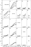
Fig. 1. Multiple logistic regression analysing the probability (Pinf) for patent Onchocerca volvulus infection in children born to O. volvulus-infected or non-infected parents from Burkina Faso (BF), Ghana (GH) and Togo (TG) before start of vector control by the Onchocerciasis Control Programme for West Africa. Children were grouped according to the parents' infection status, the country of origin (BF, GH, TG), by age and sex, and the local intensity of transmission (ATP level) of infective 3rd-stage larvae (L3) of O. volvulus. The parents' infection status was: M+F+, both mother and father O. volvulus (O.v.) microfilariae (mf)-positive; M+F−, mother O.v. mf-positive and father O.v. mf-negative; M−F+, mother O.v. mf-negative and father O.v. mf-positive; M−F−, both mother and father O.v. mf-negative. Children were grouped according to age (0–5 years, 6–10 years, 11–15 years, and 16–20 years) and sex (boy, girl). For the intensity of transmission of L3 of O. volvulus, the annual transmission potential (ATP) in the respective sentinel villages where parents and their children were living was determined, and ATP levels were grouped as low (0–10 L3/year), medium (11–100 L3/year) or high (>100 L3/year).

Table 1. Prevalence of onchocerciasis in parents and their children and intensity of transmission of infective third-stage larvae (L3) of Onchocerca volvulus before the start of vector control measures as conducted by the Onchocerciasis Control Programme (OCP) for West Africa
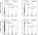
Fig. 2. Levels of Onchocerca volvulus microfilariae per skin snip in children from infected and non-infected parents from Burkina Faso, Ghana and Togo before start of vector control by the Onchocerciasis Control Programme for West Africa. Children were grouped according to the parents' infection status: M+F+, both mother and father O. volvulus (O.v.) microfilariae (mf)-positive; M+F−, mother O.v. mf-positive and father O.v. mf-negative; M−F+, mother O.v. mf-negative and father O.v. mf-positive; M−F−, both mother and father O.v. mf-negative. Data are given as geometric means of both skin snips (ss). For purposes of illustration, geometric means were transformed by logarithm to base ten. Box plots show median with the 25% and 75% quartiles and 1·5× of the interquartile range. Data were analysed by the Wilcoxon Rank Sum Test. After Bonferroni correction for multiple testing, the level of significance is α=0·017 (3 tests per age group).
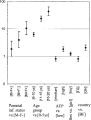
Fig. 3. Odds ratios (OR) with 95% confidence intervals for Onchocerca volvulus infection in 0 to 20-year-old children in Burkina Faso (BF), Ghana (GH) and Togo (TG) before start of vector control by the Onchocerciasis Control Programme for West Africa. Children were grouped according to their parents' infection status: M+F+, both mother and father O. volvulus (O.v.) microfilariae (mf)-positive; M+F−, mother O.v. mf-positive and father O.v. mf-negative; M−F+, mother O.v. mf-negative and father O.v. mf-positive; M−F−, both mother and father O.v. mf-negative [reference group]. Children were grouped according to age (Group 1: 0–5 years [reference group], Group 2: 6–10 years, Group 3: 11–15 years, and Group 4: 16–20 years) and sex. For the intensity of transmission of O. volvulus L3, the annual transmission potential (ATP) in the respective sentinel communities where parents and their children were living was determined, and ATP levels were grouped as low (0–10 L3/year [reference group]), medium (11–100 L3/year) and high (>100 L3/year).
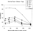
Fig. 4. Prevalence (%) of Onchocerca volvulus microfilariae and its exact 95% confidence intervals in children and their parents from Burkina Faso, Ghana and Togo during 18 years of vector control by the Onchocerciasis Control Programme for West Africa (OCP). Children were grouped according to the parents' infection status. M+F+, both mother and father O. volvulus (O.v.) microfilariae (mf)-positive; M+F−, mother O.v. mf-positive and father O.v. mf-negative; M−F+, mother O.v. mf-negative and father O.v. mf-positive; M−F−, both mother and father O.v. mf-negative. Time groups are given in months after the beginning of vector control.
 You have
Access
- 25
- Cited by