Introduction
Of the three species of tapeworm infecting equines in the northern hemisphere (Anoplocephala magna, A. perfoliata, Paranoplocephala mamillana), A. perfoliata is the most common, with worldwide records (Gasser et al., Reference Gasser, Williamson and Beveridge2005). Since the 1990s, an increased awareness of the association of A. perfoliata infection with a higher risk of clinical intestinal disease in horses has contributed to a growing interest in the use of efficient and practicable methods for the diagnosis of tapeworm infection. Although there has been considerable work on the diagnosis of A. perfoliata infection using serodiagnostic assays (Höglund et al., Reference Höglund, Ljungström, Nilsson and Uggla1995; Proudman & Trees, Reference Proudman and Trees1996a, Reference Proudman and Treesb; Maddox-Hyttel et al., Reference Maddox-Hyttel, Sørensen, Proudman, Farlam and Andersen2005; Boswinkel & Sloet van Oldruitenborgh-Oosterbaan, Reference Boswinkel and Sloet van Oldruitenborgh-Oosterbaan2007; Kjær et al., Reference Kjær, Lungholt, Nielsen, Olsen and Maddox-Hyttel2007; Skotarek, Reference Skotarek2008; Traversa et al., Reference Traversa, Fichi, Campigli, Rondolotti, Iorio, Proudman, Pellegrini and Perrucci2008), detection of A. perfoliata DNA by polymerase chain reaction (PCR) in faeces (Drogemuller et al., Reference Drogemuller, Beelitz, Pfister, Schnieder and Samson-Himmelstjerna2004; Traversa et al., Reference Traversa, Fichi, Campigli, Rondolotti, Iorio, Proudman, Pellegrini and Perrucci2008; Chlastáková et al., Reference Chlastáková, Vavrouchová, Kamler, Bodeček and Koudela2009) or coproantigen detection using an antigen-capture enzyme-linked immunosorbent assay (ELISA) (Kania & Reinemeyer, Reference Kania and Reinemeyer2005; Skotarek, Reference Skotarek2008), diagnosis under practical conditions is usually based on examination of faeces for tapeworm ova. Unfortunately, the sensitivity of coproscopic techniques is low, especially when horses harbour fewer than 100 cestodes, and the 61% and 62% found by Proudman & Edwards (Reference Proudman and Edwards1992) and Slocombe (Reference Slocombe2004), respectively, were the highest sensitivities for any procedure recorded to date (Gasser et al., Reference Gasser, Williamson and Beveridge2005).
For the detection of cestode eggs in horse faeces, simple sedimentation, direct floatation techniques with mineral salt or sugar solutions, combined sedimentation/floatation techniques (either gravitational or centrifugal forced procedures) employing either mineral salt, sugar or mixed mineral salt+sugar solutions for floatation, modified McMaster techniques and the recently described FLOTAC technique have been used (Slocombe, Reference Slocombe1979, Reference Slocombe2004; Hasslinger & Tausend, Reference Hasslinger and Tausend1989; Proudman & Edwards, Reference Proudman and Edwards1992; Hearn & Hearn, Reference Hearn and Hearn1995; Ihler et al., Reference Ihler, Rootwelt, Heyeraas and Dolvik1995; Nilsson et al., Reference Nilsson, Ljungstöm, Höglund, Lundquist and Uggla1995; Drößigk & Schuster, Reference Drößigk and Schuster1997; Höglund et al., Reference Höglund, Nilsson, Ljungström, Hellander, Osterman Lind and Uggla1998; Sangioni et al., Reference Sangioni, Vodotto, Luz Pereira and Bonzi2000; Epe et al., Reference Epe, Behrens, Samson-Himmelstjerna and Schnieder2001; Rehbein et al., Reference Rehbein, Visser and Winter2002; Boswinkel & Sloet van Oldruitenborgh-Oosterbaan, Reference Boswinkel and Sloet van Oldruitenborgh-Oosterbaan2007; Skotarek, Reference Skotarek2008; Traversa et al., Reference Traversa, Fichi, Campigli, Rondolotti, Iorio, Proudman, Pellegrini and Perrucci2008; Chlastáková et al., Reference Chlastáková, Vavrouchová, Kamler, Bodeček and Koudela2009) but only some studies compared different procedures (Beroza et al., Reference Beroza, Marcus, Williams and Bauer1986; French et al., Reference French, Chapman and Klei1994; Agneessens et al., Reference Agneessens, Debever, Engelen and Vercruysse1998; Beelitz & Gothe, Reference Beelitz and Gothe2001; Gundłach et al., Reference Gundłach, Sadzikowski and Tomczuk2003; Maddox-Hyttel et al., Reference Maddox-Hyttel, Sørensen, Proudman, Farlam and Andersen2005) with a few evaluating the findings by statistical analysis (Collobert et al., Reference Collobert, Urbain, Tariel and Lamidey1991; Meana et al., Reference Meana, Luzón, Corchero and Gómez-Bautista1998; Williamson et al., Reference Williamson, Beveridge and Gasser1998; Martins et al., Reference Martins, Verocai, Melo, Freitas, Correia, Pereira and Scott2003). Despite these limitations, simple sedimentation, direct floatation and modified McMaster techniques were generally considered to test positive for cestode eggs less frequently compared to combined sedimentation/floatation techniques.
Although several procedures to detect tapeworm eggs in horse faeces have been described, the impact of sample size and floatation solutions for the combined sedimentation/floatation technique has not been sufficiently addressed. Thus, the purpose of this work was to determine whether the processed sample weight and type of solution used for floatation have a significant effect on the detection of cestode eggs in horse faeces.
Material and methods
Faecal samples
For this study, faecal samples from 250 horses from farms in Germany with known tapeworm infection history were examined for cestode eggs using a double centrifugation/combined sedimentation–floatation technique and employing different sample weight/floatation solution combinations.
Assay procedure
Following thorough mixing of each faecal sample, three 5 g and three 15 g subsamples were weighed out. Each subsample was mixed with about 40 ml of tap water. The faecal slurry then was sieved through a tea strainer and pressed with a teaspoon to recover as much of the water as possible. The resulting suspension was poured immediately into two conical-bottom 30-ml centrifuge tubes and centrifuged at 400 g for 10 min in a swinging-bucket centrifuge. The supernatant was removed with a water-jet aspirator pump and the sediment re-suspended in a small amount of floatation solution using a vortexer. Resuspended samples were transferred to 15-ml conical-bottom centrifuge tubes. Additional floatation solution was added to the tubes until a slight convex meniscus formed, and a 22 mm × 22 mm coverslip was placed on the top of each tube, in contact with the floatation solution. Tubes with coverslips were placed in a swinging-bucket centrifuge and spun for 10 min at 200 g. After the centrifuge came to a complete stop, the coverslips, with the attached suspension, were removed and placed on a microscope slide. The preparation (two coverslips per faecal subsample placed together on one microscope slide) was examined for cestode eggs using a compound light microscope at a magnification of × 40 or × 100. Based on morphological features (Drößigk & Schuster, Reference Drößigk and Schuster1997; Rehbein et al., Reference Rehbein, Visser and Winter2002) all recovered cestode eggs were identified as Anoplocephala eggs. No efforts were made to distinguish the eggs of A. magna and A. perfoliata, which are similar in shape and size. However, differentiation is possible based on the structure of the so-called piriform apparatus (Böhm & Supperer, Reference Böhm and Supperer1958). All Anoplocephala eggs were counted per preparation to derive an egg count per sample.
For floatation, the following solutions were used: saturated NaCl solution, specific gravity (sp. g.) 1.2; concentrated sugar solution, sp. g. 1.26 (prepared by dissolving 450 g sugar in 350 ml water); and concentrated ZnSO4 solution, sp. g. 1.3.
Faeces samples were coded so that the specific combination of sample weight and floatation solution was unknown to the person examining the microscopic preparation.
In addition to the faecal egg counts, the quality of the slide preparations was estimated using criteria of ‘cleanness’ of the preparation (amount of extraneous faecal debris and air bubbles), potential influence on the eggs (e.g. distortion, deformation) and drying of the preparation.
Statistical analysis
Statistical analysis of the data was done using SPSS® version 15.0 for WINDOWS® (SPSS Inc., Chicago, Illinois, USA). McNemar's χ2-test was used to compare the percentage rates of Anoplocephala egg ‘positive’ samples between the sample weight/floatation solution combinations for dependent groups. Anoplocephala egg counts between the sample weight/floatation solution combinations were compared using Friedman's test for multiple comparisons of dependent groups and the Wilcoxon test for two dependent groups. Results were considered significant at P < 0.05.
Results
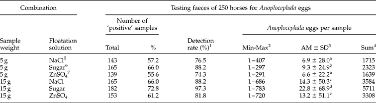
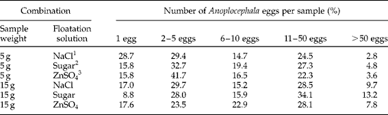
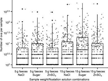
The results of the study are summarized in table 1. Table 2 and fig. 1 provide an overview of the distribution of the Anoplocephala egg counts recorded during the examination of the faecal samples of the 250 horses for the sample weight/floatation solution combinations tested.
Results of the double centrifugation technique sample weight/floatation solution combinations for the detection of Anoplocephala eggs in the faecal samples of 250 horses.

1 (Number of ‘positive’ samples/187 ( = total number of samples testing ‘positive’ for Anoplocephala eggs)) × 100.
2 Minimum–maximum number of Anoplocephala eggs in ‘positive’ samples.
3 Arithmetic mean ± standard deviation, Anoplocephala eggs per sample.
4 Total number of Anoplocephala eggs counted in 250 samples.
5 Saturated NaCl solution (sp. g. 1.2 g/ml).
6 Concentrated sugar solution (sp. g. 1.26 g/ml).
7 Concentrated ZnSO4 solution (sp. g. 1.3 g/ml).
Anoplocephala eggs counted per sample (n=250).

1 Saturated NaCl solution (sp. g. 1.2 g/ml).
2 Concentrated sugar solution (sp. g. 1.26 g/ml).
3 Concentrated ZnSO4 solution (sp. g. 1.3 g/ml).
Distribution of Anoplocephala egg counts of the tested sample weight/floatation solution combinations.

In total, the faeces from 187 horses ( = 74.8%) tested ‘positive’ for Anoplocephala eggs. Percentages of samples testing ‘positive’ for Anoplocephala ova were: 57.2% for 5 g faeces/NaCl, 66% for 15 g faeces/NaCl solution, 66% for 5 g faeces/sugar solution, 72.8% for 15 g faeces/sugar solution, 55.6% for 5 g faeces/ZnSO4 solution, and 61.2% for 15 g faeces/ZnSO4 solution.
Processing the 15 g faecal samples resulted in a significant (P < 0.05, McNemar's χ2-test) increase in the percentage of Anoplocephala egg detection compared to processing the 5 g samples for all floatation solutions. By processing 15 g faecal samples using sugar solution for floatation, 97.3% of all samples that tested ‘positive’ for Anoplocephala eggs (n = 187) were identified. There was no significant difference between the percentage of samples testing ‘positive’ using 15 g faeces/sugar solution (72.8%) and the total percentage of samples testing ‘positive’ (74.8%). Conversely, percentage rates for all other sample weight/floatation solution combinations were significantly (P < 0.0001, McNemar's χ2-test) lower than the total percentage of samples testing ‘positive’.
Anoplocephala faecal egg counts differed significantly (P < 0.0001, Friedman's test) within the 5 g and 15 g faecal sample combinations. The use of sugar solution for floatation gave significantly (P < 0.05, Wilcoxon test) higher Anoplocephala egg counts than NaCl and ZnSO4 solutions for both 5 g and 15 g faecal samples; no significant difference was found in the comparison of the results obtained for the 5 g and 15 g faecal samples processed with NaCl solution or ZnSO4 solution.
Deformation of Anoplocephala eggs was not considered of importance for any of the floatation solutions tested; however, some distorted ova were recognized with ZnSO4 solution. Considerable faecal debris was floated, and numerous bubbles were present in ZnSO4 solution preparations, making examinations time consuming, especially for the 15 g faecal samples. Preparations with NaCl solution were generally considered clear and only a few bubbles were present; however, the rapid drying of the preparations, including the formation of salt crystals, was an important disadvantage. Superior preparations were achieved with the sugar solution: sugar solution slides were cleaner than the NaCl solution slides and there was no drying of the preparations to hamper the microscopic examination.
Discussion
Only Anoplocephala cestode eggs were identified in this study. This finding is in line with previous coproscopical surveys of horses in Germany where the vast majority of cestode eggs found were identified as Anoplocephala ova and the occurrence of P. mamillana eggs was reported only infrequently (Drößigk & Schuster, Reference Drößigk and Schuster1997; Epe et al., Reference Epe, Behrens, Samson-Himmelstjerna and Schnieder2001; Rehbein et al., Reference Rehbein, Visser and Winter2002). On the basis of recent necropsy studies of horses, A. perfoliata is by far the most common tapeworm, with A. magna and P. mamillana being of rare prevalence in Germany (Çirak et al., Reference Çirak, Hermosilla and Bauer1996; Rehbein et al., Reference Rehbein, Visser and Winter2000, Reference Rehbein, Visser, Winter and Maidl2004; Beelitz & Gothe, Reference Beelitz and Gothe2001). It can therefore be assumed that the cestode eggs recovered in the present study were probably the eggs of A. perfoliata.
The present work compared the influence of two sample weights and three different solutions for floatation in a standard double centrifugation/combined sedimentation–floatation technique on the detection of Anoplocephala eggs in horse faeces. As the true infection status of the horses was not known, sensitivity of the different sample weight/floatation solution combinations employed could not be established. In addition, as field samples were used, the absolute numbers of eggs in the faeces were not known.
The major findings of this work were that processing of 15 g faecal samples resulted in a significant increase in the percentage of Anoplocephala egg detection compared to processing of 5 g samples for all floatation solutions, and that using sugar solution for floatation gave significantly higher rates of ‘positive’ samples of Anoplocephala eggs compared to the use of saturated NaCl and concentrated ZnSO4 floatation solutions. These results support the processing of larger amounts of faeces for the diagnosis of horse tapeworm infection than is commonly used in routine testing, which focuses on gastrointestinal nematode infections, to compensate for the suggested irregular shedding of proglottids and insufficient release of eggs from the proglottids during ingesta passage (Slocombe, Reference Slocombe1979; Gasser et al., Reference Gasser, Williamson and Beveridge2005). Sugar solution combinations were more effective in the floatation of Anoplocephala eggs than the mineral salt solutions tested, thus confirming the use of concentrated sugar solutions for the coproscopic diagnosis of equine cestodiasis by several authors (Beroza et al., Reference Beroza, Marcus, Williams and Bauer1986; Collobert et al., Reference Collobert, Urbain, Tariel and Lamidey1991; Proudman & Edwards, Reference Proudman and Edwards1992; French et al., Reference French, Chapman and Klei1994; Hearn & Hearn, Reference Hearn and Hearn1995; Ihler et al., Reference Ihler, Rootwelt, Heyeraas and Dolvik1995; Meana et al., Reference Meana, Luzón, Corchero and Gómez-Bautista1998; Williamson et al., Reference Williamson, Beveridge and Gasser1998; Sangioni et al., Reference Sangioni, Vodotto, Luz Pereira and Bonzi2000; Beelitz & Gothe, Reference Beelitz and Gothe2001; Rehbein et al., Reference Rehbein, Visser and Winter2002; Martins et al., Reference Martins, Verocai, Melo, Freitas, Correia, Pereira and Scott2003; Maddox-Hyttel et al., Reference Maddox-Hyttel, Sørensen, Proudman, Farlam and Andersen2005; Skotarek, Reference Skotarek2008; Traversa et al., Reference Traversa, Fichi, Campigli, Rondolotti, Iorio, Proudman, Pellegrini and Perrucci2008). In this study, the processing of 15 g faecal samples compared to 5 g samples resulted in 2.09-, 2.46- and 2.02-fold increases in faecal Anoplocephala egg counts for saturated NaCl, concentrated sugar and ZnSO4 floatation solutions, respectively. These data again support the use of a sugar solution for floatation of Anoplocephala eggs, which previously were claimed to levitate only poorly in floatation solutions (Slocombe, Reference Slocombe1979; Beroza et al., Reference Beroza, Marcus, Williams and Bauer1986). In addition, the tested sugar solution separated the Anoplocephala eggs readily from the faecal debris, and the preparations did not show crystallization.
Compared to combined sedimentation–floatation techniques employing gravitational force sedimentation or floatation (e.g. Beroza et al., Reference Beroza, Marcus, Williams and Bauer1986; Collobert et al., Reference Collobert, Urbain, Tariel and Lamidey1991; Proudman & Edwards, Reference Proudman and Edwards1992; Ihler et al., Reference Ihler, Rootwelt, Heyeraas and Dolvik1995; Meana et al., Reference Meana, Luzón, Corchero and Gómez-Bautista1998; Sangioni et al., Reference Sangioni, Vodotto, Luz Pereira and Bonzi2000; Beelitz & Gothe, Reference Beelitz and Gothe2001; Martins et al., Reference Martins, Verocai, Melo, Freitas, Correia, Pereira and Scott2003; Maddox-Hyttel et al., Reference Maddox-Hyttel, Sørensen, Proudman, Farlam and Andersen2005; Traversa et al., Reference Traversa, Fichi, Campigli, Rondolotti, Iorio, Proudman, Pellegrini and Perrucci2008), the combined sedimentation–floatation technique with two centrifugation steps of 10 min each used in this study was less time-consuming.
In conclusion, this work demonstrated that the double centrifugation/combined sedimentation–floatation technique using 15 g faecal samples and concentrated sugar solution for floatation was superior to the other combinations and offers an advantage for the detection of Anoplocephala eggs in horse faeces.