




No CrossRef data available.
Article contents
- Abstract
- INTRODUCTION
- MITOCHONDRIAL HETEROGENEITY IN THE ASEXUAL AND SEXUAL BLOOD STAGES OF PLASMODIUM FALCIPARUM
- DIFFERENTIAL EXPRESSION OF THE MITOCHONDRIAL GENE IN THE ASEXUAL AND SEXUAL BLOOD STAGES OF PLASMODIUM FALCIPARUM
- ELECTRON TRANSPORT SYSTEM AND BIOCHEMICAL FUNCTIONS OF MITOCHONDRIA OF THE ASEXUAL AND SEXUAL BLOOD STAGES OF PLASMODIUM FALCIPARUM
- OTHER METABOLIC ROLES OF THE PLASMODIUM FALCIPARUM MITOCHONDRION
- HOW MANY MITOCHONDRIAL PROTEINS ARE THERE IN PLASMODIUM FALCIPARUM?
- PLASMODIUM FALCIPARUM MITOCHONDRIA AS POTENTIAL CHEMOTHERAPEUTIC TARGETS
- CONCLUSION, FUTURE DIRECTIONS AND IMPLICATIONS FOR THE MITOCHONDRION OF PLASMODIUM FALCIPARUM
- References
The multiple roles of the mitochondrion of the malarial parasite
Published online by Cambridge University Press: 05 October 2004
Article contents
- Abstract
- INTRODUCTION
- MITOCHONDRIAL HETEROGENEITY IN THE ASEXUAL AND SEXUAL BLOOD STAGES OF PLASMODIUM FALCIPARUM
- DIFFERENTIAL EXPRESSION OF THE MITOCHONDRIAL GENE IN THE ASEXUAL AND SEXUAL BLOOD STAGES OF PLASMODIUM FALCIPARUM
- ELECTRON TRANSPORT SYSTEM AND BIOCHEMICAL FUNCTIONS OF MITOCHONDRIA OF THE ASEXUAL AND SEXUAL BLOOD STAGES OF PLASMODIUM FALCIPARUM
- OTHER METABOLIC ROLES OF THE PLASMODIUM FALCIPARUM MITOCHONDRION
- HOW MANY MITOCHONDRIAL PROTEINS ARE THERE IN PLASMODIUM FALCIPARUM?
- PLASMODIUM FALCIPARUM MITOCHONDRIA AS POTENTIAL CHEMOTHERAPEUTIC TARGETS
- CONCLUSION, FUTURE DIRECTIONS AND IMPLICATIONS FOR THE MITOCHONDRION OF PLASMODIUM FALCIPARUM
- References
Abstract
Mitochondria of the malaria parasite Plasmodium falciparum are morphologically different between the asexual and sexual blood stages (gametocytes). In this paper recent findings of mitochondrial heterogeneity are reviewed based on their ultrastructural characteristics, metabolic activities and the differential expression of their genes in these 2 blood stages of the parasite. The existence of NADH dehydrogenase (complex I), succinate dehydrogenase (complex II), cytochrome c reductase (complex III) and cytochrome c oxidase (complex IV) suggests that the biochemically active electron transport system operates in this parasite. There is also an alternative electron transport branch pathway, including an anaerobic function of complex II. One of the functional roles of the mitochondrion in the parasite is the coordination of pyrimidine biosynthesis, the electron transport system and oxygen utilization via dihydroorotate dehydrogenase and coenzyme Q. Complete sets of genes encoding enzymes of the tricarboxylic acid cycle and the ATP synthase complex are predicted from P. falciparum genomics information. Other metabolic roles of this organelle include membrane potential maintenance, haem and coenzyme Q biosynthesis, and oxidative phosphorylation. Furthermore, the mitochondrion may be a chemotherapeutic target for antimalarial drug development. The antimalarial drug atovaquone targets the mitochondrion.
Keywords
Information
- Type
- Review Article
- Information
- Copyright
- © 2004 Cambridge University Press
INTRODUCTION
Malaria remains one of the most deadly diseases in the world, causing about 500 million clinical cases annually and about 1·5–2·7 million deaths per year (Guerin et al. 2002). Plasmodium falciparum is responsible for the most severe form of human malaria. Because of the increasing prevalence of resistance to the drugs currently in use, there is an urgent need to develop new chemotherapeutic agents. The parasite mitochondrion has been suggested as a potential target for antimalarial drugs (Hudson et al. 1985; Ginsburg et al. 1986) and, in particular, the mitochondrial electron transport system, since this organelle in the parasite is underdeveloped and has relatively limited function when the parasite is in the mammalian erythrocytes. In the host erythrocytes, P. falciparum grows and differentiates as asexual blood stages from ring, trophozoite, to schizont and develops to sexual blood stages (gametocytes), under defined conditions. During the differentiation, a series of complicated processes occur, essential for the survival of the parasite (Sinden, 1983; Alano & Carter, 1990).
Several lines of evidence from ultrastructural and genetic analyses suggest that the organelle may be regulated during the switching from the asexual stage to the sexual stage in the human host bloodstream (Aikawa, 1971; Langreth et al. 1978; Sinden, 1982; Fry & Beesley, 1991; Feagin & Drew, 1995; Petmitr & Krungkrai, 1995). The process of asexual stage development from the ring to the trophozoite to the schizont stages has been well studied, aided by the feasibility of growing the parasite in in vitro culture (Trager & Jensen, 1976; Horrocks, Dechering & Lanzer, 1998). Understanding the molecular mechanisms that regulate growth, development and differentiation of the sexual blood stage would be highly desirable because this stage is critical for the parasite to be transmitted via the Anopheles mosquito vector. Interference with the sexual development of the human parasite might be an effective means of controlling the disease.
In separate investigations, detailed ultrastructural properties in the mitochondria of the sexual and asexual erythrocytic stages have been demonstrated (Aikawa, Huff & Sprinz, 1966; Aikawa, 1971; Langreth et al. 1978; Sinden, 1982; Fry & Beesley, 1991; Krungkrai, Krungkrai & Bhumiratana, 1993; Krungkrai, 1995; Krungkrai et al. 1999a,b; Krungkrai, Prapunwattana & Krungkrai, 2000). The biochemical functions of the mitochondria in these two stages of the parasite have been recently characterized (Krungkrai et al. 1999a,b; Learngaramkul et al. 1999; Krungkrai et al. 2000). It is proposed that the mitochondrion may function in electron disposal of the pyrimidine biosynthetic pathway with the mitochondrial associated enzyme dihydroorotate dehydrogenase (DHODase) catalysing dihydroorotate oxidation to orotate and generating electrons (Gutteridge, Dave & Richards, 1979; Krungkrai, Cerami & Henderson, 1991; Krungkrai, 1993a, 1995). The pyrimidine pathway is closely linked to the mitochondrial electron transport system through coenzyme Q, as demonstrated in at least three species of malarial parasites, P. falciparum (Krungkrai, 1995), P. knowlesi (Gutteridge et al. 1979) and P. berghei (Krungkrai et al. 1991). The existence of cytosolic orotate reductase, catalysing the conversion of orotate to dihydroorotate, may play a role in the regulation of pyrimidine biosynthesis in Plasmodium sp. (Krungkrai, 1993b).
More interestingly, some antimalarial drugs are known to act specifically on the mitochondrion, e.g., tetracycline (Prapunwattana, O'Sullivan & Yuthavong, 1988), primaquine (Lanners, 1991; Vaidya et al. 1993), atovaquone (Fry & Pudney, 1992; Krungkrai et al. 1997, 1998, 2003; Srivastava, Rottenberg & Vaidya, 1997; Srivastava et al. 1999; Peters et al. 2002; Kessl et al. 2003), and artemisinin derivatives (Zhao, Hanton & Lee, 1986; Kawai, Kano & Suzuki, 1993). The actions of these drugs further support the view that the mitochondrion is a chemotherapeutic target.
The mitochondrial genome of P. falciparum is a ~6·0 kb tandemly repeated linear element which is uniparentally inherited (Joseph et al. 1989; Feagin, 1992; Creasey et al. 1993). There is 90% conservation of sequence identity among the mitochondrial genomes of P. falciparum, P. vivax, P. yoelii, and P. gallinaceum (McIntosh, Srivastava & Vaidya, 1998). Detailed analyses on replication and expression have been well characterized in the mitochondrial genes of the asexual stage parasite (Feagin & Drew, 1995; Preiser et al. 1996). The mitochondrial DNA polymorphisms in P. falciparum have been compared for parasites from Africa, Asia and America (Conway et al. 2000). Relatively little is known about chromosomal and mitochondrial DNAs during gametocytogenesis, exflagellation and gametogenesis of the sexual stage parasite (Carter et al. 1989). Stage-specific expression of actin, tubulin, and rRNAs genes during gametocytogenesis of the parasite has been studied for the chromosomal genes (Wesseling et al. 1989; Rawlings et al. 1992; Waters et al. 1997).
Here the recent findings on the existence of mitochondrial heterogeneity will be reviewed based on their ultrastructural characteristics, biochemical activity and the differential expression of their genes in developing asexual and sexual blood stages of P. falciparum. The metabolic and functional roles of the mitochondrion in the parasite are proposed based mainly on biochemical assays of the isolated organelles, and partly from recent information from the completed P. falciparum genome. Finally, the organelle as a chemotherapeutic target will be considered.
MITOCHONDRIAL HETEROGENEITY IN THE ASEXUAL AND SEXUAL BLOOD STAGES OF PLASMODIUM FALCIPARUM
The in vitro culture of the P. falciparum sexual blood stage in our laboratory (Petmitr & Krungkrai, 1995) was established using the methods of Ifediba & Vanderberg (1981) and Sinden (1982), which provides sexual-stage parasites for the isolation of mitochondria. In parallel, mitochondria were isolated from asexual blood stages, mostly trophozoite stages, in which there is active synthesis of DNA, RNA and protein.
A tubular-like cristate mitochondrion has been demonstrated in the asexual stage parasite (Fig. 1). The sexual stage parasite has a mitochondrion with more numbers of the cristate structures (Fig. 2). Based on the electron density of the matrices, and the numbers and appearances of tubular-cristate structure, 3 different types of mitochondria have been identifed in these 2 blood stages (Krungkrai et al. 1999a). Figure 1 shows a clearly defined double membrane mitochondrion in the trophozoite stage of the asexual stage parasite with tubular cristae formed by infolding of the inner membrane and similar to other protozoan mitochondrion. The elongated organelle shows a limited number of cristae. All mitochondria so far examined, in the asexual blood stages have none or a few tubular cristae. They are classified as ‘type I’ mitochondria (Fig. 1B).

Fig. 1. Transmission electron micrograph of the asexual stage of Plasmodium falciparum (trophozoite stage). A single mitochondrion is observed with a clearly visible double membrane organelle, and an elongated form which is preparing for binary fission (A). A single tubular cristate structure, marked by the arrowhead, is seen in the organelle at higher magnification (B). N, nucleus; M, mitochondrion; P, pigment.

Fig. 2. Transmission electron micrograph of the sexual blood stage of Plasmodium falciparum (gametocyte stage IV). Numerous mitochondria are observed in a single parasite (A). A higher magnification of two organelles containing several cristae (arrowhead) is shown (B). M, mitochondrion; P, pigment.
The mature gametocyte stage (Fig. 2) has ~4–8 mitochondria, containing marked proliferation and pronounced infolding of the inner membrane giving rise to large numbers of tubular cristae with clear intracristal spaces. They are classified as ‘type II’ mitochondria (Fig. 2B). This type II organelle is also seen in the asexual blood stage of P. berghei (Krungkrai et al. 1993). Some mature gametocytes possess higher numbers of cristae, and contain electron-dense tubular cristae. They are classified as ‘type III’ mitochondria.
Until recently, only the organelles in the asexual blood stage parasite had been isolated from 2 species of rodent parasites (P. yoelii, P. berghei) and the human parasite P. falciparum. Most organelles are type I structures, with a definite double-membrane and are found to be metabolically active (Fry & Beesley, 1991; Krungkrai et al. 1993; Krungkrai, 1995; Takashima et al. 2001). More recently, the mitochondria from the sexual stage parasite have been isolated (Krungkrai et al. 2000). Figure 3 shows the isolated organelles, typically type II mitochondria. The different variations in the mitochondria of asexual and sexual blood stages suggest that the gametocytes might have a higher demand for energy transduction and are also more metabolically active than the asexual stages. The more active mitochondria, producing more energy in the sexual stages, may be necessary for their survival during transmission into the mosquito vector. This remains to be studied.

Fig. 3. Transmission electron micrograph of the mitochondrial organelles isolated from sexual blood stages of Plasmodium falciparum. (A) The organelles contain 6–8 tubular cristate structures (arrowhead), as was found in the intact parasites. (B) A higher magnification of an isolated organelle indicate the mitochondrion is still intact with clearly defined double membranes, marked by the double arrowheads. M, mitochondrion.
Our ultrastructural analysis has confirmed the existence of stage-specific variations in mitochondria when comparing asexual and sexual stages in the blood (Krungkrai et al. 1999a; Learngaramkul et al. 1999; Krungkrai, 2000; Krungkrai et al. 2000). Similarly, in Haemogregarina myoxocephali, an apicomplexan blood parasite, there are different numbers of mitochondria in asexual and sexual stages, 1–2 and 4–6 organelles for the asexual and sexual stages, respectively (Siddall & Desser, 1992). The ultrastructural differences in mitochondria prompted us to carry out a molecular and biochemical investigation of mitochondria isolated from these 2 developmental stages in malaria parasites.
DIFFERENTIAL EXPRESSION OF THE MITOCHONDRIAL GENE IN THE ASEXUAL AND SEXUAL BLOOD STAGES OF PLASMODIUM FALCIPARUM
The 6·0 kb mitochondrial DNA of P. falciparum encodes only 3 subunits of the electron transport chain. These are 2 subunits (cox I and cox III) of cytochrome c oxidase (complex IV) and a cytochrome b subunit (cyt b) of ubiquinol-cytochrome c reductase (cytochrome bc1 or complex III) (Feagin, 1992; Joseph et al. 1989) (Fig. 4). Most mitochondrial proteins are encoded by nuclear genes (Gardner et al. 2002; Bender et al. 2003). Interestingly, the cox II protein of complex IV is encoded by 2 independent nuclear genes, cox2a (chromosome 13) and cox2b (chromosome 14) in P. falciparum, and this is different from most eukaryotic cox II subunit which is encoded by a single gene on the mitochondrial genome (Funes et al. 2002). The cox2a and cox2b genes encode N-terminal domain and C-terminal domain of the cox II protein, respectively (Funes et al. 2002; Gardner et al. 2002). A lateral transfer of these genes from the green alga nucleus to P. falciparum nuclear genome has been suggested (Funes et al. 2002). A similar division of the cox2 gene is also seen in the unicellular alga, Chlamydomonas reihardtii (Perez-Martinex et al. 2001; Funes et al. 2002). Coordinated expression of the cox2a and cox2b genes, mitochondrial targeting and assembling of complex IV have not been studied in P. falciparum. The cyt b gene which encodes the mitochondrial cytochrome b protein of complex III does not derive from the green alga genome. Mitochondrial DNA synthesis occurs coincidentally with that of chromosomal DNA throughout the growth and multiplication cycle of the asexual parasite in its host cell (Feagin & Drew, 1995). It has a phage-like replication mechanism and undergoes extensive recombination like the mitochondrial DNA element of some other lower eukaryotes (Preiser et al. 1996). The transcripts of protein-coding genes of the mitochondrial DNA are least abundant in the ring and early trophozoite stages and most abundant in the late trophozoite and schizont stages (Feagin & Drew, 1995).

Fig. 4. A map of the organization of the 6 kb mitochondrial DNA of Plasmodium falciparum. Arrows indicate the direction of transcription. The gene regions cox I, cox III and cyt b encoding cytochrome oxidase subunits I, III and cytochrome b subunit of ubiquinol-cytochrome c reductase, respectively, are shown in shaded boxes. Fragmented large and small subunits rRNA genes are indicated as filled and open boxes respectively.
The expression profiles of both chromosomal and mitochondrial DNAs are largely unknown during gametocytogenesis, exflagellation and gametogenesis of the sexual-stage parasites. Abundant stage-specific proteins with molecular masses of 27 and 25 kDa have been shown in P. falciparum during gametocytogenesis (Carter et al. 1989). We have recently determined the copy number, the nucleotide sequence and detailed molecular characterization of the 1·3 kb cyt b gene in the mitochondrial DNA. We also determined these for the chromosomal dihydrofolate reductase (dhfr) gene, a single copy gene, in both asexual and sexual blood stages of P. falciparum, using established methods (Cowman et al. 1988; Wiesner, 1992). Increased amounts of both mitochondrial DNA templates (using quantitative polymerase chain reaction, PCR) and transcripts (using quantitative reverse transcription PCR) are found in the sexual blood stages and not in the asexual blood stages (Petmitr & Krungkrai, 1995; Learngaramkul et al. 1999). These findings imply that both gene dosage and the transcription play an important role in the expression of the mitochondrial DNA during development of the parasites in the asexual and sexual blood stages. Gene regulation by gene dosage and transcription level have also been advocated in mammalian mitochondrial biogenesis (Nagley, 1991).
Expression of the dhfr gene in the sexual blood stage is greatly reduced, compared to the relatively high expression in the asexual blood stage. In contrast to the dhfr gene, the chromosomal tubulin gene encoding the microtubule protein does not appear to be developmentally regulated as indicated when RNA from the asexual stages and from the mosquito midgut stages (i.e., gametes, zygotes) are analysed by Northern hybridization (Wesseling et al. 1989). However, there is no report of the tubulin gene expression in the sexual blood stage, which contains prominent microtubules near the parasite membrane during their development, as demonstrated by transmission electron microscopy techniques (Krungkrai et al. unpublished results). More recently, the signal-transducing protein kinases belonging to families known to regulate differentiation have been found in the sexual-stage parasite (Doerig et al. 1995; Bracchi et al. 1996; Li & Baker, 1998; Dorin et al. 1999). However, whether or not these enzymes directly influence gametocytogenesis and exflagellation remains to be determined.
It is also known that the 2 extrachromosomal genomes, the mitochondrial DNA and the apicoplast 35·0 kb circular DNA, are probably inherited only through the female gamete (Creasey et al. 1993). The molecular mechanism that regulates gene expression during the switching of the asexual stage to the gametocytic stage in the host erythrocyte remains to be elucidated (Horrocks et al. 1998; Fang et al. 2004).
ELECTRON TRANSPORT SYSTEM AND BIOCHEMICAL FUNCTIONS OF MITOCHONDRIA OF THE ASEXUAL AND SEXUAL BLOOD STAGES OF PLASMODIUM FALCIPARUM
The asexual blood stage of P. falciparum is a microaerophillic organism and requires low oxygen tension (0·5–3% oxygen) for optimal growth and development in in vitro culture (Scheibel, Ashton & Trager, 1979; Scheibel, 1988). The gametocytes also have low oxygen requirement for their development in human erythrocytes (Sinden & Smalley, 1979; Petmitr & Krungkrai, 1995; Krungkrai et al. 1999a). We investigated whether the mitochondria in the gametocytes of P. falciparum are biochemically active. The coupling of mitochondrial electron transport system (mtETS) and oxygen consumption through complex IV are well characterized in the asexual blood stages of both P. berghei and P. falciparum (Fry & Beesley, 1991; Krungkrai, 1993; Krungkrai et al. 1997, 1998, 1999a,b, 2002; Murphy et al. 1997; Krungkrai et al. 2000; Suraveratum et al. 2000). Experimental evidence indicates the biochemical functions of the mitochondria of the malarial parasite. The mitochondrial oxygen consumption of asexual and sexual blood stages of P. falciparum is similar. They have relatively low activities (~20–24 nmol/min/107 parasites), compared to human leukocytes and mouse neuroblastoma cells which show oxygen consumption of ~110 and ~130 nmol/min/107 cells, respectively. Naive human erythrocytes show no detectable mitochondrial oxygen consumption. Murphy et al. (1997) measured the rate of mitochondrial oxygen consumption in the asexual stage of P. falciparum and found it to be relatively low, which is similar to our results. However, a branched pathway with relatively low activity of alternative oxidase, transferring electrons directly from ubiquinol to oxygen, presumably located in mitochondria, has also been demonstrated in P. falciparum (Murphy et al. 1997). A gene encoding the alternative oxidase has not been detected in the P. falciparum genome, indicating the following possibilities: the gene might be lost during evolution, the sequence diversity of the gene might not be detected by homology searching, and the oxidase activity might be catalysed by another and unknown protein. This controversial finding remains to be studied. Such a gene has been found in Trypanosoma brucei and Cryptosporidium parvum (Suzuki et al. 2004).
Based on the above findings, it is suggested that the abundant mitochondria in gametocytes are biochemically active, at least in oxygen utilization. Comparison of the morphological and biochemical characteristics of the mitochondria in the asexual and sexual blood stages suggests that the metabolic activities of the developing sexual blood stages may be different from those of the asexual stages. The specific activities of the mitochondrial DHODase and the respiratory mtETS complexes III and IV were measured in mitochondria isolated from both asexual and sexual stages. The DHODase and the activities of complexes III and IV of the sexual-stage mitochondria are ~8 to 10-fold higher than those of the asexual-stage organelles, indicating that the mitochondria in the asexual stage are less metabolically active than those of the sexual blood parasite (Learngaramkul et al. 1999). Our results suggest that the P. falciparum mitochondria from both stages are still under developed forms, which exist in the uncoupling state between mitochondrial oxidation and ATP synthesis.
In the asexual stage, the major pathways of carbohydrate metabolism are essentially anaerobic (Sherman, 1979; Scheibel, 1988). It is possible that carbohydrate metabolism in the sexual-stage parasites is similar to the asexual-stage parasites. In addition, our results also demonstrate a clear difference between the parasite and the mammalian mitochondria, the latter having a tight coupling oxidative phosphorylation for ATP production with a P[ratio ]O ratio of ~2 in the presence of succinate and ADP (Krungkrai et al. 2000). In contrast, the asexual blood stage of the rodent parasite P. berghei has the ability to produce ATP through oxidative phosphorylation with a P[ratio ]O ratio of ~2, in the presence of either succinate or dihydroorotate and ADP (Uyemura et al. 2000, 2004), suggesting the coupling oxidative phosphorylation for ATP production may operate in other malaria parasites. This is consistent with our results indicating increased numbers of subunits in each mtETS complex in the rodent parasite, e.g., 2 subunits (P. falciparum) versus 4 subunits (P. berghei) of complex II (Suraveratum et al. 2000; Takeo et al. 2000; Krungkrai et al. unpublished results), or 6 subunits (P. falciparum) versus 8 subunits (P. berghei) of complex IV (Krungkrai et al. 1993, 1997, 1998).
Measurement of mitochondrial oxygen consumption of asexual (mostly trophozoites, Fig. 1) and sexual (mostly gametocytic stages IV, Fig. 2) blood stages was performed in the presence of specific inhibitors of the 4 mtETS complexes. These inhibitors were well characterized as previously described (Hatefi, 1985). By using a cell density of 1×10⁷ parasites in each oxygen consumption reaction, all mitochondrial inhibitors affecting the respiratory complexes I–IV were found to inhibit the oxygen consumption to a different degree when calculating 50% effective concentrations (EC50 values) in both asexual and sexual blood stages (Table 1). The sexual blood-stage parasite is more sensitive to rotenone and cyanide, affecting the respiratory complexes I and IV respectively, than the asexual blood-stage parasite. In contrast, both stages have a similar response to thenoyltrifluoroacetone and antimycin A, which affect the respiratory complexes II and III respectively.

In the sexual-stage parasite, the mtETS may be similar to the asexual-stage parasite in which electrons from donating enzymes, including DHODase, are transferred via coenzyme Q or ubiquinone and a series of respiratory electron transporting complexes and oxygen is the final electron acceptor (Gutteridge et al. 1979; Krungkrai et al. 1991, 1993, 1999a,b, 2000; Krungkrai, 1993a, 1995; Kita et al. 2002). It is proposed that, in P. falciparum, these asexual and sexual developmental stages have functional mitochondria with major biochemical activities in mtETS complexes I, II, III and IV, and which provide other metabolic activities operating in the organelle (Fig. 5).

Fig. 5. A diagram of the proposed arrangement of the mitochondrial electron transport components and its association with pyrimidine biosynthesis via dihydroorotate dehydrogenase in the asexual and sexual blood stages of Plasmodium falciparum. The mitochondrial electron transporting complexes are NADH-ubiquinone oxidoreductase (NADH dehydrogenase, a component of complex I), succinate-ubiquinone oxidoreductase (succunate dehydrogenase, complex II), ubiquinol-cytochrome c reductase (cytochrome bc1 or complex III) and cytochrome c oxidase (complex IV). Alternative oxidase as a cyanide (CN−) insensitive-branched pathway (shown in a broken line) and glycerol-3-phosphate (glycerol-3-P) dehydrogenase are also included in the respiratory chain. Ubiquinone and cytochrome c (Cyt c, a component of complex III), are shown serving as electron and proton transferring components. Glycolysis, links to the tricarboxylic acid (TCA) cycle, is operating in the cytosol of P. falciparum. The tree-bars indicate known active sites of inhibitors and the antimalarial drug atvaquone. The diagram is adapted from web site: http://sites.huji.ac.il/malaria.
Mitochondrial oxygen consumption in asexual and sexual stages has been measured in the presence of 5-fluoroorotate, a potent inhibitor of DHODase of the pyrimidine biosynthetic pathway of P. falciparum (Krungkrai et al. 1999a), and in the presence of the antimalarial drugs, atovaquone (Fry & Pudney, 1992; Krungkrai et al. 1997) and primaquine (Vaidya et al. 1993) that possibly act on complex III, and artemisinin that might target specifically to complex IV (Zhao et al. 1986; Kawai et al. 1993). Table 2 shows the EC50 values of 5-fluoroorotate, atovaquone, primaquine and artemisinin on the parasites' oxygen consumption in both developing stages. The 5-fluoroorotate shows strong inhibitory effects on the oxygen consumption in both the asexual- and the sexual-stage parasites, suggesting that the parasite in the sexual stage has a link to the 2 metabolic pathways, pyrimidine biosynthesis and mtETS, through the enzyme DHODase of the pyrimidine pathway, similar to the asexual blood stage (Krungkrai, 1995). The association between the pyrimidine pathway and mitochondrial ETS in the asexual-stage parasite has been recently confirmed (Krungkrai et al. 1999a,b; Takashima et al. 2001). It is concluded that in P. falciparum in asexual and sexual stages there are functioning mitochondria that contribute significantly to the de novo synthesis of pyrimidine and less to the membrane potential maintenance and the energy metabolism of the parasite as proposed in Fig. 5. The role of the mitochondria in the sexual blood stages, e.g., for gametocytogenesis in the human host, for exflagellation and gametogenesis in the mosquito vector, remains to be determined.

The findings of the effects of antimalarial drugs on oxygen consumption are directly related to the lines of evidence that these drugs act specifically on the mitochondria of the malarial parasite. Atovaquone inhibits electron transport and depolarizes the mitochondria of the asexual stages of the rodent parasite P. yoelii (Srivastava et al. 1997). It has moderate gametocytocidal activity (Fleck, Pudney & Sinden, 1996), compared to its potent blood schizontocidal activity (Krungkrai et al. 1999a). Primaquine (Vaidya et al. 1993) and artemether (Kawai et al. 1993), a derivative of artemisinin, act specifically on the mitochondria of P. falciparum by causing morphological changes. Artesunate, a derivative of artemisinin, inhibits complex IV activity in P. berghei (Zhao et al. 1986). More recently, Eckstein-Ludwig et al. (2003) have shown that artemisinin targets sarco/endoplasmic reticulum Ca2+-ATPase of P. falciparum. These multiple sites of action by artemisinin derivatives remain to be elucidated. Nevertheless, our results would suggest that the mitochondrion in both the asexual and sexual blood stages is a potential drug target.
OTHER METABOLIC ROLES OF THE PLASMODIUM FALCIPARUM MITOCHONDRION
Studies of mitochondrial functions in malaria parasites have been limited (Divo et al. 1985; Fry & Beesley, 1992; Krungkrai, 1995; Srivastava et al. 1997; Sato, Rangachari & Wilson, 2003; Sato & Wilson, 2004). Using a flow cytometric assay with a lipophilic cation-fluorescent probe, the mitochondrial membrane potential in live P. yoelii-infected erythrocytes can be measured. The membrane potential, generated by the mtETS, is constantly maintained to allow transport of metabolites and proteins across the inner mitochondrial membrane (Srivastava et al. 1997).
P. falciparum can synthesize haem de novo (Varadharajan et al. 2002). Proteins encoded by 2 nuclear genes, δ-aminolevulinate synthase and ferrochelatase, the first and the last enzymes of the de novo pathway, are localized in the mitochondrion (Wilson, Smith & Baylon, 1996; Varadharajan et al. 2002; Dhanasekaran et al. 2004). The second enzyme δ-aminolevulinate dehydratase has been found to be localized in the apicoplast organelle (Dhanasekaran et al. 2004). Compartmentalization of the de novo haem synthetic pathway in the parasite remains to be studied.
The asexual blood stages of P. falciparum rely principally on anaerobic glycolysis for ATP production. Genes encoding all of the enzymes necessary for a functional glycolytic pathway have been identified (Gardner et al. 2002). Furthermore, the genes necessary for a complete tricarboxylic acid (TCA) cycle, including a complete pyruvate dehydrogenase complex, have been identified (Fig. 6). The mitochondrial TCA cycle in these asexual stages may not be involved ATP production, but may serve mainly to synthesize metabolites for other metabolic pathways (Gardner et al. 2002; Chan & Sim, 2003; Wrenger & Muller, 2003). The succinate dehydrogenase component of the mitochondrial TCA cycle and the complex II of P. falciparum is also involved in this anaerobic metabolism by acting as quinol-fumarate reductase (Kita et al. 2002). NADH dehydrogenase acts as NADH-ubiquinol reductase, a component of complex I (Krungkrai et al. 2002). The malarial enzyme may have similar properties to other types of single polypeptide NADH dehydrogenase as found in Saccharomyces cerevisae (Luttik et al. 1998), and in T. cruzi (Fang & Beattie, 2002). The role of the mitochondrial NADH dehydrogenase in P. falciparum remains to be studied.

Fig. 6. Mitochondrial tricarboxylic acid (TCA) cycle of Plasmodium falciparum. The TCA cycle is a part of the mitochondrial metabolism shown in Fig. 5, but its function remains to be studied. It is shown here that metabolites produced by the TCA cycle are important for other metabolic pathways. The diagram is taken from web site: http://sites.huji.ac.il/malaria.
Genes encoding F0 and F1 subunits of ATP synthase (complex V) have also been identified in the P. falciparum genome. A possible functioning TCA cycle, oxidative phosphorylation and ATP synthase in the gametocytes remain to be explored.
HOW MANY MITOCHONDRIAL PROTEINS ARE THERE IN PLASMODIUM FALCIPARUM?
Gardner et al. (2002) have identified 246 (4·6% of total parasite proteins) proteins targeted to the mitochondrion after translation by the TargetP and MitoProtII methods. Using the recently developed PlasMit program for prediction of mitochondrial transit peptides, Bender et al. (2003) have predicted 381 (7·1%) mitochondrial proteins, based on 5334 annotated genes in the P. falciparum genome, close to the numbers of the mitochondrial proteins in other organisms. Most of these protein sequences are identified as hypothetical proteins (see detail in web site: http://gecco.org.chemie.uni-frankfurt.de/plasmit/). Nevertheless, homology searching has identified some components of metabolic pathways, transporter and import proteins (Table 3).

PLASMODIUM FALCIPARUM MITOCHONDRIA AS POTENTIAL CHEMOTHERAPEUTIC TARGETS
Some currently used antimalarial drugs act specifically on the mitochondria, e.g., tetracycline (Prapunwattana et al. 1988), primaquine (Lanners, 1991; Vaidya et al. 1993), atovaquone (Fry & Pudney, 1992; Krungkrai et al. 1997, 1998, 2003; Srivastava et al. 1997; Srivastava et al. 1999; Peters et al. 2002; Kessl et al. 2003) and artemisinin derivatives (Zhao et al. 1986; Kawai et al. 1993). Evidence from the action of antimalarial drugs further supports the view that the mitochondrion is a potential chemotherapeutic target. As mentioned before, in both the asexual and the sexual blood stages of P. falciparum, the DHODase of pyrimidine biosynthesis catalysing dihydroorotate oxidation and generating 2 electrons is thought to link to mtETS, containing ubiquinone (coenzyme Q), and complexes I, II, III and IV (Fig. 5). Most of these enzymes, as shown in Fig. 5, have been examined in detail by our laboratory. The predominant forms of ubiquinones in the asexual stage parasite are Q8 and Q9, whereas the major form in the mammalian host tissues is Q10 (Porter & Folkers, 1974; Ellis, 1994; de Macedo et al. 2002). The Q8–9 coenzyme performs a pivotal role in the transfer of electrons between the mtETS complexes and acts as a proton carrier that supports ATP synthesis (Hatefi, 1985). Thus the inhibition of the electron transfer at this site represents an important target for chemotherapeutic attack (Wan, Porter & Folkers, 1974).
One class of compounds extensively investigated over 50 years for antimalarial activity is the quinone analog, hydroxynaphthoquinone (Hudson, 1984; Hudson et al. 1985). One of the compounds, atovaquone which is 2-[trans-4-(4′-chlorophenyl)cyclohexyl]-3-hydroxy-1,4-naphthoquinone, is currently used as a potent antimalarial drug (TDR/WHO, 1997). Atovaquone affects multiple sites in which Q8–9 plays a significant catalytic role in the target enzyme, e.g., complex III (Krungkrai et al. 1997, 1998; Srivastava et al. 1997; Kessl et al. 2003), and DHODase (Krungkrai, 1995; Krungkrai et al. 2003). The malaria parasites have their own synthesis of ubiquinones and cannot salvage this from the host (see detail in Fig. 7). Atovaquone at micromolar levels also strongly inhibits ubiquinone biosynthesis in P. falciparum (Krungkrai et al. unpublished results), similar to its effect on Pneumocystis carinii (Kaneshiro, Sul & Hazra, 2000).

Fig. 7. Mitochondrial ubiquinone (coenzyme Q) biosynthetic pathway of Plasmodium falciparum. The de novo coenzyme Q synthetic pathway is a part of the mitochondrial metabolism shown in Fig. 5. Major forms occurring in the parasite are coenzyme Q8–9. The parasite cannot salvage coenzyme Q10, an abundant source from the host. It is also shown that metabolites derived by the de novo pathway are used for other metabolic pathways. The diagram is taken from web site: http://sites.huji.ac.il/malaria.
Many derivatives of 5-hydroxy-2-methyl-1,4-naphthoquinone have been synthesized and tested for their antimalarial activities on the in vitro growth of P. falciparum (Likhitwitayawuid et al. 1998). These include 15 quinoline quinones (Krungkrai, 2000). The lead compound 5-hydroxy-2-methyl-1,4-naphthoquinone exhibits a strong effect on the malarial mtETS complex I, II and mitochondrial oxygen consumption (Suraveratum et al. 2000; Krungkrai et al. 2002). These lines of evidence also show that the quinoline quinones will represent a new class of compounds with potent antimalarial activity.
CONCLUSION, FUTURE DIRECTIONS AND IMPLICATIONS FOR THE MITOCHONDRION OF PLASMODIUM FALCIPARUM
In conclusion, whether or not the mitochondrial heterogeneity, i.e. in ultrastructure, biochemistry of respiratory system, amplification and differential expression of the mitochondrial gene, which are observed in the asexual and sexual blood stages, have functional significance in P. falciparum growth and development remains unclear. It is necessary to study the biochemistry and physiology of the mitochondrion in more detail, for instance, membrane potential, differences in the mechanism of energy metabolism, the mitochondrial tricarboxylic acid cycle, functioning oxidative phosphorylation and ATP synthase, functional properties of the mitochondrial electron transport complexes, the absence of genes involving multi-subunits of complex I, ubiquinone biosynthesis, haem biosynthesis and the role of low oxygen tension on the survival of the parasites circulating in the human blood system (Divo et al. 1985; Ginsburg et al. 1986; Waters, Syin & McCutchan, 1989; Basco & Le Dras, 1994; Escalante et al. 1998; Lobo & Kumar, 1998; Uyemura et al. 2000, 2004; Kita et al. 2002). Basic understanding of the organelle biogenesis is still lacking. There is also evidence that the malaria parasites produce oxidative stress via the mitochondrial superoxide radical generation and is finely balanced against the parasite's antioxidant enzyme activities (Krungkrai, 1991). Also, this phenomenon exists in the mammalian mitochondria (Dileepan & Kennedy, 1985).
Based on the malarial genome databases, about 380 proteins are predicted to target to the mitochondrion post-translationally. These include the pyruvate dehydrogenase complex, the complete tricarboxylic acid cycle enzymes, many electron transport complexes and ATP synthase (Gardner et al. 2002; Bender et al. 2003). Functional analyses remain to be clarified e.g., gene deletion, RNA interference, transcriptomics and proteomics. The mitochondrion is a potential chemotherapeutic target for antimalarial drug development, for instance, the enzyme dihydroorotate dehydrogenase (Krungkrai, 1993a; Baldwin et al. 2002; Ridley, 2002). In this post-genomics era, proteomics should be performed with mitochondria from all stages of the human parasite, in the presence or absence of possible compounds affecting biogenesis and functions of the organelles.
I thank D. E. Goldberg (Washington University School of Medicine, USA) and T. Horii (Osaka University, Japan) for helpful discussions and critical reviews of the manuscript, and S. R. Krungkrai (Rangsit University, Thailand) for preparing the manuscript and figures. We also thank G. Schneider (University of Frankfurt, Germany) for providing a list of predicted mitochondrial proteins. This work was partly supported by the UNDP/World Bank/WHO Special Programme for Research and Training in Tropical Diseases (ID No. 990490), the National Science and Technology Development Agency of Thailand (Career Development Award, ID No. 01-38-007), and the Thailand Research Fund (BRG/13/2543 and BRG4580020).
References
REFERENCES
AIKAWA, M., HUFF, C. G. &
SPRINZ, H. (
1966).
Comparative feeding mechanisms of avian and primate malarial parasites.
Military Medicine
131, 969–983.CrossRefGoogle Scholar
AIKAWA, M. (
1971).
Plasmodium: the fine structure of malarial parasites.
Experimental Parasitology
30, 284–320.CrossRefGoogle Scholar
ALANO, P. &
CARTER, R. (
1990).
Sexual differentiation of malaria parasites.
Annual Reviews of Microbiology
44, 429–449.CrossRefGoogle Scholar
BALDWIN, J., FARAJALLAH, A. M., MALMQUIST, N. A., RATHOD, P. K. &
PHILLIPS, M. A. (
2002).
Malarial dihydroorotate dehydrogenase: substrate and inhibitor specificity.
Journal of Biological Chemistry
277, 41827–41834.CrossRefGoogle Scholar
BASCO, L. K. &
LE DRAS, J. (
1994).
In vitro activity of mitochondrial ATP synthetase inhibitors against Plasmodium falciparum.
Journal of Eukaryotic Microbiology
41, 179–183.CrossRefGoogle Scholar
BENDER, A., VAN DOOREN, G., RALPH, S., McFADDEN, G. I. &
SCHNEIDER, G. (
2003).
Properties and prediction of mitochondrial transit peptides from Plasmodium falciparum.
Molecular and Biochemical Parasitology
132, 59–66.CrossRefGoogle Scholar
BRACCHI, V., LANGSLEY, G., THELU, J., ELING, W. &
AMBROISE-THOMAS, P. (
1996).
PfKIN, an SNF1 type protein kinase of Plasmodium falciparum predominantly expressed in gametocytes.
Molecular and Biochemical Parasitology
76, 299–303.CrossRefGoogle Scholar
CARTER, R., GRAVES, P. M., CREASEY, A., BYRNE, K., READ, D., ALANO, P. &
FENTON, B. (
1989).
Plasmodium falciparum: an abundant stage-specific protein expressed during early gametocyte development.
Experimental Parasitology
69, 140–149.CrossRefGoogle Scholar
CHAN, M. &
SIM, T. S. (
2003).
Recombinant Plasmodium falciparum NADP-dependent isocitrate dehydrogenase is active and harbors a unique 26 amino acid tail.
Experimental Parasitology
103, 120–126.CrossRefGoogle Scholar
CONWAY, D. J., FANELLO, C., LLOYD, J. M., AL-JOUBORI, B. M. A. S., BALOCH, A. H., SOMANATH, S. D., ROPER, C., ODUOLA, A. M. J., MULDER, B., POVOA, M. M., SINGH, B. &
THOMAS, A. W. (
2000).
Origin of Plasmodium falciparum malaria is traced by mitochondrial DNA.
Molecular and Biochemical Parasitology
111, 163–171.CrossRefGoogle Scholar
COWMAN, A., MORRY, M., BIGGS, B., CROSS, G. &
FOOTE, S. (
1988).
Amino acid changes linked to pyrimethamine resistance in the dihydroorotate reductase-thymidylate synthase gene of Plasmodium falciparum.
Proceedings of the National Academy of Sciences, USA
85, 9109–9113.CrossRefGoogle Scholar
CREASEY, A. M., RANFORD-CARTWRIGHT, L. C., MOORE, D. J., WILLIAMSON, D. H., WILSON, R. J. M., WALLIKER, D. &
CARTER, R. (
1993).
Uniparental inheritance of the mitochondrial gene cytochrome b in Plasmodium falciparum.
Current Genetics
23, 360–364.CrossRefGoogle Scholar
DE MACEDO, C. S., UHRIG, M. L., KIMURA, E. A. &
KATZIN, A. M. (
2002).
Characterization of the isoprenoid chain of coenzyme Q in Plasmodium falciparum.
FEMS Microbiology Letters
207, 13–20.CrossRefGoogle Scholar
DHANASEKARAN, S., CHANDRA, N. R., SAGAR, B. K. C., RANGARAJAN, P. N. &
PADMANABAN, G. (
2004).
δ-aminolevulinic acid dehydratase from Plasmodium falciparum-indigenous vs imported.
Journal of Biological Chemistry
279, 6934–6942.CrossRefGoogle Scholar
DILEEPAN, K. N. &
KENNEDY, J. (
1985).
Complete inhibition of dihydroototate and superoxide production by 1,1,1-trifluoro-3-thenoylacetone in rat liver mitochondria.
Biochemical Journal
225, 189–194.CrossRefGoogle Scholar
DIVO, A. A., GEARY, T. G., JENSEN, J. B. &
GINSBURG, H. (
1985).
The mitochondrion of Plasmodium falciparum visualized by rhodamine 123 fluorescence.
Journal of Protozoology
32, 442–446.CrossRefGoogle Scholar
DOERIG, C., DOERIG, C., HORROCKS, P., COYLE, J., CARTON, J., SULTAN, A., ARNOT, D. &
CARTER, R. (
1995).
Pfcrk-1, a developmentally regulated cdc2-related protein kinase of Plasmodium falciparum.
Molecular and Biochemical Parasitolgy
70, 167–174.CrossRefGoogle Scholar
DORIN, D., ALANO, P., BOCCACCIO, I., CICERON, L., DOERIG, C., SULPICE, R., PARZY, D. &
DOERIG, C. (
1999).
An atypical mitogen-activated protein kinase (MAPK) homologue expressed in gametocytes of the human malaria parasite Plasmodium falciparum: identification of a MAPK significance.
Journal of Biological Chemistry
274, 29912–29920.CrossRefGoogle Scholar
ECKSTEIN-LUDWIG, U., WEBB, R. J., VAN GOETHEM, I. D., EAST, J. M., LEE, A. G., KIMURA, M., O'NEILL, P. M., BRAY, P. G., WARD, S. A. &
KRISHNA, S. (
2003).
Artemisinins target SERCA of Plasmodium falciparum.
Nature, London
424, 957–961.CrossRefGoogle Scholar
ELLIS, J. E. (
1994).
Coenzyme Q homologs in parasitic protozoa as targets for chemotherapeutic attack.
Parasitology Today
10, 296–301.CrossRefGoogle Scholar
ESCALANTE, A. A., FREELAND, D. E., COLLINS, W. E. &
LAL, A. A. (
1998).
The evolution of primate malaria based on the gene encoding cytochrome b from the linear mitochondrial genome.
Proceedings of the National Academy of Sciences, USA
95, 8124–8129.CrossRefGoogle Scholar
FANG, J. &
BEATTIE, D. S. (
2002).
Novel FMN-containing rotenone-insensitive NADH dehydrogenase from Trypanosoma brucei mitochondria: isolation and characterization.
Biochemistry
41, 3065–3072.CrossRefGoogle Scholar
FANG, J., ZHOU, H., RATHORE, D., SULLIVAN, M., SU, X. Z. &
McCUTCHAN, T. F. (
2004).
Ambient glucose concentration and gene expression in Plasmodium falciparum.
Molecular and Biochemical Parasitology
133, 125–129.CrossRefGoogle Scholar
FEAGIN, J. (
1992).
The 6-kb element of Plasmodium falciparum encodes mitochondrial cytochrome genes.
Molecular and Biochemical Parasitolgy
52, 145–148.CrossRefGoogle Scholar
FEAGIN, J. &
DREW, M. E. (
1995).
Plasmodium falciparum: alterations in organelle transcript abundance during the erythrocytic cycle.
Experimental Parasitology
80, 430–440.CrossRefGoogle Scholar
FLECK, S. L., PUDNEY, M. &
SINDEN, R. E. (
1996).
The effect of atovaquone (566C80) on the maturation and viability of Plasmodium falciparum gametocytes in vitro.
Transactions of the Royal Society of Tropical Medicine and Hygiene
90, 309–312.CrossRefGoogle Scholar
FRY, M. &
BEESLEY, J. E. (
1991).
Mitochondria of mammalian Plasmodium spp.
Parasitology
102, 17–26.CrossRefGoogle Scholar
FRY, M. &
PUDNEY, M. (
1992).
Site of action of the antimalarial hydroxynaphthoquinone, 2-[trans-4-(4′-chlorophenyl)cyclohexyl]-3-hydroxy-1,4-naphthoquinone (566C80).
Biochemical Pharmacology
43, 1545–1553.CrossRefGoogle Scholar
FUNES, S., DAVIDSON, E., REYES-PRIETO, A., MAGALLON, S., HERION, P., KING, M. P. &
GONZALEZ-HALPHEN, D. (
2002).
A green algal apicoplast ancestor.
Science
298, 2155.CrossRefGoogle Scholar
GARDNER, M. J., HALL, N., FUNG, E., WHITE, O., BERRIMAN, M., HYMAN, R. W., CARLTON, J. M., PAIN, A., NELSON, K. E., BOWMAN, S., PAULSEN, I. T., JAMES, K., EISEN, J. A., RUTHERFORD, K., SALZBERG, S. L., CRAIG, A., KYES, S., CHAN, M., NENE, V., SHALLOM, S. J., SUH, B., PETERSON, J., ANGIUOLI, S., PERTEA, M., ALLEN, J., SELENGUT, J., HAFT, D., MATHER, M. W., VAIDYA, A. B., MARTIN, D. M. A., FAIRLAMB, A. H., FRAUNHOLZ, M. J., ROOS, D. S., RALPH, S. A., McFADDEN, G. I., CUMMINGS, L. M., SUBRAMANIAN, G. M., MUNGALL, C., VENTER, J. C., CARUCCI, D. J., HOFFMAN, S. L., NEWBOLD, C., DAVIS, R. W., FRASER, C. M. &
BARRELL, B. (
2002).
Genome sequence of the human malaria parasite Plasmodium falciparum.
Nature, London
419, 498–511.CrossRefGoogle Scholar
GINSBURG, H., DIVO, A. A., GEARY, T. G., BOLAND, M. T. &
JENSEN, J. B. (
1986).
Effect of mitochondrial inhibitors on intraerythrocytic Plasmodium falciparum in in vitro cultures.
Journal of Protozoology
33, 121–125.CrossRefGoogle Scholar
GUERIN, P. J., OLLIARO, P., NOSTEN, F., DRUILHE, P., LAXMINARAYAN, R., BINKA, F., KILAMA, W. L., FORD, N. &
WHITE, N. J. (
2002).
Malaria: current status of control, diagnosis, treatment, and a proposed agenda of research and development.
Lancet Infectious Diseases
2, 564–573.CrossRefGoogle Scholar
GUTTERIDGE, W. E., DAVE, D. &
RICHARDS, W. H. G. (
1979).
Conversion of dihydroorotate to orotate in parasitic protozoa.
Biochimica et Biophysica Acta
582, 390–401.CrossRefGoogle Scholar
HATEFI, Y. (
1985).
The mitochondrial electron transport and oxidative phosphorylation system.
Annual Reviews of Biochemistry
54, 1015–1069.CrossRefGoogle Scholar
HORROCKS, P., DECHERING, K. &
LANZER, M. (
1998).
Control of gene expression in Plasmodium falciparum.
Molecular and Biochemical Parasitolgy
95, 171–181.CrossRefGoogle Scholar
HUDSON, A. T. (
1984).
Lapinone, menoctone, hydroxyquinolinequinones and similar structures. In
Antimalarial Drugs II, Handbook of Experimental Pharmacology
(ed. Peters, W. & Richards, W. H. G.), pp.
343–361.
Springer-Verlag, New York.CrossRef
HUDSON, A. T., RANDALL, A. W., FRY, M., GINGER, C. D., HILL, B., LATTER, V. S., McHARDY, N. &
WILLIAMS, R. B. (
1985).
Novel anti-malarial hydroxynaphthoquinones with potent broad spectrum antiprotozoal activity.
Parasitology
90, 45–55.CrossRefGoogle Scholar
IFEDIBA, T. &
VANDERBERG, J. P. (
1981).
Complete in vitro maturation of Plasmodium falciparum gametocytes.
Nature, London
294, 364–366.CrossRefGoogle Scholar
JOSEPH, J. T., ALDRITT, S. M., UNNASCH, T., PUIJALON, O. &
WIRTH, D. F. (
1989).
Characterization of a conserved extrachromosomal element isolated from the avian malarial parasite Plasmodium gallinaceum.
Molecular Cell Biology
9, 3621–3629.CrossRefGoogle Scholar
KANESHIRO, E. S., SUL, D. &
HAZRA, B. (
2000).
Effects of atovaquone and diospyrin-based drugs on ubiquinone biosynthesis in Pneumocystis carinii organisms.
Antimicrobial Agents and Chemotherapy
44, 14–18.CrossRefGoogle Scholar
KAWAI, S., KANO, S. &
SUZUKI, M. (
1993).
Morphologic effects of artemether on Plasmodium falciparum in Aotus Trivirgatus.
American Journal of Tropical Medicine and Hygiene
49, 812–818.CrossRefGoogle Scholar
KESSL, J. J., LANGE, B. B., MERBITZ-ZAHRADNIK, T., ZWICKER, K., HILL, P., MEUNIER, R., HUNTE, C., MESHNICK, S. &
TRUMPOWER, B. L. (
2003).
Molecular basis for atovaquone binding to the cytochrome bc1 complex.
Journal of Biological Chemistry
278, 31312–31318.CrossRefGoogle Scholar
KITA, K., HIRAWAKE, H., MIYADERA, H., AMINO, H. &
TAKEO, S. (
2002).
Role of complex II in anaerobic respiration of the parasite mitochondria from Ascaris suum and Plasmodium falciparum.
Biochimica et Biophysica Acta
1553, 123–139.CrossRefGoogle Scholar
KRUNGKRAI, J. (
1991).
Malarial dihydroorotate dehydrogenase mediates superoxide radical production.
Biochemistry International
24, 833–839.Google Scholar
KRUNGKRAI, J. (
1993 a).
Dihydroorotase and dihydroorotate dehydrogenase as a target for antimalarial drugs.
Drugs of the Future
18, 441–450.Google Scholar
KRUNGKRAI, J. (
1993 b).
A novel form of orotate reductase that converts orotate to dihydroorotate in Plasmodium falciparum and Plasmodium berghei.
Comparative Biochemistry and Physiology
104B, 267–274.Google Scholar
KRUNGKRAI, J. (
1995).
Purification, characterization and localization of mitochondrial dihydroorotate dehydrogenase in Plasmodium falciparum, human malaria parasite.
Biochimica et Biophysica Acta
1243, 351–360.CrossRefGoogle Scholar
KRUNGKRAI, J. (
2000).
Structure and function of mitochondria in human malarial pathogen Plasmodium falciparum.
Trends in Comparative Biochemistry & Physiology
6, 95–107.Google Scholar
KRUNGKRAI, J., BURAT, D., KUDAN, S., KRUNGKRAI, S. R. &
PRAPUNWATTANA, P. (
1999 b).
Mitochondrial oxygen consumption in asexual and sexual blood stages of the human malarial parasite, Plasmodium falciparum.
Southeast Asian Journal of Tropical Medicine and Public Health
30, 636–642.Google Scholar
KRUNGKRAI, J., CERAMI, A. &
HENDERSON, G. B. (
1990).
Pyrimidine biosynthesis in parasitic protozoa: purification of a monofunctional dihydroorotase from Plasmodium berghei and Crithidia fasciculata.
Biochemistry
29, 6270–6275.CrossRefGoogle Scholar
KRUNGKRAI, J., CERAMI, A. &
HENDERSON, G. B. (
1991).
Purification and characterization of dihydroorotate dehydrogenase from the rodent malaria parasite Plasmodium berghei.
Biochemistry
30, 1934–1939.CrossRefGoogle Scholar
KRUNGKRAI, J., KANCHANARITHISAK, R., KRUNGKRAI, S. R. &
ROCHANAKIJ, S. (
2002).
Mitochondrial NADH dehydrogenase from Plasmodium falciparum and Plasmodium berghei.
Experimental Parasitology
100, 54–61.CrossRefGoogle Scholar
KRUNGKRAI, J., KRUNGKRAI, S. R. &
BHUMIRATANA, A. (
1993).
Plasmodium berghei: partial purification and characterization of the mitochondrial cytochrome c oxidase.
Experimental Parasitology
77, 136–146.CrossRefGoogle Scholar
KRUNGKRAI, J., KRUNGKRAI, S. R. &
PHAKANONT, K. (
1992).
Antimalarial activity of orotate analogs that inhibit dihydroorotase and dihydroorotate dehydrogenase.
Biochemical Pharmacology
43, 1295–1301.CrossRefGoogle Scholar
KRUNGKRAI, J., KRUNGKRAI, S. R., SURAVERATUM, N. &
PRAPUNWATTANA, P. (
1997).
Mitochondrial ubiquinol-cytochrome c reductase and cytochrome c oxidase: chemotherapeutic targets in malarial parasites.
Biochemistry and Molecular Biology International
42, 1007–1014.CrossRefGoogle Scholar
KRUNGKRAI, J., KRUNGKRAI, S. R., SURAVERATUM, N. &
PRAPUNWATTANA, P. (
1998).
Mitochondrial ubiquinol-cytochrome c reductase: a chemotherapeutic target in human malarial parasites. In
Proceedings of International Congress of Parasitology IX
(ed. Tada, I., Kojima, S. & Tsuji, M.), pp.
949–952.
Bologna, Monduzzi Editore.CrossRef
KRUNGKRAI, S. R., LEARNGARAMKUL, P., KUDAN, S., PRAPUNWATTANA, P. &
KRUNGKRAI, J. (
1999 a).
Mitochondrial heterogeneity in human malarial parasite Plasmodium falciparum.
Science Asia
25, 77–83.Google Scholar
KRUNGKRAI, J., PRAPUNWATTANA, P. &
KRUNGKRAI, S. R. (
2000).
Ultrastructure and function of mitochondria in gametocytic stage of Plasmodium falciparum.
Parasite
7, 19–26.CrossRefGoogle Scholar
KRUNGKRAI, J., PRAPUNWATTANA, P., WICHITKUL, C., REUNGPRAPAVUT, S., KRUNGKRAI, S. R. &
HORII, T. (
2003).
Molecular biology and biochemistry of malarial parasite pyrimidine biosynthetic pathway.
Southeast Asian Journal of Tropical Medicine & Public Health
34 (Suppl. 2), 32–42.Google Scholar
LANGRETH, S. G., JENSEN, J. B., REESE, R. T. &
TRAGER, W. (
1978).
Fine structure of human malaria in vitro.
Journal of Protozoology
25, 443–452.CrossRefGoogle Scholar
LANNERS, H. N. (
1991).
Effect of the 8-aminoquinoline primaquine on culture-derived gametocytes of the malaria parasite Plasmodium falciparum.
Parasitology Research
77, 478–481.CrossRefGoogle Scholar
LEARNGARAMKUL, P., PETMITR, S., KRUNGKRAI, S. R., PRAPUNWATTANA, P. &
KRUNGKRAI, J. (
1999).
Molecular characterization of mitochondria in asexual and sexual blood stages of Plasmodium falciparum.
Molecular Cell Biology Research Communications
2, 15–20.CrossRefGoogle Scholar
LI, J. L. &
BAKER, D. A. (
1998).
A putative protein serine/threonine phosphatase from Plasmodium falciparum contains a large N-terminal extension and five unique inserts in the catalytic domain.
Molecular and Biochemical Parasitolgy
95, 287–295.CrossRefGoogle Scholar
LIKHITWITAYAWUID, K., KAEWAMATAWONG, R., RUANGRUNGSI, N. &
KRUNGKRAI, J. (
1998).
Antimalarial naphthoquinones from Nepenthes thorelii.
Planta Medica
64, 237–241.CrossRefGoogle Scholar
LOBO, C. A. &
KUMAR, N. (
1998).
Sexual differentiation and development in the malaria parasite.
Parasitology Today
14, 146–150.CrossRefGoogle Scholar
LUTTIK, M. A. H., OVERKAMP, K. M., KOTTER, P., DE VRIES, S., VAN DIJKEN, J. P. &
PRONK, J. T. (
1998).
The Saccharomyces cerevisiae NDE1 and NDE2 genes encode separate mitochondrial NADH dehydrogenase catalyzing the oxidation of cytosolic NADH.
Journal of Biological Chemistry
273, 24529–24534.CrossRefGoogle Scholar
McINTOSH, M. T., SRIVASTAVA, R. &
VAIDYA, A. B. (
1998).
Divergent evolutionary constraints on mitochondrial and nuclear genomes of malaria parasites.
Molecular and Biochemical Parasitolgy
95, 69–80.CrossRefGoogle Scholar
MURPHY, A. D., DOELLER, J. E., HEARN, B. &
LANG-UNNASCH, N. (
1997).
Plasmodium falciparum: cyanide-resistant oxygen consumption.
Experimental Parasitology
87, 112–120.CrossRefGoogle Scholar
NAGLEY, P. (
1991).
Coordination of gene expression in the formation of mammalian mitochondria.
Trends in Genetics
7, 1–4.CrossRefGoogle Scholar
PEREZ-MARTINEZ, X., ANTARAMIAN, A., VAZQUEZ-ACEVEDO, M., FUNES, S., TOLKUNOVA, E., D'ALAYER, J., CLAROS, M. G., DAVIDSON, E., KING, M. P. &
GONZALEZ-HALPHEN, D. (
2001).
Subunit II of cytochrome c oxidase in Chlamydomonad algae is a heterodimer encoded by two independent nuclear genes.
Journal of Biological Chemistry
276, 11302–11309.CrossRefGoogle Scholar
PETERS, J. M., CHEN, N., GATTON, M., KORSINCZKY, M., FOWLER, E. V., SAUL, A. &
CHENG, Q. (
2002).
Mutations in cytochrome b resulting in atovaquone resistance are associated with loss of fitness in Plasmodium falciparum.
Antimicrobial Agents & Chemotherapy
46, 2435–2441.CrossRefGoogle Scholar
PETMITR, S. &
KRUNGKRAI, J. (
1995).
Mitochondrial cytochrome b gene in two developmental stages of human malarial parasite Plasmodium falciparum.
Southeast Asian Journal of Tropical Medicine and Public Health
26, 600–605.Google Scholar
PORTER, T. H. &
FOLKERS, K. (
1974).
Antimetabolites of coenzyme Q. Their potential application as antimalarials.
Angewandte Chemie International Edition in English
13, 559–618.Google Scholar
PRAPUNWATTANA, P., O'SULLIVAN, W. J. &
YUTHAVONG, Y. (
1988).
Depression of Plasmodium falciparum dihydroorotate dehydrogenase activity in in vitro culture by tetracycline.
Molecular and Biochemical Parasitology
27, 119–124.CrossRefGoogle Scholar
PREISER, P. R., WILSON, R. J., MOORE, P. W., McCREADY, S., HAJIBAGHERI, M. A., BLIGHT, K. J., STRATH, M. &
WILLIAMSON, D. H. (
1996).
Recombination associated with replication of malarial mitochondrial DNA.
EMBO Journal
15, 684–693.Google Scholar
RAWLINGS, D. J., FUJIOKA, H., FRIED, M., KEISTER, D. B., AIKAWA, M. &
KASLOW, D. C. (
1992).
Alpha-tubulin II is a male-specific protein in Plasmodium falciparum.
Molecular and Biochemical Parasitology
56, 239–250.CrossRefGoogle Scholar
RIDLEY, R. G. (
2002).
Medical need, scientific opportunity and the drive for antimalarial drugs.
Nature, London
415, 686–693.CrossRefGoogle Scholar
SATO, S., RANGACHARI, K. &
WILSON, R. J. M. (
2003).
Targeting GFP to the malarial mitochondrion.
Molecular and Biochemical Parasitology
130, 155–158.CrossRefGoogle Scholar
SATO, S. &
WILSON, R. J. M. (
2004).
The use of DsRED in single- and dual-color fluorescence labeling of mitochondrial and plastid organelles in Plasmodium falciparum.
Molecular and Biochemical Parasitology
134, 175–179.CrossRefGoogle Scholar
SCHEIBEL, L. W. (
1988).
Plasmodial metabolism: Carbohydrate. In
Malaria, Vol. I
(ed. Wernsdorfer, W. H. & McGregor, I.), pp.
171–217.
Churchill Livingstone, New York.
SCHEIBEL, L. W., ASHTON, H. S. &
TRAGER, W. (
1979).
Plasmodium falciparum: microaerophillic requirements in human red blood cells.
Experimental Parasitology
47, 410–418.CrossRefGoogle Scholar
SHERMAN, I. W. (
1979).
Biochemistry of Plasmodium (malaria parasites).
Microbiological Reviews
43, 453–495.Google Scholar
SIDDALL, M. E. &
DESSER, S. S. (
1992).
Ultrastructure of gametogenesis and sporogony of Haemogregarina (sensu lato) myoxocephali (Apicomplexa: Adeleina) in the marine leech Malmiana scorpii.
Journal of Protozoology
39, 545–554.CrossRefGoogle Scholar
SINDEN, R. E. &
SMALLEY, M. E. (
1979).
Gametocytogenesis of Plasmodium falciparum in vitro: the cell-cycle.
Parasitology
79, 277–296.CrossRefGoogle Scholar
SINDEN, R. E. (
1982).
Gametocytogenesis of Plasmodium falciparum in vitro: an electron microscopic study.
Parasitology
84, 1–11.CrossRefGoogle Scholar
SINDEN, R. E. (
1983).
Sexual development of malarial parasites.
Advances in Parasitology
22, 154–216.CrossRefGoogle Scholar
SRIVASTAVA, I. K., MORRISEY, J. M., DARROUZET, E., DALDAL, F. &
VAIDYA, A. B. (
1999).
Resistance mutations reveal the atovaquone-binding domain of cytochrome b in malaria parasites.
Molecular Microbiology
33, 704–711.CrossRefGoogle Scholar
SRIVASTAVA, I. K., ROTTENBERG, H. &
VAIDYA, A. B. (
1997).
Atovaquone, a broad spectrum antiparasitic drug, collapses mitochondrial membrane potential in a malarial parasite.
Journal of Biological Chemistry
272, 3961–3966.CrossRefGoogle Scholar
SURAVERATUM, N., KRUNGKRAI, S. R., LEARNGARAMKUL, P., PRAPUNWATTANA, P. &
KRUNGKRAI, J. (
2000).
Purification and characterization of Plasmodium falciparum succinate dehydrogenase.
Molecular and Biochemical Parasitology
105, 215–222.CrossRefGoogle Scholar
SUZUKI, T., HASHIMOTO, T., YABU, Y., KIDO, Y., SAKAMOTO, K., NIHEI, C., HATO, M., SUZUKI, S., AMANO, Y., NAGAI, K., HOSOKAWA, T., MINAGAWA, N., OHTA, N. &
KITA, K. (
2004).
Direct evidence for cyanide-insensitive quinol oxidase (alternative oxidase) in apicomplexan parasite Cryptosporidium parvum: phylogenetic and therapeutic implications.
Biochemical and Biophysical Research Communications
313, 1044–1052.CrossRefGoogle Scholar
TAKASHIMA, E., TAKAMIYA, S., TAKEO, S., MI-ICHI, F., AMINO, H. &
KITA, K. (
2001).
Isolation of mitochondria from Plasmodium falciparum showing dihydroorotate dependent respiration.
Parasitology International
50, 273–278.CrossRefGoogle Scholar
TAKEO, S., KOKAZE, A., NG, C. S., MIZUCHI, D., WATANABE, J., TANABE, K., KOJIMA, S. &
KITA, K. (
2000).
Succinate dehydrogenase in Plasmodium falciparum mitochondria: molecular characterization of the SDHA and SDHB genes for the catalytic subunits, the flavoprotein (Fp) and iron-sulfur (Ip) subunits.
Molecular and Biochemical Parasitology
107, 191–205.CrossRefGoogle Scholar
TRAGER, W. &
JENSEN, J. B. (
1976).
Human malaria in continuous culture.
Science
193, 673–677.CrossRefGoogle Scholar
TROPICAL DISEASE RESEARCH (
1997).
Thirteenth Programme Report: Progress 1995–1996.
Geneva, World Health Organization.
UYEMURA, S. A., LUO, S., MORENO, S. N. J. &
DOCAMPO, R. (
2000).
Oxidative phosphorylation, Ca2+ transport, and fatty acid-induced uncoupling in malaria parasites mitochondria.
Journal of Biological Chemistry
275, 9709–9715.CrossRefGoogle Scholar
UYEMURA, S. A., LUO, S., VIEIRA, M., MORENO, S. N. &
DOCAMPO, R. (
2004).
Oxidative phosphorylation and rotenone-insensitive malate- and NADH-quinone oxidoreductases in Plasmodium yoelii yoelii mitochondria in situ.
Journal of Biological Chemistry
279, 385–393.CrossRefGoogle Scholar
VAIDYA, A. B., LASHGARI, M. S., POLOGE, L. G. &
MORRISEY, J. (
1993).
Structural features of Plasmodium cytochrome b that may underline susceptibility to 8-aminoquinolines and hydroxynaphthoquinones.
Molecular and Biochemical Parasitology
58, 33–42.CrossRefGoogle Scholar
VARADHARAJAN, S., DHANASEKARAN, S., BONDAY, Z. Q., RANGARAJAN, P. N. &
PADMANABAN, G. (
2002).
Involvement of δ-aminolaevulinate synthase encoded by the parasite gene in de novo haem synthesis by Plasmodium falciparum.
Biochemical Journal
367, 321–327.CrossRefGoogle Scholar
WAN, Y. P., PORTER, T. H. &
FOLKERS, K. (
1974).
Antimalarial quinones for prophylaxis based on a rationale of inhibition of electron transfer in Plasmodium.
Proceedings of the National Academy of Sciences, USA
71, 952–956.CrossRefGoogle Scholar
WATERS, A. P., SYIN, C. &
McCUTCHAN, T. F. (
1989).
Developmental regulation of stage-specific ribosome populations in Plasmodium.
Nature, London
342, 438–440.CrossRefGoogle Scholar
WATERS, A. P., VAN SPAENDONK, R. M., RAMESAR, J., VERVENNE, R. A., DIRKS, R. W., THOMPSON, J. &
JANSE, C. J. (
1997).
Species-specific regulation and switching of transcription between stage-specific ribosomal RNA genes in Plasmodium berghei.
Journal of Biological Chemistry
272, 3583–3589.CrossRefGoogle Scholar
WESSELING, J. G., DIRKS, R., SMITS, M. A. &
SCHOENMAKERS, J. G. G. (
1989).
Nucleotide sequence and expression of a beta-tubulin gene from Plasmodium falciparum.
Gene
83, 301–309.CrossRefGoogle Scholar
WIESNER, R. J. (
1992).
Direct quantification of picomolar concentrations of mRNAs by mathematical analysis of a reverse transcription/exponential polymerase chain reaction assay.
Nucleic Acids Research
20, 5863–5864.CrossRefGoogle Scholar
WILSON, C. M., SMITH, A. B. &
BAYLON, R. V. (
1996).
Characterization of δ-aminolevulinate synthase gene homologue in P. falciparum.
Molecular and Biochemical Parasitology
79, 135–140.CrossRefGoogle Scholar
WRENGER, C. &
MULLER, S. (
2003).
Isocitrate dehydrogenase of Plasmodium falciparum.
European Journal of Biochemistry
270, 1775–1783.CrossRefGoogle Scholar
ZHAO, Y., HANTON, W. K. &
LEE, K. H. (
1986).
Antimalarial agents, 2. Artesunate, an inhibitor of cytochrome oxidase activity in Plasmodium berghei.
Journal of Natural Products
49, 139–142.Google Scholar
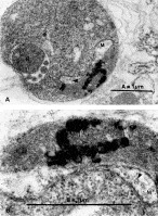
Fig. 1. Transmission electron micrograph of the asexual stage of Plasmodium falciparum (trophozoite stage). A single mitochondrion is observed with a clearly visible double membrane organelle, and an elongated form which is preparing for binary fission (A). A single tubular cristate structure, marked by the arrowhead, is seen in the organelle at higher magnification (B). N, nucleus; M, mitochondrion; P, pigment.
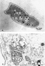
Fig. 2. Transmission electron micrograph of the sexual blood stage of Plasmodium falciparum (gametocyte stage IV). Numerous mitochondria are observed in a single parasite (A). A higher magnification of two organelles containing several cristae (arrowhead) is shown (B). M, mitochondrion; P, pigment.
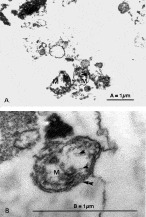
Fig. 3. Transmission electron micrograph of the mitochondrial organelles isolated from sexual blood stages of Plasmodium falciparum. (A) The organelles contain 6–8 tubular cristate structures (arrowhead), as was found in the intact parasites. (B) A higher magnification of an isolated organelle indicate the mitochondrion is still intact with clearly defined double membranes, marked by the double arrowheads. M, mitochondrion.

Fig. 4. A map of the organization of the 6 kb mitochondrial DNA of Plasmodium falciparum. Arrows indicate the direction of transcription. The gene regions cox I, cox III and cyt b encoding cytochrome oxidase subunits I, III and cytochrome b subunit of ubiquinol-cytochrome c reductase, respectively, are shown in shaded boxes. Fragmented large and small subunits rRNA genes are indicated as filled and open boxes respectively.
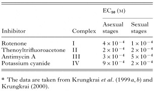
Table 1. The inhibition (EC50 values) of compounds known to affect mitochondrial oxygen consumption in both asexual and sexual blood stages of Plasmodium falciparum*
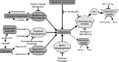
Fig. 5. A diagram of the proposed arrangement of the mitochondrial electron transport components and its association with pyrimidine biosynthesis via dihydroorotate dehydrogenase in the asexual and sexual blood stages of Plasmodium falciparum. The mitochondrial electron transporting complexes are NADH-ubiquinone oxidoreductase (NADH dehydrogenase, a component of complex I), succinate-ubiquinone oxidoreductase (succunate dehydrogenase, complex II), ubiquinol-cytochrome c reductase (cytochrome bc1 or complex III) and cytochrome c oxidase (complex IV). Alternative oxidase as a cyanide (CN−) insensitive-branched pathway (shown in a broken line) and glycerol-3-phosphate (glycerol-3-P) dehydrogenase are also included in the respiratory chain. Ubiquinone and cytochrome c (Cyt c, a component of complex III), are shown serving as electron and proton transferring components. Glycolysis, links to the tricarboxylic acid (TCA) cycle, is operating in the cytosol of P. falciparum. The tree-bars indicate known active sites of inhibitors and the antimalarial drug atvaquone. The diagram is adapted from web site: http://sites.huji.ac.il/malaria.
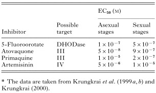
Table 2. The inhibition (EC50 values) of antimalarial drugs affecting the mitochondrial oxygen consumption in both asexual and sexual blood stages of Plasmodium falciparum*

Fig. 6. Mitochondrial tricarboxylic acid (TCA) cycle of Plasmodium falciparum. The TCA cycle is a part of the mitochondrial metabolism shown in Fig. 5, but its function remains to be studied. It is shown here that metabolites produced by the TCA cycle are important for other metabolic pathways. The diagram is taken from web site: http://sites.huji.ac.il/malaria.
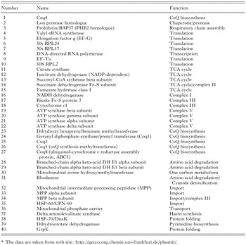
Table 3. List of predicted mitochondrial proteins and biochemical functions*

Fig. 7. Mitochondrial ubiquinone (coenzyme Q) biosynthetic pathway of Plasmodium falciparum. The de novo coenzyme Q synthetic pathway is a part of the mitochondrial metabolism shown in Fig. 5. Major forms occurring in the parasite are coenzyme Q8–9. The parasite cannot salvage coenzyme Q10, an abundant source from the host. It is also shown that metabolites derived by the de novo pathway are used for other metabolic pathways. The diagram is taken from web site: http://sites.huji.ac.il/malaria.
 You have
Access
- 75
- Cited by