A haemodynamically significant patent ductus arteriosus causes left-sided chamber dilation due to left-to-right shunting of blood with increased volume returning to the left atrium and left ventricle. The mitral valve annulus can be stretched by progressive chamber enlargement and sometimes result in mitral valve regurgitation. It is not known whether severe mitral valve regurgitation can resolve solely by closure of the patent ductus arteriosus.
Case report
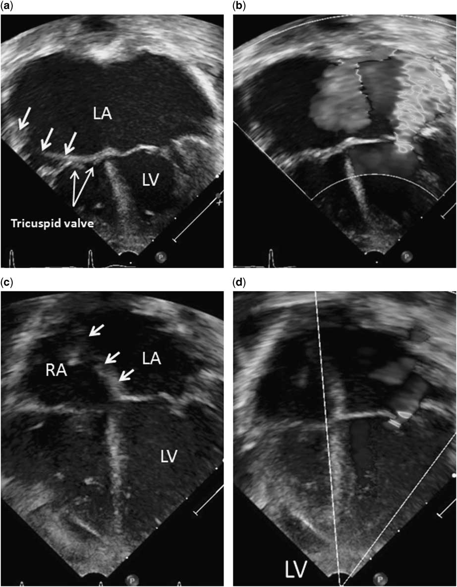
A 5-month-old premature infant, born at 25 weeks of gestation, was transferred from an outside hospital because of worsening respiratory distress. Her weight was 5.1 kg. Her oxygen saturation was 98% on mechanical ventilation with FiO2 50%. Cardiac examination revealed a grade 2/6 continuous murmur at the right upper sternal border and a grade 3/6 systolic murmur at the apex. There were bounding pulses felt at all extremities. Echocardiography showed massive left atrial and left ventricular dilatation with the atrial septum bowing rightward to the level of the tricuspid valve annulus. The mitral valve leaflets did not coapt well, resulting in severe mitral valve regurgitation (Fig 1a and b and Supplementary Video). The mitral valve apparatus was morphologically normal on echocardiography. There was a large patent ductus arteriosus with bidirectional shunt flow. The estimated right ventricular systolic pressure was elevated at 55 mmHg, consistent with pulmonary hypertension from the widely patent ductus arteriosus. Clinically, she had intermittent pulmonary hypertension crises with desaturation. Surgical ligation of the patent ductus arteriosus with mitral valve repair was considered; however, the risk of right heart failure associated with pulmonary hypertension was a considerable concern. The consensus opinion was to assess her pulmonary vascular resistance in the catheterisation laboratory and its response to pulmonary vasodilators and test occlusion of the ductus, with consideration of transcatheter closure of the patent ductus arteriosus.
Transthoracic echocardiography before ( a and b ) and after ( c and d ) closure of the patent ductus arteriosus. ( a and b ) Apical four-chamber view showing severely dilated left atrium (LA) and left ventricle (LV). The atrial septum (3 arrows) is significantly deviated to the right. There is severe mitral valve regurgitation. Mitral valve leaflets do not coapt well because of stretched mitral valve annulus. ( c and d ) 12 months after closure of the patent ductus arteriosus, LA and LV normalise with trace mitral valve regurgitation and reversal of atrial septal deviation (arrow). RA=right atrium.

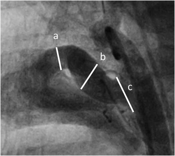
Cardiac catheterisation was performed under general anaesthesia. The baseline haemodynamic data were obtained under FiO2 30% and nitric oxide 5 ppm. Her right ventricular systolic pressure was 42 mmHg (70% systemic), and her mean pulmonary artery pressure was 23 mmHg. Oxygen saturations in the descending aorta and the left ventricle were 89 and 100%, respectively. The Qp:Qs ratio was 0.8, indicating a net right-to-left shunt at the ductal level. Pulmonary and systemic vascular resistances were 3.5 and 4.3 indexed Woods unit, respectively, resulting in a pulmonary/systemic vascular resistance ratio of 0.8. These data indicated severe pulmonary vascular disease at baseline conditions. Under combined 100% oxygen and inhaled nitric oxide 20 ppm, the Qp:Qs ratio increased to 1.9. Although the systemic vascular resistance increased to 5.6 index Woods unit, the pulmonary vascular resistance decreased to 1.7 indexed Woods unit. The pulmonary/systemic vascular resistance ratio dropped to 0.3, indicating the reactivity of pulmonary vasculature. Angiography in the descending aorta showed a large type C patent ductus arteriosus with the ductal ampulla measuring 7.3 mm, the pulmonary artery end 4.3 mm, the largest diameter of the tubular part 5.5 mm, and the ductal length 14.7 mm (Fig 2). After test balloon occlusion of the patent ductus arteriosus was well tolerated, an 8/6-mm Amplatzer duct occluder (St. Jude Medical, Plymouth, MN, United States of America) was deployed in the standard manner using a 6-Fr Amplatzer delivery sheath. After device closure, the mean pulmonary artery pressure was unchanged at 23 mmHg.
Angiographic images in the straight lateral view before the closure of patent ductus arteriosus. A large type C patent ductus arteriosus ((a) 4.3 mm; (b) 5.5 mm; (c) 7.3 mm) is shown.

Her clinical status gradually improved with treatment of the pulmonary hypertension. At 4 weeks after closure of the patent ductus arteriosus, left-sided chamber sizes significantly regressed with only mild mitral valve regurgitation. At the last follow-up at 12 months of age, there was only trace mitral valve regurgitation (Fig 1c and d).
Discussion
This case demonstrates the reversibility of even severe mitral valve regurgitation associated with a large left-to-right shunt, which saved the small infant an operation and mitral valvuloplasty. Chronic left-to-right shunting had progressed to left-sided chamber dilation with a stretched mitral valve annulus, which caused severe mitral valve regurgitation. We hypothesise that her pulmonary vascular disease acutely exacerbated her underlying chronic lung disease and patent ductus arteriosus. Pulmonary vascular resistance was elevated but remained reactive, modified by treatment of pulmonary hypertension. Transcatheter closure of the patent ductus arteriosus was performed and resulted in resolution of the mitral regurgitation without surgical repair. When mitral valve regurgitation is functional, elimination of volume overload by closure of the patent ductus arteriosus allows the mitral valve annulus to re-model and improve coaptation with a reduction in mitral regurgitation.
The haemodynamic changes of patent ductus arteriosus closure can lead to variable responses to ventricular function. Left ventricular systolic function can decrease acutely after closure of the patent ductus arteriosus, despite regression of left ventricular dilation. This dysfunction is usually transient as it tends to improve and normalise over several weeks.Reference Alvarez, Borgia and Guccione 1 – Reference Tilahun and Tefera 3
Treatment of severe pulmonary hypertension is challenging in the presence of a large patent ductus arteriosus. Haemodynamic data under combined oxygen and inhaled nitric oxide testing help identify appropriate candidates for surgery.Reference Balzer, Kort and Day 4 The most reliable criterion for surgery is pulmonary/systemic vascular resistance ratio <0.33 in the combined oxygen and inhaled nitric oxide testing. Balloon test occlusion is a valuable tool to identify patients who can tolerate patent ductus arteriosus closure.Reference Niu, Mallory and Justino 5 Systolic pulmonary artery pressure-to-systolic blood pressure ratio <0.8 is favourable for patent ductus arteriosus closure during balloon test occlusion. The weight of our patient was 5.1 kg. Transcatheter closure of patent ductus arteriosus can be effectively and safely performed even in small infants weighing <6 kg.Reference Dimas, Takao and Ing 6 Furthermore, cardiac catheterisation allows the vasoreactivity testing and balloon test occlusion of patent ductus arteriosus.
In conclusion, transcatheter closure of patent ductus arteriosus is a reasonable first approach for closure of large, hypertensive ductuses, even in the presence of severe mitral valve regurgitation. This may obviate the need for surgical mitral valve repair in such children.
Acknowledgements
We acknowledge that Dr. Thomas J. Forbes provided his expert opinion on this presented case.
Financial Support
This research received no specific grant from any funding agency or from commercial or not-for-profit sectors.
Conflicts of Interest
None.
Ethical Standards
The authors assert that all procedures reported comply with the ethical standards of the Helsinki convention.
Supplementary materials
For supplementary material referred to in this article, please visit http://dx.doi.org/10.1017/S1047951116000998