


Introduction
Acute liver failure (ALF) is defined as an injury resulting in the acute development of hallmark features including jaundice, liver synthetic failure (international normalized ratio [INR] > 1.5) and impairment of mental status. Reference Trey and Davidson1 Current formal diagnostic criterion is accepted as hepatic failure within 26 weeks of acute liver injury, without preexisting disease. Reference Wlodzimirow, Eslami, Abu-Hanna, Nieuwoudt and Chamuleau2,Reference Karvellas, Artru and Coilly3 This definition has been extended to include acute injury in certain chronic liver diseases, including Wilson’s disease, acute Budd-Chiari, autoimmune hepatitis or hepatitis B virus reactivation without cirrhosis. Reference Karvellas, Artru and Coilly3
ALF remains an infrequent but important cause of mortality and morbidity both globally and within North America. Incidence in developed countries is estimated to be 1–6 per million, Reference Bower, Johns, Margolis, Williams and Bell4 accounting for 6% of liver-related deaths Reference Germani, Theocharidou and Adam5 and holding an estimated 30% mortality. Reference Stravitz and Lee6 The most prevalent cause of ALF is drug-induced liver injury and viral infection in developed and developing countries, respectively. Reference Stravitz and Lee6
ALF does not occur as an isolated hepatic injury and should be viewed as a systemic disease, often accompanied by peripheral vasodilation, systemic inflammation and subsequent organ failure. Reference Bernal and Wendon7 The most frequent cause of death in ALF is multiorgan failure due to sepsis, followed by brain herniation due to high intracranial pressure (ICP). Reference Stravitz and Lee6,Reference Bernal, Hall, Karvellas, Auzinger, Sizer and Wendon8 Encephalopathy is a hallmark feature of ALF, indicating the transition from injury to failure. Encephalopathy denotes an altered mental state due to an underlying medical cause. This may occur in ALF due to systemic metabolic disturbances or cerebral edema but can also develop acutely due to hemorrhage or ischemia.
Increased ICP is common in ALF, though the incidence has decreased. In a UK cohort study from 2009 to 2018, the overall incidence of ALF was reported to be 9%, with decreasing incidence over that time period from 13% in 2009 to 3% in 2018. Reference Murphy, El-Dalil and Patel9 Mortality from elevated ICP in ALF patients has similarly decreased, with accompanying improvement in transplant-free survival (TFS). Reference MacDonald, Speiser and Ganger10,Reference Bernal, Hyyrylainen and Gera11 In acetaminophen-related ALF, comparison of groups from 1998–2007 and 2008–2018 demonstrated a decrease in incidence of elevated ICP from 52% to 30% and of mortality from 11.6% to 4.5%. Reference MacDonald, Speiser and Ganger10 Despite the declining incidence of neurological complications, cerebral edema and elevated ICP remain poor prognostic markers in ALF. Reference Bernal, Hyyrylainen and Gera11,Reference Larsen and Wendon12 Clinicians caring for ALF patients must be wary of the development and management of increased ICP in this population.
Though neurological complications and HE are similarly observed in patients with chronic liver disease (cirrhosis), the review will focus on the neurological presentation and management of patients with ALF.
Pathogenesis and Pathophysiology
The skull is a fixed and rigid bony structure that holds three main components: brain tissue (∼80%), CSF (∼10%) and blood (∼10%), commonly referred to as the Monro-Kellie doctrine. Reference Mokri13 Since the total intracranial volume is constant, under physiological conditions, any increase in the volume of one component is compensated by a decrease in one or both others. However, under pathophysiological conditions, when compensatory mechanisms become exhausted, the nonlinear relationship between intracranial volume and ICP explains the risk that a further small increase in volume can cause a disproportionately large rise in ICP.
Brain edema
Brain edema can be induced via cytotoxic or vasogenic mechanisms. An accumulation of brain water resulting from alterations in cellular metabolism, with an intact blood-brain barrier (BBB), is defined as cytotoxic edema. The BBB is a highly selective and regulated physiological multicellular vascular structure that protects the brain from endogenous and exogenous neurotoxins. Composed of endothelial cells with tight intercellular junctions surrounded by a basal lamina, astrocytes and pericytes, molecules (amino acids, metabolites, ions, etc.) are enabled to cross through specific channels and transporters. Vasogenic edema occurs following physical breakdown and/or metabolic dysregulation of the BBB, which causes an increase in molecules and solutes into the brain, followed by brain water uptake. Advanced MRI techniques, such as diffusion-weighted imaging, allow for a noninvasive method to differentiate between vasogenic and cytotoxic brain edema. By calculating the apparent diffusion coefficient (ADC), the diffusivity of water molecules can be quantified. Given that primarily low ADC values were found in patients with ALF, these MRI studies support a cytotoxic origin of brain edema, corresponding to an increase in intracellular water and astrocytic swelling. Reference Rai, Nath, Saraswat, Purwar, Rathore and Gupta14–Reference Saksena, Rai and Saraswat16
Ammonia
Ammonia exists in equilibrium between unionized form (NH3) and ionized form (NH4+), with the equilibrium between the two being defined by the Henderson–Hasselbach equation. At a physiological pH (7.4), 98% of ammonia is present as NH4+ and only 2% as NH3. The gaseous NH3 can cross all plasma membranes through passive diffusion and NH4+ and, due to similar ionic properties to those of K+, can also cross biological membranes through K + channels as well as K + cotransporters. Reference Moser17,Reference Aickin, Deisz and Lux18 Therefore, these properties imply that blood-derived ammonia, both as NH3 and NH4+, can easily cross the BBB. Therefore, conditions of hyperammonemia lead to elevated and toxic levels of ammonia in the brain causing deleterious effects. Reference Bosoi and Rose19 Increased brain ammonia has long been considered to be the main pathogenic factor in hepatic encephalopathy (HE) since ammonia homeostasis is maintained primarily by the liver via the hepatocyte-specific urea cycle. Reference Cooper and Plum20 Within the brain, the astrocyte bears the brunt in attempting to detoxify toxic levels of ammonia since it houses glutamine synthetase, an enzyme that catalyzes the condensation of glutamate and ammonia to form glutamine. Consequently, an accumulation of intracellular astrocytic glutamine leads to osmotic imbalance and cellular edema. Reference Rama Rao, Jayakumar and Norenberg21 For this, elevated brain levels due to ALF-induced hyperammonemia are the primary pathogenic factor involved in the development of brain edema and have been reported to be associated with high ICP. Reference Bernal, Hall, Karvellas, Auzinger, Sizer and Wendon8,Reference Clemmesen, Larsen, Kondrup, Hansen and Ott22,Reference Tofteng, Hauerberg, Hansen, Pedersen, Jørgensen and Larsen23
Lactate
In patients with ALF, it has been shown that increases in extracellular lactate correlate with rises in intracranial pressure. Reference Tofteng and Larsen24 Lactate:pyruvate ratio has been elevated in the extracellular space of patients with ALF, which reflects compromised oxidative metabolism. Reference Bjerring, Hauerberg and Frederiksen25 It has been shown that elevated ammonia can redirect the citric acid cycle, leading to less ATP production and energy impairment and swelling of astrocytes. Reference Drews, Zimmermann and Westhoff26 Interestingly, lactate is not only a product of anaerobic glycolysis but also a metabolite used by neurons as an energetic substrate. Reference Descalzi, Gao, Steinman, Suzuki and Alberini27 An increase in cerebral lactate, due to increased glycolytic activity and/or energy failure, can osmotically induce an increase in water influx in the brain and thus lead to brain edema. Reference Bosoi, Zwingmann and Marin28
Inflammation
Severe systemic inflammation (infection) could also have an impact on the development of high ICP since a systemic inflammatory state accentuates brain inflammation by activating immune cells like microglia and astrocytes, releasing inflammatory mediators (cytokines, reactive oxygen species) that can breach the BBB, increasing BBB permeability, causing impairment in cerebral autoregulation. Reference Rama Rao, Jayakumar and Norenberg21
Clinical Diagnosis of Intracranial Hypertension in ALF
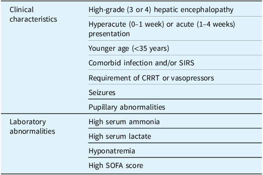
Patients with increased risk of high ICP and cerebral edema include those with hyperacute (0–1 week) and acute (1–4 weeks) presentations, most typically seen in patients with acetaminophen poisoning and hepatitis A/B/E infection. Reference Bernal, Hall, Karvellas, Auzinger, Sizer and Wendon8,Reference O’Grady, Schalm and Williams29–Reference Bernal, Auzinger, Dhawan and Wendon32 Though the risk of elevated ICP is highest in hyperacute liver failure, it otherwise carries the best overall prognosis. Reference Shingina, Mukhtar and Wakim-Fleming33 Increased risk of high ICP is seen in patients under 35 years of age, Reference Bernal, Hall, Karvellas, Auzinger, Sizer and Wendon8 high serum ammonia (>150 μmol/l), Reference Bernal, Hall, Karvellas, Auzinger, Sizer and Wendon8,Reference Clemmesen, Larsen, Kondrup, Hansen and Ott22 high arterial lactate, Reference Bernal, Wang and Maggs34 high Sequential Organ Failure Assessment scores, Reference Kitzberger, Funk and Holzinger35 comorbid infection or systemic inflammatory response syndrome, Reference Rolando, Wade, Davalos, Wendon, Philpott-Howard and Williams36,Reference Vaquero, Polson and Chung37 requirement for continuous renal replacement therapy (CRRT) or vasopressors, Reference Bernal, Hall, Karvellas, Auzinger, Sizer and Wendon8 seizures or pupillary abnormalities Reference Bernal, Hall, Karvellas, Auzinger, Sizer and Wendon8,Reference Stravitz, Kramer and Davern38–Reference Scott, Kronsten, Hughes and Shawcross40 and high-grade HE (West Haven grade 3–4) Reference Bernal, Hall, Karvellas, Auzinger, Sizer and Wendon8,Reference O’Grady, Alexander, Hayllar and Williams30,Reference Blei and Larsen41 (Table 1). The risk of cerebral edema is 25–35% in patients with grade 3 HE and 65–75% in grade 4 HE. Reference Munoz, Moritz, Bell, Northrup, Martin and Radomski42
Features associated with elevated risk of high ICP in acute liver failure patients

*CRRT = continuous renal replacement therapy; SIRS = systemic inflammatory response syndrome; SOFA = Sequential Organ Failure Assessment.
The development of high ICP may rapidly develop into herniation, hydrocephalus, ischemia or hemorrhage. Early diagnosis and treatment of elevated ICP is important in reducing morbidity and mortality. Reference Ware, D’Agostino and Combes43 Management of ALF should include frequent monitoring through frequent clinical assessment and adjunct tools.
Change in neurological status, including abnormal posturing, change in level of consciousness, seizure activity or change in pupillary or vestibulo-ocular reflexes, should evoke suspicion for high ICP. Reference Mohsenin44 Clinical monitoring in isolation is insufficient in detecting high ICP. A Kings College Hospital cohort study conducted in ALF demonstrated that 64% of high-grade (3 or 4) HE patients had increased ICP when tested with invasive monitoring, but only 29% showed clinical signs. Reference Bernal, Hall, Karvellas, Auzinger, Sizer and Wendon8 The same cohort examined patients with a serum ammonia >200 μmol/L, of which 55% of patients developed high ICP. Levels above 150 μmol/L are correlated with increased risk of brain herniation. Reference Clemmesen, Larsen, Kondrup, Hansen and Ott22,Reference Kumar and Sharma45 In the acute care setting, neuroimaging is more technically challenging but remains useful for initial assessment, specifically in detecting ischemia or hemorrhage. Some studies suggest that neuroimaging is insensitive in detecting high ICP and is impractical for continued monitoring. Reference Munoz, Robinson and Northrup46,Reference Wijdicks, Plevak, Rakela and Wiesner47 A cohort study examining the use of CT imaging in ALF patients found that CT scans were performed in 11.5% of patients. Of these patients, 43% had findings consistent with cerebral edema. It was also helpful in identifying patients with ischemia and hemorrhage, identified in 8.6% and 7% of scanned patients, respectively. Reference Cardoso, Lee and Karvellas48 This suggests that CT imaging may have value in the diagnosis of intracranial hemorrhage (ICH) in patients with ALF, in addition to identifying acute complications including ischemia or hemorrhage. The development of high ICP may be predicted with arterial ammonia levels and can be useful in predictive management.
Monitoring Intracranial Pressure in ALF
Monitoring of ICP is important for assessment of underlying edema and can help direct targeted management. There exist both invasive and noninvasive modalities of ICP monitoring. Invasive monitoring measures ICP via catheterization using intraventricular or intraparenchymal placement. Epidural or subdural monitoring may also be seen but is used less frequently. Reference Mehtani, Garg, Kajal, Soni and Premkumar49 It is the gold standard for ICP monitoring and provides accurate real-time measurement of ICP. In practice, it has demonstrated limited clinical benefit and is confounded by an associated risk of 1–10% ICH. Reference Bernal, Auzinger, Sizer and Wendon50–Reference Keays, Alexander and Williams55 The real-time monitoring unique to invasive ICP remains helpful in ALF, where life-threatening increased ICP may develop rapidly without associated clinical signs. Reference Mohsenin44 In contrast, noninvasive techniques have no associated risk for hemorrhage but are less reliable and do not provide continuous measurement. ICP monitoring should be considered in patients with high-grade HE (grade 3–4) or clinical signs or at higher risk of evolving elevation in ICP. Reference Bernal and Wendon7,Reference Stravitz, Kramer and Davern38 The European Association for the Study of Liver (EASL) guidelines recommend consideration of invasive ICP monitoring in patients with elevated risk of high ICP, including patients with hyperacute/acute liver failure, serum ammonia between 150 and 200 μmol/L that is unresponsive to initial renal replacement therapy (RRT) or fluid therapy, evidence of renal impairment or pressor dependence. Reference Bernal, Hall, Karvellas, Auzinger, Sizer and Wendon8,Reference Montagnese, Rautou and Romero-Gómez56 In the absence of demonstrated improved outcomes with the use of invasive monitoring, other guidelines have recommended against the use of invasive ICP monitoring, Reference Nanchal, Subramanian and Karvellas57 or retain an equivocal stance regarding the utility of invasive ICP monitoring. Overall, invasive ICP monitoring may be considered in certain clinical presentations and is best performed in centers experienced in this methodology. This may necessitate early transfer to tertiary centers for observation and careful selection for invasive monitoring as required. ALF patients should always be considered for early transfer to a tertiary care center, ideally with liver transplantation capability. Reference Shingina, Mukhtar and Wakim-Fleming33,Reference Wendon, Cordoba and Dhawan58 This permits expert monitoring and accessibility of clinical tools including invasive or noninvasive ICP monitoring, viscoelastic testing and access to transplant if necessary and available. If transfer is not immediately possible, experts recommend early consultation of specialized centers. Subsequent transfer to a transplant capable center would subsequently be prompted by any evidence of extrahepatic organ dysfunction, including metabolic acidosis, hypoglycemia, elevation of creatinine, oliguria or INR >1.5. Reference Wendon, Cordoba and Dhawan58
Recent trends have demonstrated declining use of invasive ICP monitoring. Reference Murphy, El-Dalil and Patel9,Reference Rabinowich, Wendon, Bernal and Shibolet59 From 2009 to 2018, there was a decrease in the use of invasive ICP monitoring from 20% to 1%. Reference Murphy, El-Dalil and Patel9 There is notable heterogeneity secondary to variability in the healthcare system and center-specific expertise. Reference Gurakar, Conde Amiel and Ozturk60,Reference Rajajee, Fontana, Courey and Patil61 Reduced invasive monitoring may be in part due to the overall declining incidence of high ICP seen in ALF. Reference Murphy, El-Dalil and Patel9,Reference Bernal, Hyyrylainen and Gera11,Reference Karvellas, Fix, Battenhouse, Durkalski, Sanders and Lee53 Additionally, studies examining invasive monitoring techniques have repeatedly demonstrated an absence of survival benefit. Reference Murphy, El-Dalil and Patel9,Reference Vaquero, Fontana and Larson51,Reference Karvellas, Fix, Battenhouse, Durkalski, Sanders and Lee53,Reference Keays, Alexander and Williams55,Reference Lidofsky, Bass and Prager62,Reference Daas, Plevak and Wijdicks63 An observational cohort study examining ALF patients with grade 3–4 HE over a seven-year period noted ICP monitoring in 28% of the cohort, of which only 51% had a measured ICP over 25 mmHg. The use of invasive ICP monitoring did not impact 21-day mortality, though when stratified by the cause of ALF, non-acetaminophen-related ALF patients were found to have increased 21-day mortality with the use of ICP monitoring. Reference Karvellas, Fix, Battenhouse, Durkalski, Sanders and Lee53
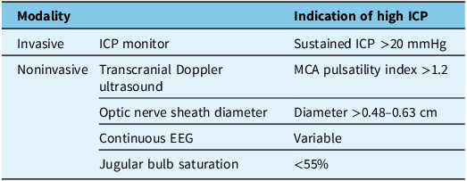
Described noninvasive monitoring techniques include transcranial Doppler (TCD) ultrasound and optic nerve sheath diameter (ONSD) (Table 2). These techniques are typically employed in patients at high risk of bleeding. Reference Sidi and Mahla64
Neuromonitoring of ICP in acute liver failure

EEG = electroencephalography; ICP = intracranial pressure; MCA = middle cerebral artery.
In ALF, cerebral vasculature demonstrates impaired ability to autoregulate, resulting in changes to cerebral perfusion pressure (CPP) and cerebral blood flow (CBF). TCD quantifies CBF velocities and therefore provides indirect evidence of cerebral hypoperfusion and the presence of high ICP. Reference Aggarwal, Brooks, Kang, Linden and Patzer65 The pulsatility index correlates with the downstream vascular resistance, providing an estimate of CPP, which has been correlated with ICP. Reference Bellner, Romner, Reinstrup, Kristiansson, Ryding and Brandt66 However, it remains susceptible to confounding variables, including blood pressure, cerebral autoregulation, body temperature or viscosity. Reference Bershad, Anand and Desantis67 The pulsatility index is calculated as (systolic – diastolic velocity)/mean velocity. A pulsatility index greater than 1.2 in the middle cerebral artery is associated with increased patient mortality risk. Reference Kawakami, Koda and Murawaki68–Reference Picard, Sigaut and Roux70 Drawbacks in the use of TCD include user variability and technical difficulty, limiting usage in up to 20% of patients. Reference Naqvi, Yap, Ahmad and Ghosh71
ONSD reflects the transmission of ICP into the perineural subarachnoid space. Reference Hansen and Helmke72,Reference Hansen and Helmke73 Through ultrasound, the clinician measures the diameter of the optic nerve at a fixed distance posterior to the globe. Reference Rajajee, Vanaman, Fletcher and Jacobs74 ONSD greater than 0.48–0.63 cm has been correlated with high ICP, with measured ICP greater than 20 mmHg. Reference Robba, Santori and Czosnyka75 Elevated ONSD has been correlated with higher grade HE and is found to be higher in non-survivors. Reference Cardoso, Pereira, Moreno, Karvellas and Germano76 However, the validity of ONSD has remained unclear in clinical practice. Reference Robba, Santori and Czosnyka75
Continuous electroencephalography (CEEG) measures cortical electrical activity. This is valuable in detecting the presence of seizures, including subclinical events or status epilepticus. In the context of ALF, CEEG may also reflect the progression of HE. CEEG in ALF may demonstrate slowing marked by low alpha rhythm frequency with bilateral theta activity. These findings may progress to demonstrate theta–delta frequency, triphasic patterns or sleep activity disruption. In severe cases, this may progress to electrographic silence. Reference Ellis, Wendon and Williams77 Structural changes, including ischemia or hemorrhage, may be reflected by the emergence of periodic lateralized epileptiform discharges. The presence of clinical or subclinical seizures, seen in 6–30% of patients with ALF, may result in progression of edema and ischemia. Reference Ellis, Wendon and Williams77–Reference Chen, Farrque, Karakis, Gupta, Rodriguez Ruiz and Kandiah79 Identification and treatment with appropriate anti-seizure medications may prevent or reduce the burden of cerebral edema. Increased ICP results in decreased CBF, which can be reflected with CEEG, with subsequent changes in morphology, amplitude and frequency. Reference Newey, Sarwal and Hantus80,Reference Sanz-García, Pérez-Romero and Pastor81 Electroencephalography can be used to calculate the pressure index, which demonstrates a significant negative correlation with ICP. Reference Chen, Wang, Mao, Dong and Yang82 This may be valuable in demonstrating real-time monitoring of changes in ICP, but further studies are required.
Jugular bulb oxygen saturation is not a measurement of ICP but provides an assessment of CBF autoregulation by providing an approximation of cerebral metabolism and oxygenation. Reference Shawcross and Wendon39,Reference Larsen, Ejlersen, Clemmesen, Kirkegaard and Hansen83 Jugular bulb saturation below 55% is associated with elevated ICP. Reference Bernal and Wendon84 Other noninvasive monitoring techniques include near-infrared spectroscopy, which is not yet validated in ALF. Reference Kandiah and Kumar85
A 2018 study examining noninvasive ICP monitoring examined 41 patients with ALF, of which 27 received invasive ICP monitoring, 23 received optic nerve ultrasound and 21 underwent TCD flow velocity monitoring. In patients with concurrent invasive and noninvasive testing, ICP > 20 mmHg was found to be not reliably detected with ONSD (area under the receiver operator curve [AUROC] 0.59) nor TCD pulsatility index (AUROC 0.55), though TCD calculated from flow velocities was found to be reliable (AUROC 0.90). Reference Rajajee, Williamson, Fontana, Courey and Patil86 However, other studies have noted poor reliability in noninvasive measures to detect high ICP, and therefore, these modalities do warrant further investigation. Reference Rajajee, Williamson, Fontana, Courey and Patil86
Coagulation in ALF
In cases where ICP monitoring is indicated, clinicians are often reluctant to use invasive measures due to the risk of hemorrhage. ALF results in dysfunction in many physiological functions, which are then met by compensatory mechanisms. There is a deficiency of hepatic coagulation factors, which are then compensated with the generation of pro-hemostatic factors, which may lead to relative hypercoagulability. Despite the deficiency in coagulation factors, bleeding events are rare. These episodes are seen in 10% of patients with ALF and are typically gastrointestinal in origin, with ICH only reported in 1% of patients. Reference Stravitz, Ellerbe and Durkalski87 With the low bleeding risk and coexisting relative hypercoagulability, prophylactic transfusion of blood products including fresh frozen plasma (FFP) is unnecessary. Inappropriate use of FFP or other blood products may lead to adverse events including transfusion reactions, thrombosis or death. Reference Shingina, Mukhtar and Wakim-Fleming33
Testing of coagulation in ALF is poorly reflected in standard measures (INR), which is insensitive to natural anticoagulants and only measures procoagulant pathways; therefore, it may not necessarily reflect an imbalance in these factors, and thrombin production may be normal despite abnormalities seen in conventional testing. Reference Lisman, Caldwell and Burroughs88,Reference Tripodi, Salerno and Chantarangkul89 Low fibrinogen levels and dysfibrinogenemia are often seen in ALF, and a functional fibrinogen deficit should trigger the use of substitutes such as cryoprecipitate or fibrinogen concentrate. Platelet count is closely correlated with hemostasis in ALF, and spontaneous bleeding complications are more clearly correlated with platelet count than with INR. Reference Stravitz, Ellerbe and Durkalski87
Given the tumultuous balance between the pro-hemostatic and pro-hemorrhagic states, viscoelastic testing including rotational thromboelastometry (ROTEM) and thromboelastography (TEG) may be a key clinical tool in managing hemostasis in ALF. Viscoelastic testing provides an assessment of both procoagulant and anticoagulant proteins, fibrinogen, platelets and red blood cells, which provides information regarding multiple components, including coagulation cascade activation, clot formation, fibrin formation, clot strength and degree of fibrinolysis. Reference Stravitz90 As opposed to conventional coagulation tests, ROTEM and TEG provide an assessment of overall function and reflect the complex nature of hemostasis in ALF. To mitigate bleeding risk for invasive procedures such as insertion of invasive ICP monitoring tools, it is recommended to pursue viscoelastic testing for guidance in coagulation management, with a target fibrinogen level generally accepted to be above 1.5 g/L. Reference Nadim, Durand and Kellum91
Neuroprotection and High ICP
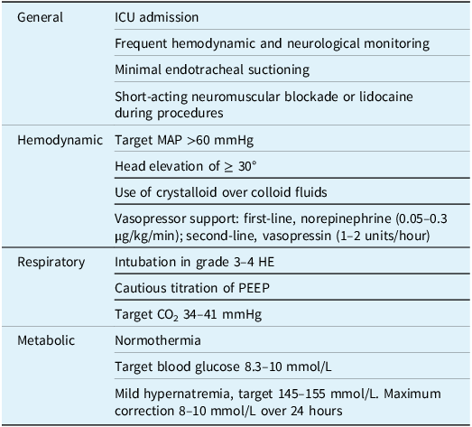
Neuroprotective principles encompass measures taken to protect cerebral function by preventing neuronal loss. This encompasses the core principles of intensive care, with measures taken to maintain CBF, oxygen delivery and metabolic balance (Table 3). Neuroprotection in ALF is of critical importance in reducing mortality and improving long-term neurological outcomes in both patients at risk and with established intracranial hypertension.
Neuroprotective measures in acute liver failure

*HE = hepatic encephalopathy; ICU = intensive care unit; MAP = mean arterial pressure; PEEP = positive end-expiratory pressure.
ALF patients are at high risk for deterioration. Guidelines recommend admission to the intensive care unit for any patient at high risk for decompensation, including patients with West Haven grade 2–4 HE and patients with additional risk factors for the development of high ICP (Table 1). Reference Shingina, Mukhtar and Wakim-Fleming33,Reference Montagnese, Rautou and Romero-Gómez56,Reference Lee, Stravitz and Larson92 The intensive care setting facilitates hemodynamic monitoring and an environment for further neuroprotection to prevent the development of secondary injury or increased ICP. Reference Bernal and Wendon7,Reference Stravitz, Kramer and Davern38
Hemodynamic management
Patients with ALF develop systemic hypotension due to vasodilation. This results in decreased CPP and subsequent secondary injury due to ischemia, but systemic hypertension may result in cerebral edema. Reference Ede, Gimson, Bihari and Williams93 Blood pressure targets are disputed in the literature, with guidelines generally recommending a target mean arterial pressure (MAP) of > 60 mmHg. Reference Shingina, Mukhtar and Wakim-Fleming33,Reference Wendon, Cordoba and Dhawan58 Patients with known elevated ICP have a target MAP of 70–80 mmHg with a central venous pressure < 20 mmHg to permit venous return. Reference Wendon, Cordoba and Dhawan58,Reference Lee, Stravitz and Larson92,Reference Cardoso, Marcelino, Bagulho and Karvellas94–Reference Jörres96 Volume repletion is recommended early. EASL 2017 guidelines indicate little literature to suggest the use of specific fluid but generally recommend the use of crystalloid over colloid fluids. Reference Wendon, Cordoba and Dhawan58 Norepinephrine is the recommended first-line vasopressor after appropriate fluid resuscitation with a starting dose of 0.05 μg/kg/min, with low-dose vasopressin (1–2 units/hour) as a second-line agent, if norepinephrine requirements are greater than 0.2–0.3 μg/kg/min. Reference Shingina, Mukhtar and Wakim-Fleming33,Reference Nanchal, Subramanian and Karvellas57,Reference Wendon, Cordoba and Dhawan58,Reference Dellinger, Rhodes and Evans97,Reference Shi, Hamzaoui, De Vita, Monnet and Teboul98 Venous drainage is facilitated through the elevation of the patient’s head to ≥30°, though elevation beyond 30° may compromise CPP. Reference Davenport, Will and Davison99
Respiratory management
Airway protection is beneficial in cases of decreased level of consciousness. Guidelines recommend intubation in patients with high-grade HE (West Haven grade 3–4). Reference Shingina, Mukhtar and Wakim-Fleming33,Reference Wendon, Cordoba and Dhawan58 EASL guidelines specifically recommend against noninvasive ventilation in patients at risk of HE or significant metabolic disturbance, given the high risk for progression in neurological impairment. Reference Wendon, Cordoba and Dhawan58 Positive end-expiratory pressure (PEEP) can be beneficial in ALF patients with lung injury but consequentially may increase ICP and impair venous return. Reference Nanchal, Subramanian and Karvellas57,Reference Laffey, Bellani and Pham100 PEEP levels should be titrated cautiously, though there is minimal effect on ICP with PEEP < 15. Reference Kandiah and Kumar85 As recommended by EASL, CO2 levels should be targeted between 34 and 41 mmHg to avoid hypercarbia and hypocarbia. Reference Wendon, Cordoba and Dhawan58
Sedation
Noxious and environmental stimuli may also increase intracranial pressure and should be minimized as much as possible. Reference Bernal, Auzinger, Sizer and Wendon50,Reference Lee, Stravitz and Larson92 Procedures such as endotracheal suctioning, which results in elevated intrathoracic pressures, may result in venous outflow obstruction and indirectly increase intracranial pressure; therefore, minimizing these procedures is of additional importance. Reference Lee, Stravitz and Larson92,Reference Citerio, Vascotto, Villa, Celotti and Pesenti101 Short-term neuromuscular blockade or lidocaine during procedures may be useful in minimizing elevated pressures. Sedation may interfere with serial assessment; therefore, continuous sedation is not recommended and, if required, should be achieved with short-acting medications such as propofol or dexmedetomidine. Reference Wendon, Cordoba and Dhawan58 Seizures should be treated, though prophylactic anti-seizure medications do not reduce the seizure incidence or mortality in ALF. Reference Bhatia, Batra and Acharya102
Metabolic management
Nutritional support is recommended in patients with ALF, with preference for enteral nutrition when possible. Reference Shingina, Mukhtar and Wakim-Fleming33,Reference Wendon, Cordoba and Dhawan58 Guidelines recommend the delay of protein support until ammonia levels stabilize, which often occurs after 24 hours. Reference Bischoff, Bernal and Dasarathy103 Hypoglycemia is frequently observed in ALF and may contribute to clinically observed encephalopathy. Guidelines recommend frequent blood glucose monitoring, and supplementation is recommended to a target of 8.3–10 mmol/L. Reference Shingina, Mukhtar and Wakim-Fleming33,Reference Wendon, Cordoba and Dhawan58 Rapid bolus correction should be avoided when possible due to the risk of osmotic shifts resulting in fluid movement into the intravascular and cerebral space, posing risk for edema.
Hyponatremia is a frequent complication of ALF and should be avoided. Treatment with large volume hypotonic fluids used in volume resuscitation may result in hyponatremia and subsequent worsening edema. Reference Bernal and Wendon7,Reference Fernández, Bassegoda, Toapanta and Bernal104 Hypertonic saline (HTS) is often infused to prevent increased ICP. Rapid correction also poses risk of osmotic demyelination; therefore, guidelines suggest maximum correction of 8–10 mmol/L over 24 h. Reference Shingina, Mukhtar and Wakim-Fleming33,Reference Wendon, Cordoba and Dhawan58 Mild hypernatremia is often targeted, with sodium maintained between 145 and 155 mmol/L. Reference Murphy, Auzinger, Bernal and Wendon54,Reference Wendon, Cordoba and Dhawan58
Practice recommendations by the American College of Gastroenterology recommend the use of CRRT in patients with HE meeting West Haven grade 2 or higher. Reference Shingina, Mukhtar and Wakim-Fleming33 This is discussed in further detail below. There is no data to support the use of ammonia-lowering drugs such as lactulose or rifaximin. Reference Stravitz and Lee6 Lactulose should be avoided in patients with high-grade HE due to poor effect and risk of ileus. Reference Fernández, Bassegoda, Toapanta and Bernal104,Reference Maiwall, Kulkarni, Arab and Piano105
Management of Increased Intracranial Pressure
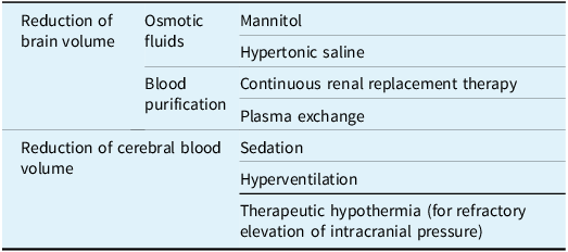
Once high ICP has developed, management targets the reduction of brain volume and cerebral blood volume while maintaining adequate CBF and CPP (Table 4). The results of a large multicenter international survey demonstrated variability in target endpoints. Most centers targeted a CPP of 50–60 mmHg, with others targeting 60–70 mmHg. Within this group, most centers targeted ICP below 20–25 mmHg, while others targeted below 25–30 mmHg when monitored through invasive modalities. Reference Rabinowich, Wendon, Bernal and Shibolet59 Other studies have recommended similar targets. Reference Lee, Stravitz and Larson92,Reference Polson and Lee106
Intracranial pressure management in acute liver failure

Mannitol
Reduction of brain volume is facilitated as a first-line measure with either mannitol or HTS. The subsequent osmotic dehydration shifts fluids into the intravascular space and reduces cerebral edema. Reference Wendon, Cordoba and Dhawan58,Reference Winkler and Munoz-Ruiz107,Reference Suarez108 Mannitol is the most widely accepted first-line therapy in elevated ICP. Reference Rabinowich, Wendon, Bernal and Shibolet59,Reference Lee, Stravitz and Larson92 A controlled trial demonstrated a significant reduction of ICP with the use of mannitol (1 g/kg) in ALF; however, no significant effect was observed in patients with renal failure Reference Canalese, Gimson, Davis, Mellon, Davis and Williams109 or with severe elevation of ICP (> 60 mmHg). Reference Hanid, Davies and Mellon110 EASL 2017 guidelines recommend a bolus of 150 mL of 20% mannitol to be administered over 20 minutes. Reference Wendon, Cordoba and Dhawan58 Serum osmolality should be measured regularly. Reference Garcia-Morales, Cariappa, Parvin, Scott and Diringer111 The administration of mannitol may be repeated 1–2 times if serum osmolality remains below 320 mOsm/L. Reference Lee, Stravitz and Larson92 Repeated use of mannitol may result in rebound ICP elevation, intravascular volume depletion and renal failure. Reference Diringer and Zazulia112,Reference Wakai, Roberts and Schierhout113 Concurrent ultrafiltration may be required to maintain volume status and prevent increased central venous pressure due to expansion of circulating blood volume. Reference Lee, Stravitz and Larson92 The prophylactic administration of mannitol has not been studied and is not recommended. Reference Lee, Stravitz and Larson92
Hypertonic saline
HTS has demonstrated similar efficacy to mannitol. Reference Harutjunyan, Holz, Rieger, Menzel, Grond and Soukup114,Reference Battison, Andrews, Graham and Petty115 In a prospective randomized controlled trial, HTS resulted in a reduction of the incidence of intracranial hypertension and reduced measured ICP. Reference Murphy, Auzinger, Bernal and Wendon54 Certain guidelines recommend prophylactic use of HTS in patients at higher risk of HE. Reference Lee, Stravitz and Larson92 The use of HTS carries risk due to osmotic shifts causing dehydration, hypernatremia and pontine/extrapontine myelinolysis. Reference Suarez108,Reference Qureshi and Suarez116,Reference Martin117 To avert this, serum osmolality should be serially monitored and maintained at< 320 mOsm/L. Reference Suarez108,118 The 2017 EASL guidelines recommend maintenance of serum sodium at 140–145 mmol/L with additional bolus (200 mL of 2.7% HTS or 20 mL of 30% HTS) only when intracranial hypertension has developed. Reference Wendon, Cordoba and Dhawan58 Hyponatremia itself is not a contraindication to the use of HTS in ALF, though clinicians should remain cautious. To reduce the risk of osmotic demyelination, US Acute Liver Failure Study Group guidelines recommend a slow rate of correction, which is inversely proportional to the known preceding duration of hyponatremia. Reference Lee, Stravitz and Larson92 HTS may also be useful in preventing rapid shifts of osmolality in CRRT, preventing subsequent cerebral edema and neurological deterioration. A study by Liotta et al. noted that ALF patients who received concurrent HTS with CRRT were less likely to demonstrate radiographic evidence of cerebral edema or a decrease in osmolality. Reference Liotta, Romanova and Lizza119 Concurrent infusion of HTS may be cautiously applied to patients undergoing CRRT, though infusions should remain gradual to prevent dramatic shifts.
Renal replacement therapy
Early use of CRRT has shown benefit in the management of hyperammonemia, even in the absence of acute kidney injury. Reference Germani, Theocharidou and Adam5,Reference MacDonald, Speiser and Ganger10,Reference Karvellas, Leventhal and Rakela120–Reference Cardoso, Gottfried, Tujios, Olson and Karvellas122 Early use of CRRT in patients with arterial ammonia >150 μmol/L is associated with improved neurological status, reduced mortality and TFS. Reference Cardoso, Gottfried, Tujios, Olson and Karvellas122,Reference Warrillow, Fisher and Bellomo123 There is no clear superior method between continuous venovenous hemodialysis, continuous venovenous hemofiltration or continuous venovenous hemodiafiltration, as long as therapy remains continuous rather than intermittent. Reference Fisher, Baldwin, Fealy, Naorungroj and Bellomo124 Multiple clinical guidelines have suggested the early use of CRRT in hyperammonemia in ALF, even in the absence of standard RRT indications. Reference Shingina, Mukhtar and Wakim-Fleming33,Reference Nanchal, Subramanian and Karvellas57,Reference Wendon, Cordoba and Dhawan58 Intermittent hemodialysis should not be used in ALF as, although ammonia clearance is greater, it may exacerbate high ICP and neural injury due to resulting systemic hypotension and dialysis disequilibrium. Reference Wendon, Cordoba and Dhawan58
Plasma exchange
There is evidence to support the use of plasmapheresis in ALF. A study by Larsen et al. compared high-volume plasmapheresis to standard medical therapy, which demonstrated increased TFS, though there was no difference in ICP or the observed change in ICP across groups. Reference Larsen, Schmidt and Bernsmeier125 A study examining standard volume plasma exchange again not only noted higher TFS but also noted improvement in arterial ammonia and ICP measured noninvasively through ONSD. Reference Maiwall, Bajpai and Singh126 Plasma exchange may be considered in ALF patients with vasoplegia refractory to CRRT, at a standard volume replacement.
Therapeutic hypothermia
The clinical rationale in the use of hypothermia in ALF relies on the suspected physiological benefit due to decreased cerebral metabolic rate, decreased accumulation of brain lactate and intracellular glutamine, normalization of CBF and shift to an anti-inflammatory cytokine environment. Reference Stravitz and Larsen127 There is no evidence of the benefit of therapeutic hypothermia in the prevention of high ICP in ALF. Reference Karvellas, Todd Stravitz, Battenhouse, Lee, Schilsky and Group128,Reference Bernal, Murphy and Brown129 However, there is utility in the use of therapeutic hypothermia in patients with refractory intracranial hypertension. Reference Karvellas, Todd Stravitz, Battenhouse, Lee, Schilsky and Group128,Reference Bernal, Murphy and Brown129 ALF patients with refractory elevation in ICP treated with hypothermia to 32–33 °C core temperature demonstrated reduction in CBF and improved cerebral perfusion. Reference Jalan, O.D., Deutz, Lee and Hayes130 Therapeutic hypothermia remains a recommendation for patients with refractory elevation in ICP, but otherwise, the target core body temperature should remain 35–36 °C. Reference Bernal and Wendon7,Reference Wendon, Cordoba and Dhawan58
Sedation
Propofol is the first-line sedative agent in ALF that has demonstrated benefit in the prevention and control of high ICP in ALF. Reference Wijdicks and Nyberg131 It is commonly used in ALF due to its neuroprotective and short duration of action. Propofol reduces ICP by decreasing neuronal activity, which decreases subsequent metabolic demand. This causes proportionate physiologic decrease in CBF, subsequently reducing cerebral blood volume and reducing ICP. Reference Shawcross and Wendon39,Reference Wijdicks, Plevak, Rakela and Wiesner47,Reference Ravussin, Guinard, Ralley and Thorin132,Reference Hartung133 Propofol is additionally neuroprotective by increasing the seizure threshold. Reference Shawcross and Wendon39 Unlike barbiturates, the pharmacokinetics of propofol are unchanged in ALF, making it a more reliably used sedative agent.
Barbiturates including thiopental and pentobarbital may similarly be used to decrease ICP, as they also decrease cerebral metabolic rate. Reference Forbes, Alexander and O’Grady134,Reference Raghavan and Marik135 The reduction in ICP from the use of barbiturates does not persist, and therefore, this class of medication is used only as a rescue therapy. It is additionally associated with cardiac depression and systemic hypotension, which may decrease cerebral perfusion. Reference Forbes, Alexander and O’Grady134
Hyperventilation
Hyperventilation can cause brief vasoconstriction, briefly improving CBF that has been impaired due to cerebral vasodilation seen in ALF. Reference Strauss, Hansen, Knudsen and Larsen136 However, the consequential vasoconstriction also presents the risk of cerebral ischemia and therefore is recommended as a temporary rescue therapy. Reference Wendon and Lee137,Reference Ellis and Wendon138 Spontaneous hyperventilation generally does not require treatment. Reference Lee, Stravitz and Larson92 Forced hyperventilation is still used as a short-term rescue therapy in patients with evidence of life-threatening elevation of ICP and herniation and, in such a situation, would only be recommended for a limited period. Reference Bernal and Wendon7,Reference Jalan139 EASL 2017 guidelines recommend the use of hyperventilation to a target arterial PaCO2 of 25–30 mmHg only in the case of resistant elevation in ICP. Reference Wendon, Cordoba and Dhawan58 Ventilation targets in ALF should generally remain pCO2 35–40 or to the patient’s prior non-intubated pCO2. Reference Fernández, Bassegoda, Toapanta and Bernal104,Reference Baquerizo, Anselmo and Shackleton140
Transplant
Medical management of intracranial hypertension may act as a bridge to transplant. Reference Stravitz and Lee6,Reference Nanchal, Subramanian and Karvellas57,Reference Kok and Karvellas141 Patients deemed ineligible for transplant should still receive appropriate medical management and may demonstrate spontaneous recovery in the absence of transplant. Reference Bernal, Hyyrylainen and Gera11,Reference Rajajee, Fontana, Courey and Patil61 In practice, the presence of cerebral edema does not preclude patients from undergoing a liver transplant. Patients with cerebral edema identified on CT imaging have demonstrated similar transplant selection rates to those without evidence of cerebral edema. Reference Cardoso, Lee and Karvellas48 Intensive care and early transplant are important variables in patient outcome. Complications of cerebral edema remain one of the leading causes of mortality in patients on the waitlist for liver transplant. Reference Karvellas, Leventhal and Rakela120 Guidelines suggest that a transplant may be futile in patients with irreversible brain injury, including absent cerebrovascular blood flow, cerebral herniation or fixed nonreactive pupils. Reference Fernández, Bassegoda, Toapanta and Bernal104
Conclusion
Increased ICP is frequently seen in the setting of ALF. Early diagnosis and management of high ICP are critical, and there are a variety of invasive and noninvasive monitoring tools available. Invasive ICP monitoring has historically been limited by the risk of hemorrhage due to suspected coagulopathy in ALF. Viscoelastic testing provides a comprehensive measurement of hemostasis in ALF and may be used to guide decisions regarding invasive monitoring.
Management of high ICP includes strategies to reduce cerebral volume and cerebral metabolic rate. Implementation of these strategies is often stepwise, requiring continued monitoring of ICP.
Author contributions
NP drafted the manuscript and significantly revised the manuscript. CFR significantly revised the final manuscript. CJK conceived the idea for the review and significantly revised the final manuscript. All authors approve the final manuscript.
Funding statement
None.
Competing interests
NP reports no disclosures. CFR reports receiving consulting fees from Genfit, Merz Therapeutics, Norgine, Alfasigma, Intercept, Gilead Sciences, AbbVie, Ipsen, Falk Foundation e.V., CSL Behring and Boehringer Ingelheim. CFR reports receiving research support from Genfit, Satellite Bio. C.L., Merz Therapeutics, Norgine and Schwiete Foundation. CJK reports receiving consulting fees from Genfit, Vantive, Mallinckrodt Pharmaceuticals, Morphocell Inc. and DSMB committee service for Miromatrix.

 Open access

