



LEARNING OUTCOMES
After reading this article you will be able to:
-
• review the pathophysiology of psychosis and TRS
-
• describe current neuroimaging findings in relation to TRS
-
• recognise how neuroimaging can assist in further understanding TRS, with focus on earlier diagnosis and optimisation of treatment.
Psychosis is characterised by a significant impairment in an individual’s perception of reality and commonly presents with features such as hallucinations, delusions and disorganised thought processes. It may occur in the context of primary psychiatric disorders, such as schizophrenia and bipolar affective disorder, or as a secondary consequence of substance use or underlying medical conditions.
Schizophrenia encompasses a spectrum of subtypes, including treatment-resistant schizophrenia (TRS), which affects approximately 20–30% of individuals diagnosed with the disorder and is associated with a markedly poorer prognosis, including cognitive and social impairments, as well as a heightened risk of death by suicide (Parkes Reference Parkes, Mantell and Oloyede2022). TRS is defined by an inadequate response to at least two different antipsychotic medications, each prescribed at therapeutic doses for an adequate duration and with confirmed adherence (Correll Reference Correll and Howes2021). Clozapine, an atypical antipsychotic, is widely regarded as the gold-standard treatment for TRS. However, its use remains limited, primarily because of concerns about adverse drug reactions and the need for regular plasma level monitoring.
Although the exact pathophysiology of TRS remains unclear, evidence suggests that early onset of psychotic symptoms is linked to a higher risk of developing the condition (Smart Reference Smart, Kępińska and Murray2019). In light of this, the INTEGRATE (International Guidelines for the Algorithmic Treatment of Schizophrenia) study advocates for the early identification of antipsychotic non-response and timely initiation of clozapine therapy (McCutcheon Reference McCutcheon, Pillinger and Varvari2025). Complementary efforts have also focused on improving the timely and accurate diagnosis of TRS. The TRRIP criteria (Time, Resistance, Refractoriness, Inadequate improvement, Pharmacological factors) offers a structured diagnostic framework (Box 1). Adherence to this framework can support accurate diagnosis and effective management, reduce unnecessary medication trials and minimise variability in clinical approaches (Howes Reference Howes, McCutcheon and Agid2017).
The TRRIP framework for identifying treatment-resistant schizophrenia

A summary of pathophysiology: underpinning mechanism(s)
The development and maintenance of psychosis is a complex and multifactorial process incorporating several biological mechanisms. There are several hypotheses postulating the aetiopathogenesis of schizophrenia, including TRS. These are reviewed below.
The dopamine hypothesis
Perhaps the most well-known model, the dopamine (or monoamine) hypothesis, is considered a key configuration for understanding schizophrenia. This hypothesis suggests that an aberration of dopamine transmission within some of the key primary dopaminergic pathways contributes to the emergence of psychotic features. In the mesolimbic pathway, there is evidence of dopaminergic overactivity – due to dysregulation of striatal dopamine synthesis, release or receptor sensitivity. Overactivity within this pathway leads to the emergence of the ‘classic’ positive features of psychosis, such as hallucinations and delusions. Underpinning this are atypical salience mechanisms, which appear to place undue emphasis on irrelevant stimuli, bringing new meaning and contributing to the alternative, subjective reality of those experiencing psychosis (Howes Reference Howes and Kapur2009). In contrast to the hyperactivity of the mesolimbic pathway, the mesocortical pathway is subject to an underactivity of dopamine. This contributes to the development of negative and cognitive features of schizophrenia.
The widespread functions facilitated by these two dysfunctional dopaminergic pathways is one of the possible reasons for the heterogeneous presentation of psychosis and schizophrenia seen in clinical practice (Howes Reference Howes and Kapur2009). It is noteworthy that evidence of dopaminergic dysfunction, as proven via neuroimaging using 18F-fluorodopa (F-DOPA) positron emission tomography (PET), is seen in both established illness and first-episode psychosis (Jauhar Reference Jauhar, Veronese and Nour2019). However, although there is much focus on dopaminergic activity, there is evidence that dopaminergic dysfunction may be more characteristic of those who are treatment responsive, compared with those who are resistant to conventional treatment approaches (Vanes Reference Vanes, Mouchlianitis and Collier2018a). There is accumulating evidence indicating that, in TRS, striatal dopaminergic activity appears unremarkable, whereas glutamate levels in the anterior cingulate cortex (ACC) are elevated (Demjaha Reference Demjaha, Egerton and Murray2014). Increased glutamate metabolites in the ACC have been linked to poor response to dopaminergic agents (Egerton Reference Egerton, Griffiths and Casetta2023), suggesting that glutamatergic dysfunction may underlie resistance to conventional dopaminergic therapies. This observation challenges the classic dopamine hypothesis in TRS, instead implicating broader neurotransmitter imbalances – particularly involving glutamate and gamma-aminobutyric acid (GABA) – in its pathophysiology.
Such findings regarding neurotransmitter imbalances align with the developmental risk factor model, which posits that cumulative early life insults, including prenatal stress, aberrant synaptic pruning, and genetic or environmental perturbations, disrupt neurotransmitter homeostasis beyond dopaminergic systems, thereby contributing to the emergence of treatment-resistant symptomatology (Murray Reference Murray, Bhavsar and Tripoli2017). These early neurodevelopmental disturbances, which impair the maturation of the prefrontal cortex (PFC) and hippocampi, contribute to deficits in cognitive and emotion regulation. Supporting this, neuroimaging studies consistently report structural abnormalities, such as enlargement of the lateral ventricles and pallidum when compared to healthy controls (van Erp Reference van Erp, Walton and Hibar2018). These components of the corticolimbic circuit can be examined at structural, functional and molecular levels to yield insight into schizophrenia’s neurobiology and inform novel experimental interventions (Gee Reference Gee, Dazzan and Grace2025). Overall, converging evidence supports conceptualising TRS as a mechanistically distinct subtype, warranting the exploration of glutamatergic and other non-dopaminergic treatment strategies (Table 1).
Dopaminergic pathways – origin, terminus and functions

The glutamatergic–GABAergic hypothesis
A less widely known, but increasingly studied, framework for understanding schizophrenia is the glutamate hypothesis, which posits that dysfunction in the glutamatergic system plays a central role in the pathophysiology of psychosis, particularly in the context of TRS. This hypothesis highlights an imbalance between excitatory glutamatergic and inhibitory GABAergic neurotransmission as a potential mechanism underlying the disorder. At the core of this model is the proposed hypofunction of N-methyl-d-aspartate (NMDA) receptors, which are essential for synaptic plasticity, learning and neurodevelopment. It is theorised that impaired NMDA receptor activity, particularly due to deficient glutamatergic signalling, results in an excitation/inhibition (E/I) imbalance, leading to widespread downstream neural disruptions. A key consequence of this disruption involves the dysregulation of GABAergic parvalbumin-expressing interneurons, which are critical for maintaining cortical E/I balance. Their dysfunction has been linked to abnormal activity in the hippocampus and PFC, regions strongly implicated in the manifestation of the positive, negative and cognitive symptoms of psychosis, including TRS (Gee Reference Gee, Dazzan and Grace2025).
Empirical support for the glutamate hypothesis includes observational studies in which administration of NMDA receptor antagonists (such as phencyclidine or ketamine) induce psychosis-like symptoms in healthy individuals. Rather than questioning the dopamine hypothesis, the glutamate model may complement it, suggesting an integrated neurochemical framework in which glutamatergic dysfunction contributes to dopaminergic dysregulation, which may represent a final common pathway for psychosis (Howes Reference Howes and Kapur2009). Despite the growing body of evidence, pharmacological interventions targeting the glutamatergic system have thus far yielded inconsistent clinical efficacy (Begni Reference Begni, Marchesin and Riva2025). Nonetheless, glutamate-modulating agents, such as D-serine, may be a promising area of therapeutic development. These compounds aim to restore NMDA receptor functionality and potentially alleviate the negative and cognitive symptoms of schizophrenia.
Neuroimaging research has contributed valuable insights to this hypothesis. Early PET studies reported reduced NMDA receptor binding in individuals with schizophrenia, although subsequent work using alternative glutamatergic tracers has produced mixed results. More recently, proton magnetic resonance spectroscopy (1H-MRS), a neuroimaging technique that quantifies in vivo brain metabolites using 3T magnetic resonance imaging (MRI), found no significant difference in occipital cortex glutamate levels between individuals with ultra-TRS and healthy controls (Lopes Reference Lopes, Rae and Meyer2025), although further 1H-MRS studies have identified reduced PFC GABA and glutamate levels in those with schizophrenia (Gee Reference Gee, Dazzan and Grace2025). However, Egerton et al (Reference Egerton, Griffiths and Casetta2023) have demonstrated that TRS, identifiable via poor response to conventional antipsychotic drugs, is associated with elevated glutamate metabolites in the ACC.
It may be argued that targeted glutamatergic treatments are beneficial for those with TRS by engaging non-dopaminergic pathways, especially given that TRS is defined by a lack of response to conventional dopaminergic treatments. However, optimism has been tempered by recent findings from Boehringer Ingelheim’s Phase III CONNEX trial, which investigated iclepertin, a selective α7 nicotinic acetylcholine receptor agonist, for cognitive enhancement in schizophrenia. The trial was terminated prematurely after primary and secondary trial end-points were not met (Boehringer Ingelheim 2025).
Cholinergic hypothesis
The cholinergic hypothesis centres on dysregulation of the cholinergic system being integral to the development and maintenance of psychosis. This is reportedly driven by dysregulation of both muscarinic acetylcholine receptors (mAChRs) and nicotinic acetylcholine receptors. The hypothesis, which is not new, has come to the fore over recent decades owing to the evidence that mAChR agonists such as xanomeline appear to reduce the clinical features of psychosis. This is considered promising, particularly given the impact of xanomeline on negative and cognitive features, which are often difficult to treat with standard dopaminergic agents (Begni Reference Begni, Marchesin and Riva2025). This has led to the development of a xanomeline–trospium chloride combination, licensed under the trade name Conbenfy by the Food and Drug Administration (FDA) in the USA. Xanomeline–trospium chloride has a novel mechanism as a combined agent. Xanomeline offers central M1 and M4 muscarinic receptor agonism, whereas trospium chloride provides muscarinic antagonism, primarily in peripheral tissues. Trospium chloride therefore prevents the peripheral adverse effects of xanomeline. This is possible owing to its hydrophilicity, which prevents penetration across the blood–brain barrier while permitting activity in peripheral tissues (Smith Reference Smith, Augustine and Dorrough2024).
This drug represents enhanced clinical options for the 20–30% of people with schizophrenia who are resistant to the mainstay options of dopamine-based medications (Nair Reference Nair, Shergill and Whiskey2025). Its approval by the FDA is a significant milestone as it introduces a non-dopaminergic pharmacological treatment for psychosis and schizophrenia. In addition, given its non-dopaminergic mechanism, xanomeline–trospium chloride is not associated with the archetypical adverse drug reactions often associated with treating psychosis and schizophrenia, such as extrapyramidal side-effects, metabolic syndrome and hormonal abnormalities (Smith Reference Smith, Augustine and Dorrough2024). The promise of xanomeline–trospium is leading the way for other non-dopaminergic medications, which are currently in development (Begni Reference Begni, Marchesin and Riva2025; McCutcheon Reference McCutcheon, Pillinger and Varvari2025). This may be of particular benefit for those with TRS, who, as a criterion for the diagnosis, have not responded adequately to dopaminergic interventions previously.
Why TRS?
As mentioned above, TRS can be diagnosed when there is evidence of no response or suboptimal response to at least two types of antipsychotic medication; this diagnostic process is aided by the TRRIP criteria. Although the majority of people with schizophrenia do respond to commonly used antipsychotic agents, a significant proportion do not. TRS is evidenced to be associated with poorer outcomes, longer-term disability and a higher risk of relapse and death by suicide (Parkes Reference Parkes, Mantell and Oloyede2022). Although treatment responsive schizophrenia involves striatal dopaminergic hyperactivity, TRS has a distinct neurobiological signature. This model can be characterised by ACC glutamatergic dysregulation, aberrant synaptic plasticity pathways, hippocampal–striatal circuitry disruption and altered neurodevelopmental signalling. These findings appear to support a dual-pathway model, in which TRS reflects a non-dopaminergic subtype of illness. In addition, TRS may – in turn – be further categorised into primary and secondary TRS. Primary TRS presents with treatment resistance from the outset, whereas secondary TRS shows early response to antipsychotic treatment, yet this reduces over time to become resistant (Correll Reference Correll and Howes2021).
It has been proposed that other subtypes of TRS may also occur, including one stemming from dopamine supersensitivity (hypersensitivity) (Suzuki Reference Suzuki, Kanahara and Yamanaka2015). This proposal posits that chronic antipsychotic use or underlying pathology sensitises dopamine D2 receptors, leading to psychotic relapse and treatment resistance. Suzuki et al (Reference Suzuki, Kanahara and Yamanaka2015) propose that approximately 70% of those with TRS experience dopamine supersensitivity. The dopamine hypersensitivity hypothesis remains largely speculative, with more consistent data supporting presynaptic dopamine dysregulation and broader neurobiological mechanisms (including glutamate, inflammation, and connectivity influences).
Xanomeline–trospium chloride, as a non-dopaminergic option, may offer distinct advantage to individuals hypersensitive to dopamine (Smith Reference Smith, Augustine and Dorrough2024). However, in the UK, clozapine is currently the only licensed treatment for TRS. Clozapine acts primarily by antagonising serotonergic (5-HT2A) and dopaminergic receptors, but also interacts with muscarinic acetylcholine, glutamatergic, alpha-adrenergic and histaminergic receptors. Its high affinity for 5-HT2A receptors potentially contributes to its efficacy in TRS, with some evidence suggesting that its affinity for D4 receptors may also play a role.
Clozapine is superior to other antipsychotics in reducing positive symptoms, relapse rates and the need for inpatient care; however, its use is often delayed because of the need for regular monitoring and the risk of serious adverse effects, such as blood dyscrasias (including agranulocytosis), seizures and myocarditis (Parkes Reference Parkes, Mantell and Oloyede2022). The underutilisation of clozapine often leads to significant delays, often years, before the medication is initiated (Parkes Reference Parkes, Mantell and Oloyede2022). This underutilisation may be compounded by regional variations in practice (Whiskey Reference Whiskey, Barnard and Oloyede2021). The INTEGRATE study recommends prompt use of clozapine to minimise this delay, alongside lifestyle interventions to reduce the cardiometabolic side-effects (McCutcheon Reference McCutcheon, Pillinger and Varvari2025). Despite the potential risks associated with clozapine therapy, gradual titration and careful monitoring can lead to positive outcomes and improved patient satisfaction (Krivoy Reference Krivoy, Joyce and Tracy2019, Reference Krivoy, Whiskey and Webb-Wilson2021; Correll Reference Correll and Howes2021; Parkes Reference Parkes, Mantell and Oloyede2022).
Although clozapine has proven effectiveness, there is evidence of clozapine-resistant schizophrenia in one-third to half of people treated with the drug: this may indicate a distinct neurobiological subtype of TRS, potentially linked to hypofrontality (Pang Reference Pang, Chun and Wong2023).
Imaging in psychosis
Neuroimaging in schizophrenia has various uses, for example in diagnostic processes, treatment response measurement, research and predictive value (Papanastasiou Reference Papanastasiou and Shergill2021). The diagnostic potential involves the identification of both structural and functional brain abnormalities. Individuals with schizophrenia demonstrate a thinning of the cortex, with reduced cortical surface area, particularly in the frontotemporal regions (van Erp Reference van Erp, Walton and Hibar2018; Barry Reference Barry, Vanes and Andrews2019) Additionally, there is evidence of reduced hippocampal volume and density, when compared with the general population (Nelson Reference Nelson, Kraguljac and Maximo2022). Other notable aberrations include reduced volume in various elements of the limbic system, including the amygdaloid complex and thalami, as well as enlarged lateral ventricles (van Erp Reference van Erp, Hibar and Rasmussen2016). Further evidence elicited from MRI studies reveals that those with marked cognitive disturbances have shown reduced volume in prefrontal, parietal and perisylvian areas; these features appear associated with worsened prognosis at 2-year follow-up (Koutsouleris Reference Koutsouleris, Buciuman and Neuner2025). It is noteworthy that these findings are not replicated in those considered at ultra-high risk of developing psychosis but who did not show cognitive disturbances (Koutsouleris Reference Koutsouleris, Buciuman and Neuner2025).
When employed to measure treatment response, neuroimaging can be used to explore the mechanisms of action of various medications and to track changes in brain structure and function over the course of treatment. In research, neuroimaging contributes to exploring the pathophysiology of psychosis and the identification of novel treatment options. The predictive value of neuroimaging aids estimation of prognosis and our understanding of associations between psychotic symptoms and impairment in specific neural regions (Keshavan Reference Keshavan, Collin and Guimond2020; Pines Reference Pines, Frandsen and Drew2025). Moreover, evidence of elevated glutamate metabolites in the ACC may serve as a prognostic indicator of TRS. The emerging field of ultra-low-field MRI (ULF-MRI) shows promise in advancing these efforts, with enhanced affordability and accessibility, although it is currently limited to structural studies (Cooper Reference Cooper, Hayes and Corcoran2024).
Although neuroimaging in TRS seeks to elucidate the underlying neural network dysfunctions and associated pathophysiology, the benefits of neuroimaging may remain largely within the realm of research at present. For most practising clinicians, these developments have limited immediate clinical relevance, as treatment decisions continue to rely primarily on phenomenological assessment and pragmatic pharmacological strategies. However, strengthening the evidence base is crucial to enable eventual translation into clinical practice.
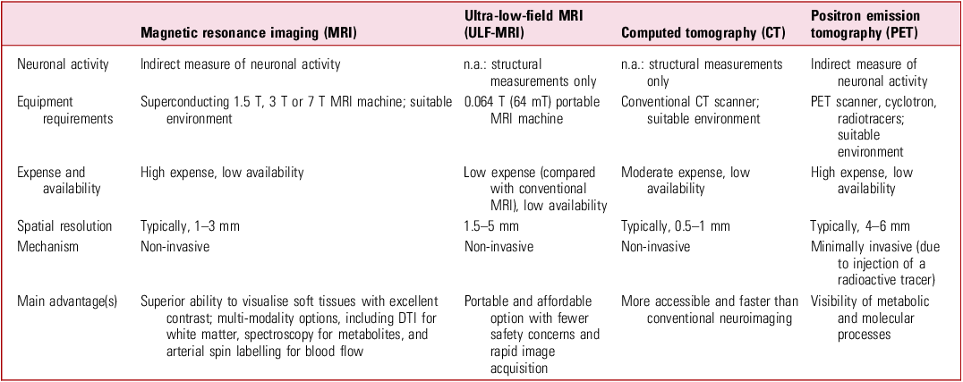
Table 2 compares the capabilities of various forms of neuroimaging.
An overview of brain neuroimaging capabilities

n.a., not applicable; DTI, diffusion tensor imaging.
Overview of neuroimaging findings
Advanced imaging techniques, including MRI and PET, provide valuable insights into the brain changes that underlie schizophrenia. Structural MRI studies have shown widespread grey matter reductions in schizophrenia, particularly in the prefrontal cortex, temporal lobes and hippocampi – areas implicated in memory, emotion regulation and cognitive processes such as language. Although grey matter is affected, white matter appears more resilient to these changes, with some evidence of plasticity that may mitigate cognitive decline to a degree (Emsley Reference Emsley, du Plessis and Phahladira2023). Longitudinal meta-analyses indicate that grey matter atrophy is progressive, but particularly noticeable in the early stages of illness, emphasising the importance of reducing the duration of untreated psychosis (Yang Reference Yang, Jing and Naici2021). The left hemispheric structures are primarily affected by this atrophy. It has been argued that many of the abnormalities identified in chronic illness are also present in those experiencing a first episode of psychosis; this could indicate that this neurobiological basis is a primary pathological feature rather than a consequence secondary to chronic illness or the impact of treatment. However, exposure to antipsychotic drugs is an important consideration, with higher cumulative exposure linked to greater grey matter atrophy (Yang Reference Yang, Jing and Naici2021). Clozapine therapy, employed in the management of TRS, is demonstrated to be implicated in decreases of grey matter volume in a multitude of areas, including frontotemporal regions, parietal and occipital cortices, the striatum and cerebellum (Sun Reference Sun, Zelaya and Sendt2024).
Furthermore, there is evidence that the detection of lesions in the hippocampal areas may precipitate psychotic features that would not suitably respond to dopaminergic antipsychotic medications (Pines Reference Pines, Frandsen and Drew2025). Identification of these lesions (which affect functional circuitry on the posterior hippocampal–subicular axis) via neuroimaging can offer potential for non-dopaminergic therapeutics by way of neuromodulatory interventions such as transcranial magnetic stimulation (Pines Reference Pines, Frandsen and Drew2025).
Functional neuroimaging has advanced our understanding of the neural mechanisms underlying schizophrenia. There is considerable evidence of hypofrontality – reduced perfusion of the parenchyma – in the PFC, as well as altered metabolism in the frontal cortex, with deficits of cognitive control and executive functioning (Townsend Reference Townsend, Pillinger and Selvaggi2023). As mentioned above, F-DOPA is a radiotracer utilised in PET imaging; F-DOPA studies have revealed that striatal dopamine is higher in those with psychosis, even during the first episode of illness, compared with controls; moreover, imaging has also shown that elevated dopamine synthesis is associated with responsiveness to standard antipsychotic drugs (Jauhar Reference Jauhar, Veronese and Nour2019). This may be a potential biomarker in future practice; however, the aberration of dopamine activity is heterogeneous, outlining the need for a bespoke, personalised approach (Giacomel Reference Giacomel, Martins and Nordio2025).
Neuroimaging in TRS
Neuroimaging has emerged as a valuable tool in the investigation and diagnosis of TRS, offering the ability to identify both structural and functional abnormalities through advanced neuroradiological techniques (Vanes Reference Vanes, Mouchlianitis and Collier2018a, Reference Vanes, Mouchlianitis and Wood2018b; Mouchlianitis Reference Mouchlianitis, Vanes and Tracy2023). Modalities such as functional magnetic resonance imaging (fMRI) and PET have provided critical insights into dopaminergic and glutamatergic dysfunctions, thereby informing targeted therapeutic strategies and enhancing our understanding of the neurobiological mechanisms underlying resistance to conventional antipsychotic treatments.
PET and single-photon emission computed tomography (SPECT) studies have demonstrated reduced cerebral blood flow (CBF) in individuals with TRS relative to healthy controls. This reduction in CBF extends beyond the traditional concept of hypofrontality, encompassing not only the PFC but also parietal and subcortical regions (Sun Reference Sun, Zelaya and Sendt2024). Furthermore, clozapine therapy appears to exacerbate this phenomenon, with additional reductions in CBF observed specifically in the ACC (Sun Reference Sun, Zelaya and Sendt2024). Intriguingly, elevated baseline CBF in subcortical structures – particularly the striatum and hippocampi – has been associated with a more favourable response to clozapine treatment (Sun Reference Sun, Zelaya and Sendt2024). Resting-state fMRI studies have also revealed disrupted connectivity patterns in TRS, including reduced functional connectivity between the ventral striatum and the substantia nigra (White Reference White, Wigton and Joyce2016), and cognitive activation studies have shown differential cognitive control in TRS (Horne Reference Horne, Vanes and Verneuil2021). Structural neuroimaging findings further support this altered neurobiology (Vanes Reference Vanes, Mouchlianitis and Wood2018b), with evidence of reduced cortical mantle thickness in several regions, notably the left hemispheric temporal cortex and bilateral cingulate cortices, when comparing individuals with TRS with those who have adequately responded to standard antipsychotic therapies (Barry Reference Barry, Vanes and Andrews2019).
Collectively, these findings underscore the potential of neuroimaging to inform a more individualised, precision medicine approach to the diagnosis and management of TRS, with potential core pathognomic features observed in TRS but not in typical treatment-responsive schizophrenia (Sun Reference Sun, Zelaya and Sendt2024; Giacomel Reference Giacomel, Martins and Nordio2025).
Conclusions
TRS continues to pose a significant clinical challenge, characterised by persistent symptoms, functional impairment and poor long-term outcomes. Despite advances in antipsychotic therapy, a substantial proportion of individuals remain unresponsive to standard dopaminergic and psychosocial interventions. This review has highlighted the complex and multifactorial pathophysiology of TRS, involving dopaminergic, glutamatergic, cholinergic and neurodevelopmental mechanisms, with probable interplay. Additionally, findings such as impaired CBF and disrupted connectivity between the ventral striatum and substantia nigra may be considered potential pathognomonic markers of TRS.
Further fractionation of the schizophrenia spectrum, informed by contemporary neuroimaging, may offer a valuable shift in how TRS is identified and managed. In particular, glutamatergic dysfunction, specifically elevated glutamate metabolite levels in the ACC, is emerging as a distinguishing feature. Neuroimaging could help refine diagnostic criteria and facilitate earlier, more targeted interventions.
The development of non-dopaminergic treatments, such as xanomeline–trospium chloride, is a promising step towards more personalised and mechanistically targeted therapies. These alternatives may benefit individuals who respond poorly to conventional dopaminergic medications or experience significant adverse effects. Diagnostic frameworks like TRRIP and the INTEGRATE algorithm provide a foundation for earlier recognition of TRS, timely initiation of clozapine and incorporation of neuroimaging into routine assessment.
Multimodal imaging techniques offer valuable insights into the structural, functional and molecular underpinnings of TRS. When combined with pharmacological data, these tools can enhance understanding of how specific molecular targets influence broader brain circuitry (Gee Reference Gee, Dazzan and Grace2025). Functional neuroimaging methods, such as PET and MRI, have expanded our knowledge of TRS and its neurobiological basis; this approach offers considerable potential for guiding precision medicine approaches (Orlov Reference Orlov, O’Daly and Tracy2017). Notably, some neuroimaging findings appear specific to TRS and are absent in treatment-responsive schizophrenia, potentially strengthening the case for including neuroimaging in the initial evaluation of psychosis. Moreover, recent developments of ULF-MRI offer potential to improve clinical practice through improved accessibility, lower cost and ease of use, although it should be noted that this technology cannot currently provide functional utility.
Future research should aim to refine biomarkers, broaden therapeutic targets beyond the dopaminergic system and integrate neuroimaging into both diagnostic and treatment paradigms. By adopting a multifaceted, neurobiologically informed approach, clinicians and researchers can work towards reducing the burden of TRS and improving outcomes for affected individuals.
MCQs
Select the single best option for each question stem
-
1 Which of the following best describes the purpose of the TRRIP framework in the management of TRS?
-
a To aid accurate diagnosis of TRS and reduce unnecessary medication prescribing
-
b To aid selection of first-line antipsychotic medications in schizophrenia
-
c To identify individuals who are likely to develop psychosis in adolescence
-
d To provide a stepwise approach for medication augmentation in the management of TRS
-
e To provide a structured method for early psychosocial therapies.
-
-
2 Which neuroimaging finding is most consistently associated with TRS?
-
a Elevated glutamate metabolites in the anterior cingulate cortex
-
b Enlarged hippocampal volume compared with healthy controls
-
c Increased cortical thickness in the prefrontal cortex
-
d Increased dopaminergic activity in the ventral striatum
-
e Reduced grey matter volume in the insular cortex.
-
-
3 How might the current literature best summarise TRS?
-
a TRS is defined by treatment failure with clozapine and typically resolves with long-acting injectable treatment
-
b TRS primarily reflects progressive dopaminergic hyperactivity and is typically responsive to higher-dose D2 antagonists
-
c TRS is a neurobiologically distinct subtype of schizophrenia, marked by persistent symptoms despite adequate trials of at least two antipsychotics
-
d TRS is best explained by excessive synaptic pruning and reduced hippocampal volume, with dopaminergic dysfunction only occasionally observed
-
e TRS is diagnosis only after neuroimaging confirmation of elevated glutamate in the ACC.
-
-
4 Resting-state fMRI studies in individuals with TRS most commonly reveal which of the following findings?
-
a Elevated occipital cortex GABA concentrations
-
b Hyperactivity in the mesolimbic pathway
-
c Increased connectivity between the hippocampus and amygdaloid complex
-
d Reduced connectivity between the ventral striatum and substantia nigra
-
e Reduced default mode network coherence with preserved salience network connectivity.
-
-
5 Which of the following statements best highlights a key limitation of the dopamine hypothesis in explaining TRS?
-
a Dopaminergic overactivity is linked to treatment responsiveness rather than resistance
-
b Dopaminergic dysfunction is only observed in individuals with bipolar disorder
-
c It does not account for hallucinations or delusions in schizophrenia
-
d It explains the majority of structural brain abnormalities seen in TRS
-
e It predicts a linear progression of dopaminergic dysfunction over the course of the illness.
-
MCQ answers
-
1 a
-
2 a
-
3 c
-
4 d
-
5 a
Data availability
Data availability is not applicable to this article as no new data were created or analysed in this study.
Author contributions
All authors contributed equally to the conception, design and writing of this article.
Funding
This research received no specific grant from any funding agency, commercial or not-for-profit sectors.
Declaration of interest
None.




eLetters
No eLetters have been published for this article.