Introduction
Ramadan is a month of day-time fasting that occurs annually in Islamic culture. The timing of Ramadan varies as it follows the lunar calendar; therefore it can fall in any season. Geographical and seasonal differences lead to wide variation in fasting hours ranging from 5 to 20 h/day. Although pregnant women are allowed to defer fasting until birth and weaning have occurred, most women in Saudi Arabia prefer to fast with their family and to share the spiritual practice.Reference Alwasel, Abotalib and Aljarallah 1 Inadequate nutrition during pregnancy is associated with fetal programming of adult diseases such as hypertension,Reference Van Abeelen, Veenendaal and Painter 2 diabetes mellitusReference Sánchez-Muniz, Gesteiro, Espárrago Rodilla, Rodríguez Bernal and Bastida 3 and cancer.Reference Abiri, Kelishadi, Sadeghi and Azizi-Soleiman 4 The fetal programming hypothesis proposes that these diseases originate from adaptations that occur during early development, which may be vascular, metabolic and/or endocrine in nature. They permanently change the physiology of the offspring in adult life.Reference Barker, Osmond, Winter, Margetts and Simmonds 5
A number of studies have provided evidence that maternal malnutrition contributes to abnormal structure and function of the kidney, which is linked to the development of hypertension.Reference Bagby 6 , Reference Tain, Hsu, Chan and Huang 7 When nutrients are restricted in utero, their distribution to the kidney is reduced to ensure appropriate development of the brain and heart: this is denoted as a trade-off. As a result of this trade-off between the brain and kidney during organogenesis, the number of nephrons is diminished, thus increasing the risk of chronic kidney disease.Reference Koleganova, Piecha and Ritz 8 Maternal food restriction reduces nephron number,Reference Brennan, Kaufman and Reynolds 9 , Reference Hoppe, Evans, Bertram and Moritz 10 decreases the glomerular filtration rateReference Woods, Ingelfinger, Nyengaard and Rasch 11 and alters renal sodium transporters in the resulting offspring.Reference Luzardo, Silva and Einicker-Lamas 12 These changes in the kidney are linked to the development of hypertension in humansReference Schreuder, Langemeijer, Bökenkamp, Delemarre-Van de Waal and Van Wijk 13 and in experimental animal models.Reference Harrison and Langley-Evans 14 In humans, nephrogenesis begins during week 9 of gestation and is completed by 32–36 weeks.Reference Luzi, Bori and Iammarino 15 In rats, nephrogenesis begins at gestational day 13 and continues until approximately postnatal day 10.Reference Hartman, Lai and Patterson 16 It has been postulated that maternal nutrient deficiency may lead to retardation of nephrogenesis and delayed maturation of the glomeruli.Reference Nascimento, Ceciliano, Aguila and Mandarim-de-Lacerda 17 Investigators have utilized different levels of maternal malnutrition, including global food restriction,Reference Pennington, Harper and Sigafoos 18 low iron,Reference Lisle, Lewis and Petry 19 low protein dietsReference Barros, De Brito Alves, Nogueira, Wanderley and Costa-Silva 20 and other dietary regimes. Although most studies have shown that different types of maternal malnutrition induce permanent negative effects on the kidney, the mechanisms underlying this remain controversial.
Despite the fact that over 1 billion Muslims fast during Ramadan every year, relatively few studies have investigated the effects of fasting on physiology. Some studies have shown that intermittent fasting and periodic fasting have positive impact on various organ systems such as the brainReference Anson, Guo and de Cabo 21 and heart.Reference Horne, May and Anderson 22 In contrast, fasting was found to reduce blood glucose, body weight and body mass index in adults.Reference Mansi 23 Fasting during pregnancy results in poor weight gain,Reference Khoshdel, Kheiri and Hashemi-Dehkordi 24 shorter gestational length,Reference Firouzbakht, Kiapour and Jamali 25 smaller placenta,Reference Alwasel, Harrath and Aljarallah 26 and occasionally, smaller baby size.Reference Alwasel, Abotalib and Aljarallah 1 No studies have investigated the effects of maternal fasting on kidney development of the offspring. Therefore, we hypothesized that fasting during pregnancy may reduce the availability of essential nutrients for the developing fetus and, subsequently, the fetal kidney might be vulnerable to permanent structural changes. The aim of this study was to introduce a new model to evaluate structural changes in the kidneys of rat offspring following maternal Ramadan-type fasting (RTF).
Methods
Animals and housing conditions
In total, 52 young adult virgin female Wistar rats weighing 230–250 g were obtained from the Central Animal House of Pharmacy College at King Saud University, and were maintained and monitored in a specific pathogen-free environment. All animal treatments were conducted according to the standards set forth in the guidelines for the care and use of experimental animals by the Committee for the Purpose of Control and Supervision of Experiments on Animals and the National Institutes of Health (NIH). The Research Ethics Committee in the Zoology Department, College of Science, King Saud University approved the protocol used in this study. All animals were allowed to acclimatize in polycarbonate cages inside a well-ventilated room for 1 week before experimentation. The animals were maintained under standard laboratory conditions (temperature of 23°C, relative humidity of 60–70%, and 12-h light–12-h dark cycle). Standard rat chow (containing 20% protein, 6% ash, 4% crude fats, 3.5% crude fibers, 1% calcium, 0.6% phosphorus, 0.5% salt, 70 IU/kg vitamin E, 20 IU/kg vitamin A, 2.2 IU/kg vitamin D and 2850 kcal/kg) was purchased from the Grain Silos & Flour Mills Organization (GSFMO), Riyadh, Saudi Arabia. The animals were provided with food and water ad libitum.
Mating
Animals were mated in wire-mesh floored mating cages. Mating was confirmed by the appearance of a white vaginal plug on the cage floor. This was counted as day 0 of gestation. After that, all pregnant females were transferred to husbandry cages.
Feeding regimen and experimental design
Pregnant females were randomly assigned into two groups. The control group (C, n=26) received food and water ad libitum from day 0 of conception until birth. The second group was exposed to fasting for 16 h to mimic Ramadan fasting (RTF, n=26), beginning on day 0 when conception was confirmed. In this model, food and water were withdrawn from 4 pm until 8 am, and over the next 8 h, rats were provided with food and water ad libitum. Maternal food intake and body weight were recorded daily until the end of pregnancy. After birth, both groups were provided with food and water ad libitum. Pup number, sex and weight were recorded on the day of birth. Litters were culled to four males and four females, wherever possible, in order to standardize milk availability. Mothers and offspring were monitored on a daily basis until the end of weaning.
Sample collection
Pregnant dams at gestational days 18 (GD 18) and 20 (GD 20) (n=8 per group) were anaesthetized with Inactin (100 mg/kg, i.p.). Fetuses were dissected and weighed before fetal kidneys were harvested. Blood samples from pregnant rats at GD 18 and GD 20 were withdrawn from the heart and placed into heparinized tubes. Litters of control and RTF dams (n=10 litters/group) were euthanized on postnatal day 1, 28 and 112 (one male rat from each litter per time point), and kidneys were immediately immersed and fixed in 10% neutral buffer formalin (pH 7.4) for subsequent use in different examinations.
Blood biochemistry
Plasma was separated by centrifugation at 800 g for 10 min, and the resulting plasma was stored at −80°C for subsequent use in blood biochemistry analyses. Plasma total protein was estimated according to the Biuret method, in which protein in alkaline solution form with copper(II) ions a colored complex, highly stable, which is spectrophotometrically measurable by commercially available diagnostic kits, according to the manufacturer’s instructions (Quimica Clinica Aplicada S.A., Spain). Estimation of plasma glucose based on the oxidation of glucose to gluconic acid is catalyzed by glucose oxidase producing hydrogen peroxide. The hydrogen peroxide reacts with 4-aminoantipyrine and p-hydroxybenzoic acid in the presence of peroxidase to give a quinone derivative, whose coloration was measurable by commercially available diagnostic kits, according to the manufacturer’s instructions (Quimica Clinica Aplicada S.A., Spain) which is proportional to the glucose concentration in the sample.
Renal histopathology
After kidney fixation, all samples (n=10) were placed in tissue cassettes. The cassettes were then placed in an automated histology tissue processor (Tissue-Tek VIP 5, Sakura, USA) and processed overnight for wax infiltration. The tissue cassettes were removed from the processor and embedded in paraffin wax using Tissue-Tek TEC 5 (Sakura, USA). Sections (5–7 µm) were obtained using an automated microtome (Leica RM 2125 RM, Leica Microsystems, Nussloch, Germany), floated on warm (30°C) water and placed onto glass slides. Sections were stained with hematoxylin and eosin (H&E) using an automated staining machine (Multistainer Leica ST5020, Leica, Germany) and cover slipped (Leica CV5030, Leica, Germany). A light microscope (Olympus BX 50, Japan) with a digital high-resolution camera (Olympus DP 70, Japan) was used for imaging.
Cortex, medulla and nephrogenic zone assessment
For the measurement of the cortex, medulla and the nephrogenic zone in day 1 kidneys (n=7), 4 µ sections from the central region of the kidney were H&E stained and examined by optical microscopy. For each section, three microphotographs were obtained at a magnification of 40×. Three measurements for each parameters were recorded and measured using image analysis software (Imagej v. 1.46 r for Windows; NIH, USA). An average was determined for each kidney. The nephrogenic zone was defined as the area in the outer renal cortex exhibiting developing nephrons in the form of comma and S-shaped bodies. The renal cortex was defined as the area from the cortico-medullary junction to the superficial edge of the outer section.
Glomerulosclerosis index and interstitial fibrosis
Kidney specimens (n=6) were sectioned at 4 µm, and stained with periodic acid–Schiff (PAS) for demonstration of glomerulosclerosis and interstitial fibrosis. Histological changes were assessed semi-quantitatively. PAS-stained sections were examined using a Nikon Eclipse E600 light microscope. In total, 100 glomeruli per section were randomly selected and the degree of glomerular damage assessed using a semiquantitative scoring method: grade 0, normal glomeruli; grade 1, sclerotic area up to 25% (minimal sclerosis); grade 2, sclerotic area 25–50% (moderate sclerosis); grade 3, sclerotic area 50–75% (moderate-severe sclerosis); grade 4, sclerotic area 75–100% (severe sclerosis).
The glomerulosclerotic index (GSI) was calculated using the following formula:
 $${\rm GSI}\, {\equals}\, \left( {{\rm 1}{\times}n{\rm 1}} \right){\plus}\left( {{\rm 2}{\times}n{\rm 2}} \right){\plus}\left( {{\rm 3}{\times}n{\rm 3}} \right){\plus}\left( {{\rm 4}{\times}n{\rm 4}} \right)\,/\,n{\rm 0}{\plus}n{\rm 1}{\plus}n{\rm 2}{\plus}n{\rm 3}{\plus}n{\rm 4}$$
where nx is the number of glomeruli in each grade of glomerulosclerosis.Reference Saito, Sumithran, Glasgow and Atkins 27
Additionally, interstitial fibrosis was assessed at 400× magnification on PAS-stained sections using 10 randomly selected fields for each animal and scored by the following criteria: 1, area of damage <25%; 2, 25–50%; 3, 50–75%; and 4, 75–100% as previously described by Leelahavanichkul et al.Reference Leelahavanichkul, Yan and Hu 28
TUNEL assay
Kidneys (n=5) were harvested following dissection from newborn rats, paraffin sections were prepared at a thickness of 3 μm, mounted on slides, and then deparaffinized, rehydrated and washed in PBS. Paraffin sections were permeabilized in 0.1% Triton X-100, with 0.1% sodium citrate, and treated with pepsin (0.3% in HCl, pH 2) for 5 min at 37°C. Slides were placed in plastic jars containing 0.1 citrate buffer, pH 6, for microwave irradiation at 750 W for 45 s, and then rinsed twice with 1× PBS. TUNEL staining was performed using the In Situ Cell Death Detection Kit, TMR red 12156792910 (Roche Diagnostics, Mannheim, Germany) following the manufacturer’s instructions. Samples were rinsed twice with 1× PBS, stained with Hoechst, washed in TE buffer for 10 min and mounted in 50% glycerol/TE. Sections were observed with a Nikon TE 2000 fluorescent microscope and images were acquired using a Nikon DS-cooled camera head DS-5Mc connected to a Nikon DS camera control unit DS-L1. Immunostaining of the control and RTF groups was quantified using ImageJ software (Imagej v. 1.46 r for Windows; NIH, USA).
Morphometrical and stereological measurements
Kidney samples (n=8) from 28-day-old rats, were extensively sectioned at thickness of 20 μm. During sectioning, every 30th and 31st section (a section pair) were collected and mounted on slides, with the first collected pair being chosen randomly between one and 30. All sections were placed in a 60°C oven for 48 h, deparaffinized in xylol, dehydrated in a series of ethanol and stained with H&E. All stereological analyses were carried out using a microscope (Olympus, BX-51) equipped with a digital camera (Olympus DP, 70) and a pro Scan III motorized stage (Prior Scientific, Rockland, MA, USA) connected to a computer (Dell Optiplex GX110; Dell, Round Rock, TX, USA) with two monitors. The system was controlled by the newCAST stereological software package (version 2.1.5.8, Visiopharm, Horsholm, Denmark) and the main objectives used were 1.25× and 10× dry lenses.
Nephron number
Glomerular number was counted in every 20 μm kidney section pair using a modified version of the physical fractionator technique as described by Schreuder et al.Reference Schreuder, Nyengaard, Fodor, van Wijk and Delemarre-van de Waal 29 Approximately eight pairs of consecutive kidney sections per rat were used. Glomerular number was estimated using the following formula:
 $$N\, {\equals}\, {1 \over {{\rm SSF}}}{\times}{1 \over {{\rm ASF}}}{\times}{\rm }{{{\rm \Sigma Q}{\minus}} \over 2}$$
where, N is the total number of glomeruli, SSF the section sampling fraction (=1/30) and ASF the area sampling fraction, which is calculated as the counting frame area (~3,75,000 µm2) divided by the step lengths in the x- and y-direction (1580 µm). ΣQ− refers to the sum of glomeruli counted per kidney.
The total volume of the kidney was calculated from the weight using a kidney tissue density of 1.05 g/cm2.Reference Nyengaard 30 Two different point-counting test grids were used to estimate the volume density of glomeruli in the kidney as follows:
 $${\rm Vv}\left( {{\rm glom}\,/\,{\rm kid}} \right)\, {\equals}\, {{{\rm \Sigma }P\left( {{\rm glom}} \right) \cdot p({\rm kidney})} \over {{\rm \Sigma }P\left( {{\rm kidney}} \right) \cdot p\left( {{\rm glom}} \right){\rm }}},$$
where ΣP(glom) is the total number of points of the counting grid that hit glomeruli (renal corpuscles), p(kidney)=4 the number of points in the counting grid used to count kidney tissue, ΣP(kidney) the total number of points of the counting grid that hit kidney tissue and p(glom)=81 the number of points in the counting grid used to count glomeruli. Total glomerular volume was calculated by multiplying the total kidney volume with the glomerular volume density. Mean glomerular volume was then obtained by dividing the total glomerular volume with the total number of glomeruli per kidney. Total and mean glomerular volume are affected to an unknown degree by tissue shrinkage following paraffin embedding.
Statistical analysis
Data were first tested for normality and homogeneity of variance prior to further statistical analyses. Data were normally distributed and are expressed as means±standard errors (s.e.m.). Maternal body weight and food intake were compared using a two-way repeated measures analysis of variance; all other comparisons were made using two-tailed Student’s unpaired t-tests (SPSS software, version 17). Differences were considered statistically significant at P<0.05.
Results
Model characterization
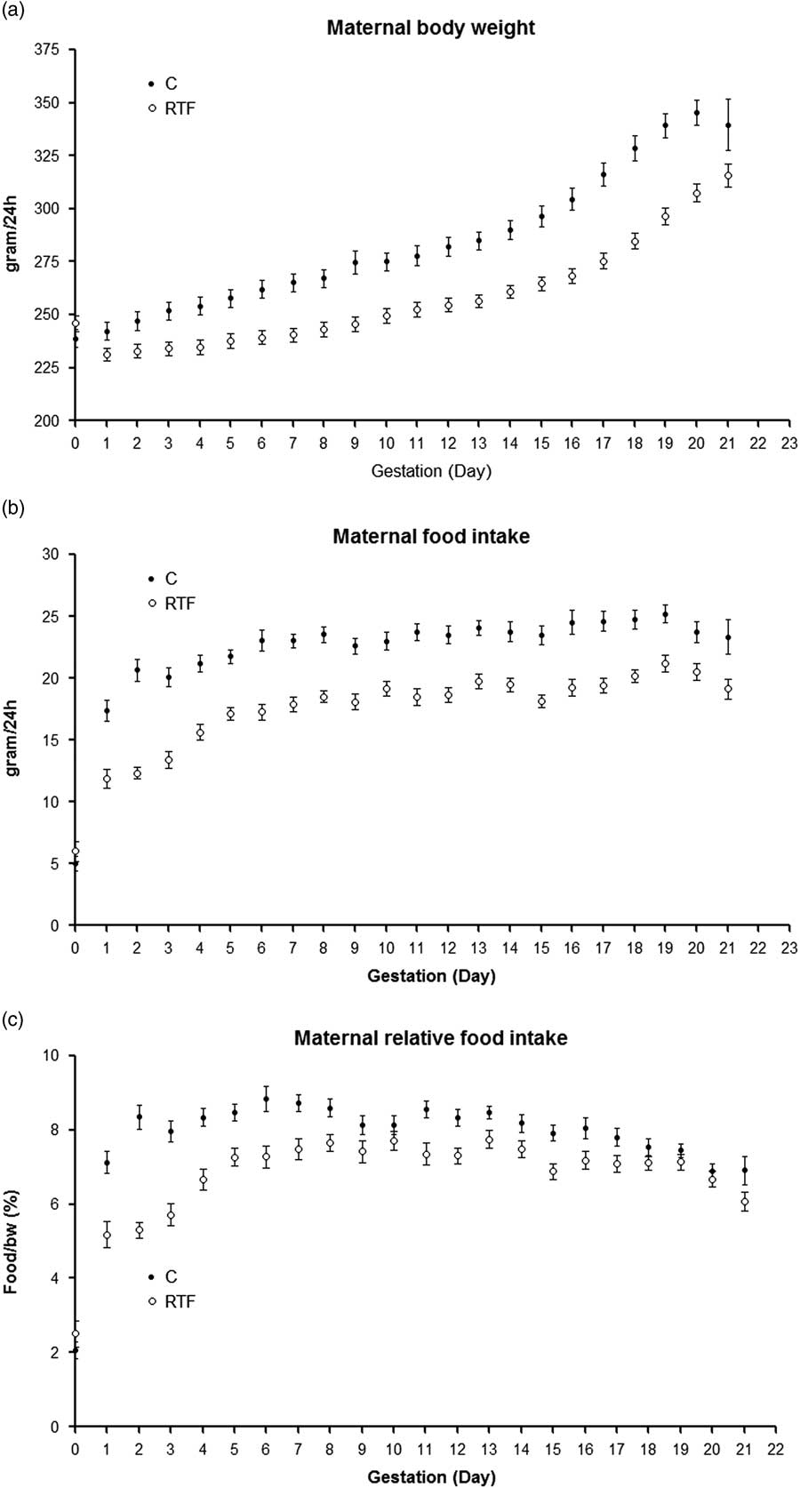
Body weight was significantly (P<0.001) lower in RTF dams compared with C animals (Fig. 1a). Although animals in the RTF group had plenty of food during the day time, they ate significantly (P<0.001) less food compared with the C group, a phenomenon that was observed on all gestation days (Fig. 1b). This represent a reduction of 180 kcal during the whole gestation (C, 1305±35.4 v. RTF, 1125±23.7 kcal, P<0.001). The calculation showed that relative food intake was significantly lower (P˂0.05) in RTF dams (Fig. 1c). The total body weight gain throughout pregnancy was 114 g in the C group v. 71 g in the RTF group, resulting in a significant reduction (P<0.001) in total weight gain in response to fasting.
(a) Body weight, (b) food intake and (c) relative food intake were recorded daily during gestation. Maternal body weight, food intake and relative food intake were significantly lower in the Ramadan-type fasting (RTF) (P<0.001, P<0.001 and P<0.05, respectively) group compared with control (C) animals on all gestation days. Statistical comparisons were performed by repeated measures ANOVA. Data are presented as means±s.e.m.

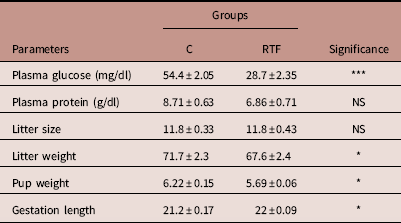
Analysis of blood samples obtained on GD 20 showed that the plasma glucose level was significantly lower in rats exposed to RTF than the C group. Plasma protein level was also decreased in RTF pregnant rats; however, this reduction did not reach statistical significance (Table 1). Maternal fasting did not affect litter size of RTF (Table 1). Additionally, no differences were noted in maternal nursing behavior between the RTF and C mothers, and no pups were eaten by their mothers in either group.
Characteristics of the Ramadan fasting model

C, control; RTF, Ramadan-type fasting; NS, not significant.
Data are presented as the mean±s.e.m.; n=16 per group. Maternal glucose level, litter weight, and pup weight were significantly lower in the RTF group. Maternal fasting resulted in a longer gestation period in the RTF group. There were no significant differences in maternal plasma protein and litter size between groups.
*P<0.05; ***P<0.001.
Litter weight was significantly reduced by ~8% in the RTF group. The average RTF pup weight was significantly lower than that of the control pups (Table 1). Gestation length was slightly but significantly increased in the fasting group.
Kidney development
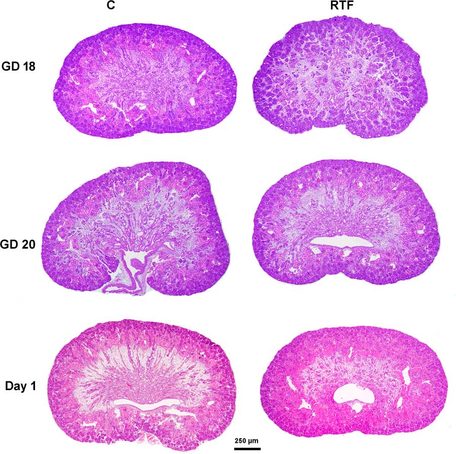
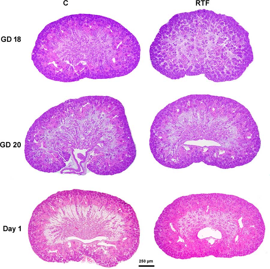
Generally, kidneys derived from the RTF group (Fig. 2) were less developed in comparison with age-matched controls at GD 18, GD 20 and birth, examined at 40× magnification. The kidney from RTF animals at GD 18 appeared to have a greater proportion of mesenchymal-like connective tissue compared with the controls. At GD 20, the cortex and medulla were well differentiated in the control kidneys but were less-differentiated in RTF kidneys, and the latter seemed to have fewer medullary rays. RTF kidneys at birth exhibited a wider nephrogenic zone and had a greater proportion of connective tissue and relatively few medullary rays in comparison with the controls.
Representative micrographs of kidney cross-sections from the control (C) and Ramadan-type fasting (RTF) groups at gestational day (GD) 18, GD 20 and day 1 stained with hematoxylin and eosin (H&E). RTF kidneys are immature, as illustrated by the higher level of connective tissue at GD 18, fewer medullary rays at GD 20 and wider nephrogenic zone at birth. H&E (40×).
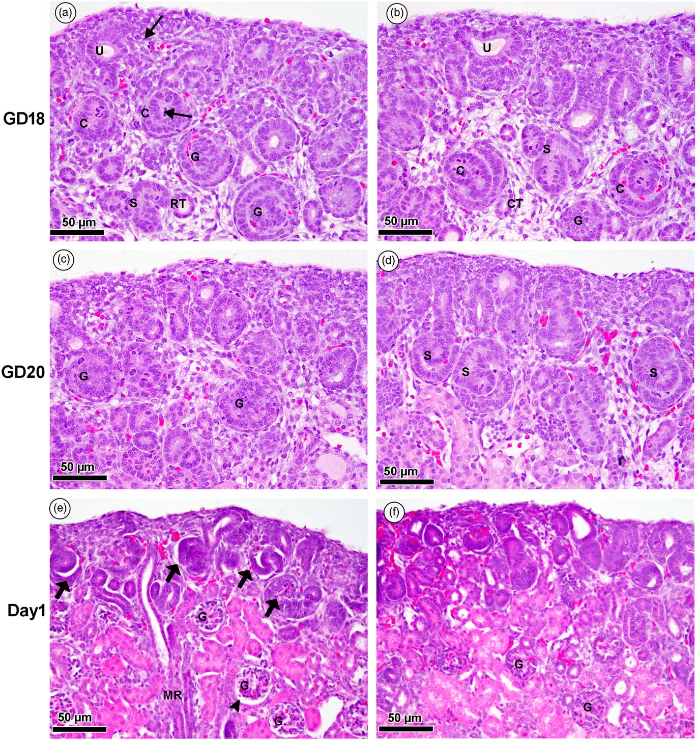
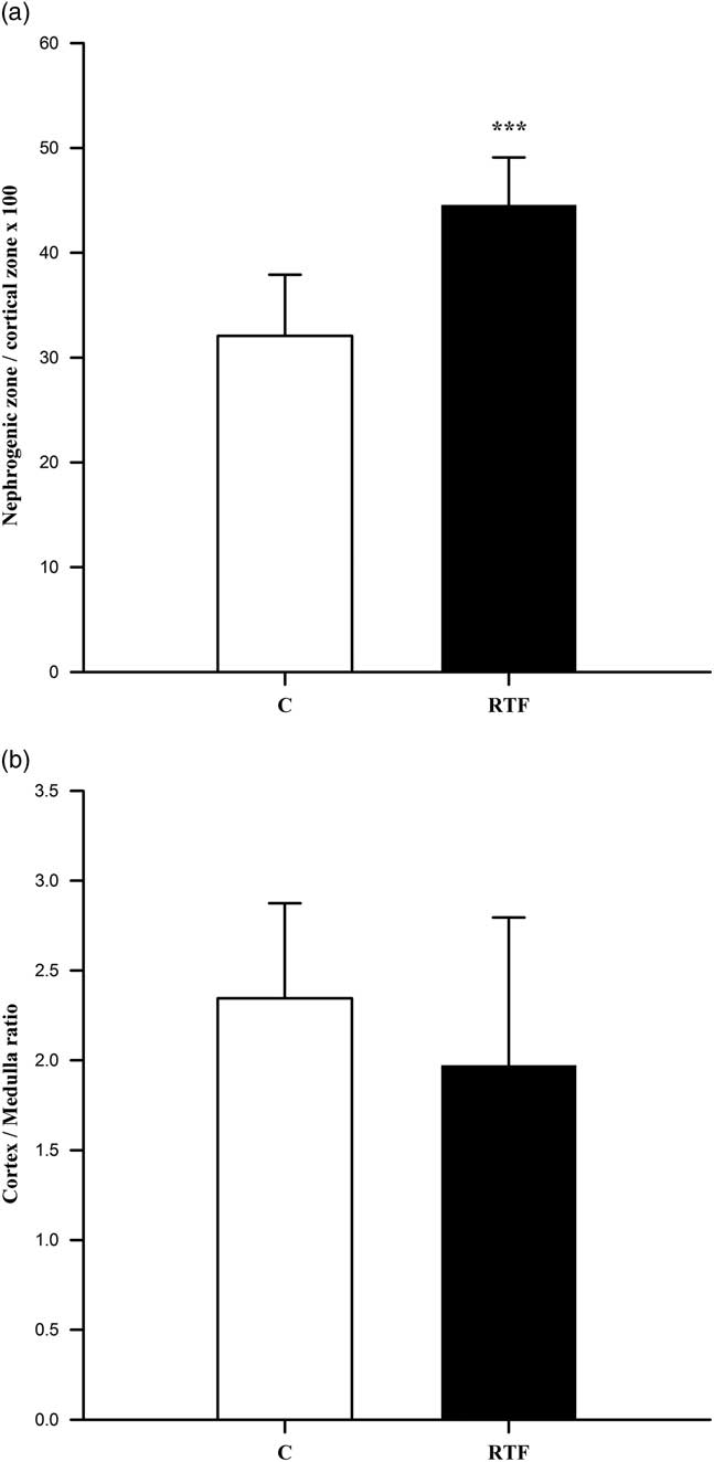
At high magnification (400×), control GD 18 kidneys exhibited extensive glomerulogenesis at various stages, with the normal development of peripherally induced, developing nephron sites and ureteric buds. The control nephrogenic developmental stages were identifiable as comma- and S-shaped, pre-capillary and immature glomeruli (Fig. 3a). Few mature glomeruli were observed in the RTF group since most glomeruli were less well differentiated in the form of S- and comma-shaped bodies. There were also more interstitial mesenchymal cells in the interstitial area (Fig. 3b). Kidneys from the GD 20 control group (Fig. 3c) had immature and mature glomeruli under the capsule, whereas the metanephroi of the RTF group showed predominantly immature S-shaped bodies (Fig. 3d). Many well-developed glomeruli were observed in the cortico-medullary region of the kidney at day 1 in the control group (Fig. 3e), and the renal cortex showed two distinct cortical zones; the nephrogenic zone containing immature forms of renal development and the juxtamedullary zone containing mature renal corpuscles and convoluted tubules. The medullary rays were present in both zones. At day 1, the RTF kidneys showed retarded nephron maturation and many immature glomeruli in the nephrogenic zone (Fig. 3f). The semiquantitative analysis indicated that the cortex/medulla ratio was not different between RTF and control groups, however, the nephrogenic zone/cortex zone ratio was significantly higher in RTF kidney compared with control (C, 32.1±2.2 v. RTF, 44.5±21.7; P<0.001) (Fig. 4).
Light micrographs of kidney tissue from the control (a, c, e) and Ramadan-type fasting (RTF) (b, d, f) groups at gestational day (GD) 18, GD 20 and day 1. Developing nephrons at various stages are seen in the renal cortex. At GD 18, the arrow indicates a cluster of mesenchymal cells around the ureteric duct (U). S, S-shaped body; C, comma-shaped body with mitotic figure (arrow); G, glomerulus at the capillary loop stage, the parietal epithelium is cuboidal and the renal tubule (RT). Note that kidneys from the RTF group have more mesenchymal-like connective tissue (CT) with fewer well-differentiated glomeruli in the form of S- and comma-shaped bodies. GD 20 metanephros sections from control animals depict stages of glomerular maturity with more glomeruli (G). Although the metanephros of the RTF group at GD 20 shows considerable growth and differentiation, the most predominant nephron stages are the S-shaped bodies (S). Section from the renal cortex of a control kidney at day 1 reveals the presence of two cortical zones; the nephrogenic zone, containing immature forms of renal developmental stages (arrows) and the juxtamedullary zone, containing mature glomeruli (G) and Bowman’s space (head arrow). The medullary rays (MR) are located in both zones. The RTF group shows delayed glomerular maturation (G) and fewer (immature) glomeruli in the nephrogenic zone (hematoxylin and eosin).

Quantitative analysis of percent of nephrogenic zone (a) and cortex/medulla ratio (%) (b) from control (C) and Ramadan-type fasting (RTF) kidneys at day 1. Data are presented as means±s.e.m., n=7. *** indicates highly significant vs C group at P<0.001.

Kidney apoptosis
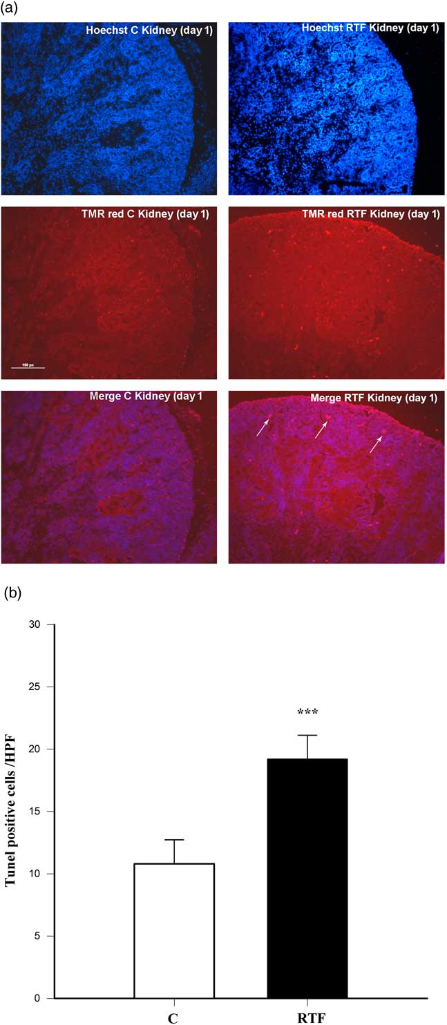
Apoptosis was assessed by TUNEL assay, in which positive staining indicates DNA fragmentation indicative of apoptosis. The incidence of TUNEL-positive staining was higher in the RTF group compared with the controls (Fig. 5a). The results showed that RTF significantly increased apoptotic nuclei by 77% compared with control (C, 10.8±0.85 v. RTF, 19.2±0.86; P<0.001) (Fig. 5b).
Representative photomicrographs of TUNEL staining of apoptotic cells (arrows) in nephrogenic zone of kidney tissues on day 1 from control (C) and Ramadan-type fasting (RTF) animals, scale bar=100 µm (a). Accumulative data for five individual rats from each group indicated a significant increase (P<0.001) of apoptosis in a RTF kidney compared with a control kidney at birth (b). *** indicates highly significant vs C group at P<0.001.

Stereology
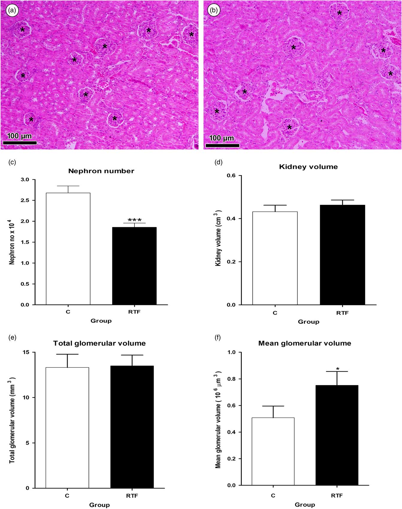
Histological examination of the renal cortex from 28-day-old rats revealed no qualitative differences in either the glomeruli or distal and proximal convoluted tubules between the RTF and control groups (Fig. 6a). However, stereology revealed glomerular enlargement and fewer nephrons in kidneys from the RTF group (Fig. 6b). There was a significant reduction in nephron number (~31% fewer) in kidneys from the RTF group compared with those from the control group (C, 26,800±1680 v. RTF, 18,500±1000; P<0.001; Fig. 6c). Conversely, the mean glomerular volume was significantly greater in kidneys from the RTF group compared with those from the C group (C, 0.50±0.088×106 µm3 v. RTF, 0.75±0.1×106 µm3; P=0.197; Fig. 6f). Thus, the total volume of all glomeruli in RTF kidneys was not different from that of the control animals as shown in Fig. 6e. Although nephron number and mean glomerular volume were altered in the RTF group, the overall volume of the kidneys were not different between the RTF and C groups (C, 0.432±0.0299 cm3 v. RTF, 0.463±0.0228 cm3; P=0.454; Fig. 6d).
Representative micrographs of kidney tissue from the control (a) and Ramadan-type fasting (RTF) (b) groups at day 28, depicting a low nephron number with glomerular enlargement (stars) in the RTF group (hematoxylin and eosin, 200×). The nephron number in rat kidneys was significantly (P<0.001) reduced in the RTF rats compared with the control rats (c). There was no significant difference in kidney volume between the two groups (d). There was no significant difference in total glomerular volume between RTF and control (C) offspring (e). Mean glomerular volume was significantly higher in the RTF group compared with the C group (f) (n=8 per group). * indicates significant vs C group P at<0.05.

Histopathology
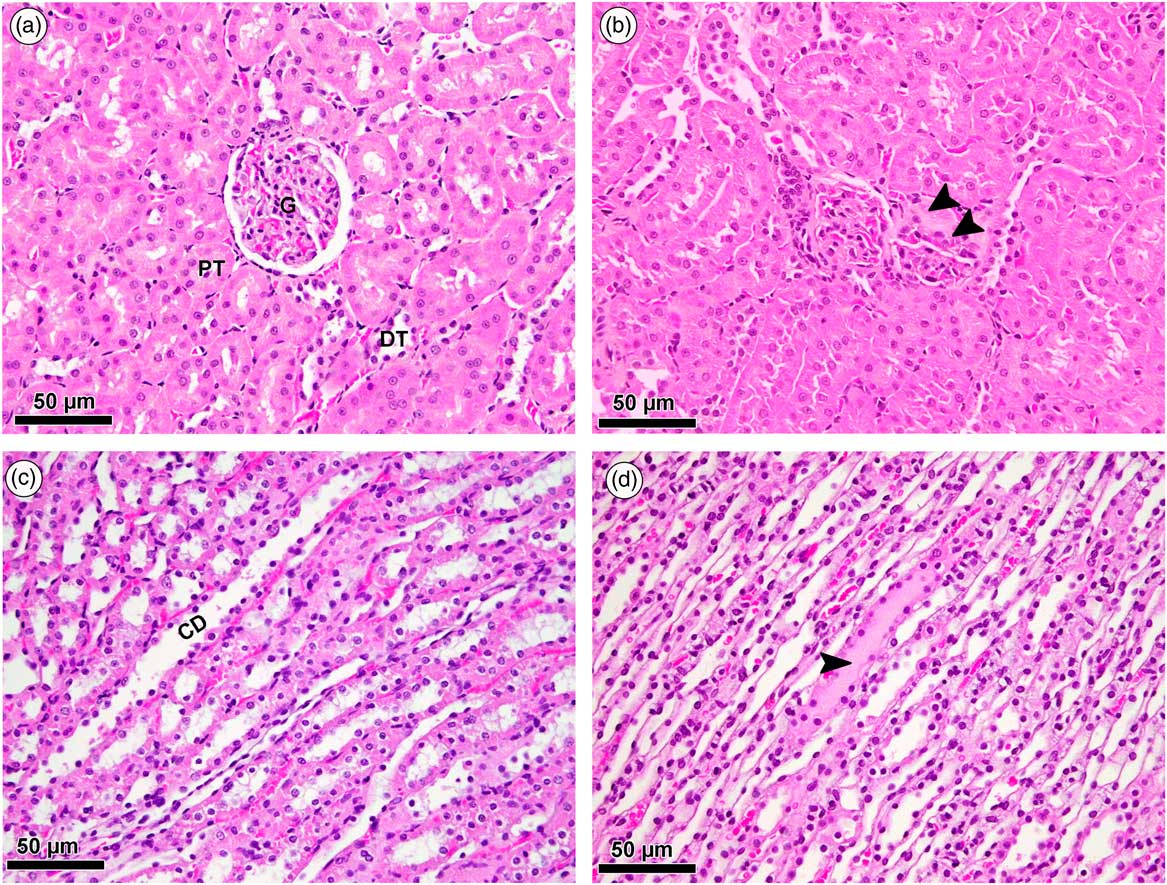
Sections of renal cortex from 112-day-old rats revealed normal structure of the proximal convoluted tubules, distal convoluted tubules, Bowman’s capsule and glomeruli in the control group (Fig. 7a). RTF kidneys showed more tubular atrophy and interstitial fibrosis with enlarged lobulated glomeruli in the renal cortex (Fig. 7b). Renal medulla from the control group (Fig. 7c) showed normal microscopic architecture with collecting ducts, thin limb loops of Henle and thick descending limbs, whereas the medulla from RTF offspring exhibited changes, including tubular dilatation and infiltration of proteinaceous casts (Fig. 7d).
Light micrographs of renal cortex from the control (a) and Ramadan-type fasting (RTF) groups (b) at day 112. Section from the renal cortex of the control group reveals the normal appearance of the proximal convoluted tubules (PT), distal convoluted tubules (DT), Bowman’s capsule and glomerulus (G). The RTF renal cortex shows mild glomerulosclerosis with segmental sclerosis (head arrows), thickening of Bowman’s capsule and hyaline changes. Light micrographs of renal medulla from the control (c) and RTF groups (d) at day 112. The renal medulla from control rats shows normal collecting ducts (CD), thin limb loop of Henle, and thick descending limb. Medulla of RTF offspring shows tubular dilatation and infiltration of proteinaceous casts (head arrow) (hematoxylin and eosin).

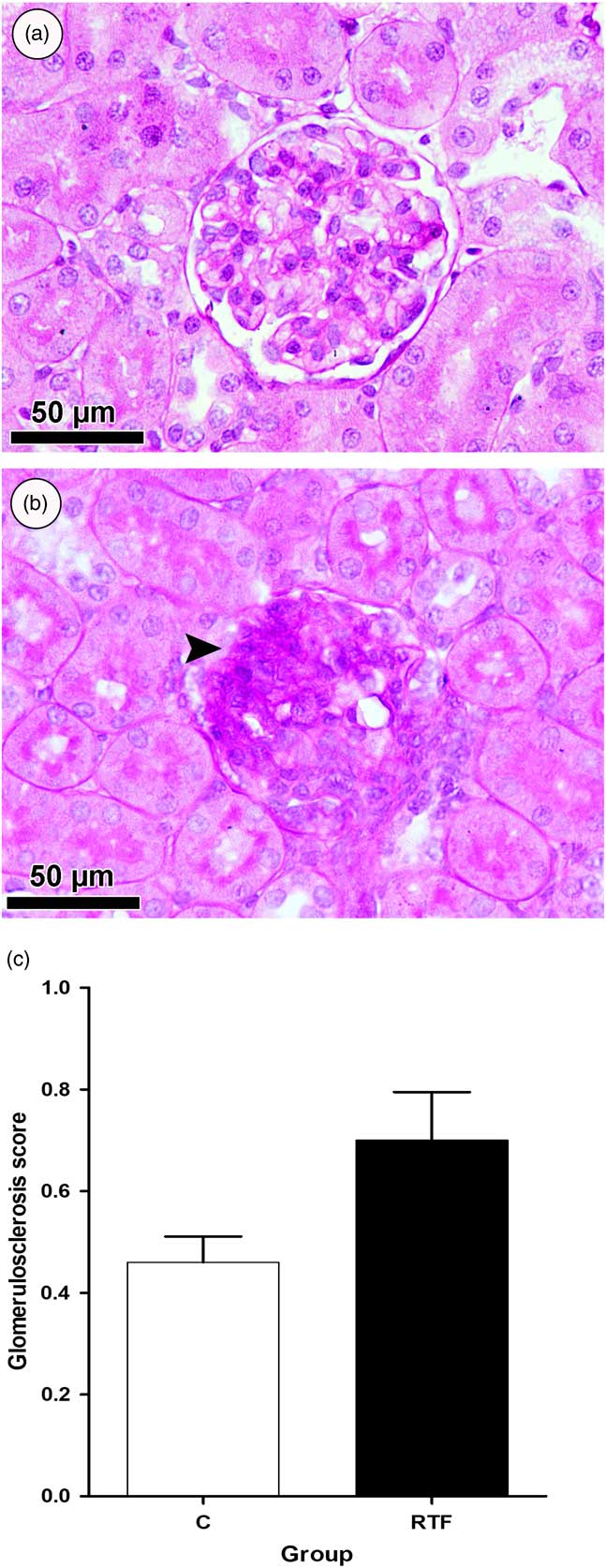
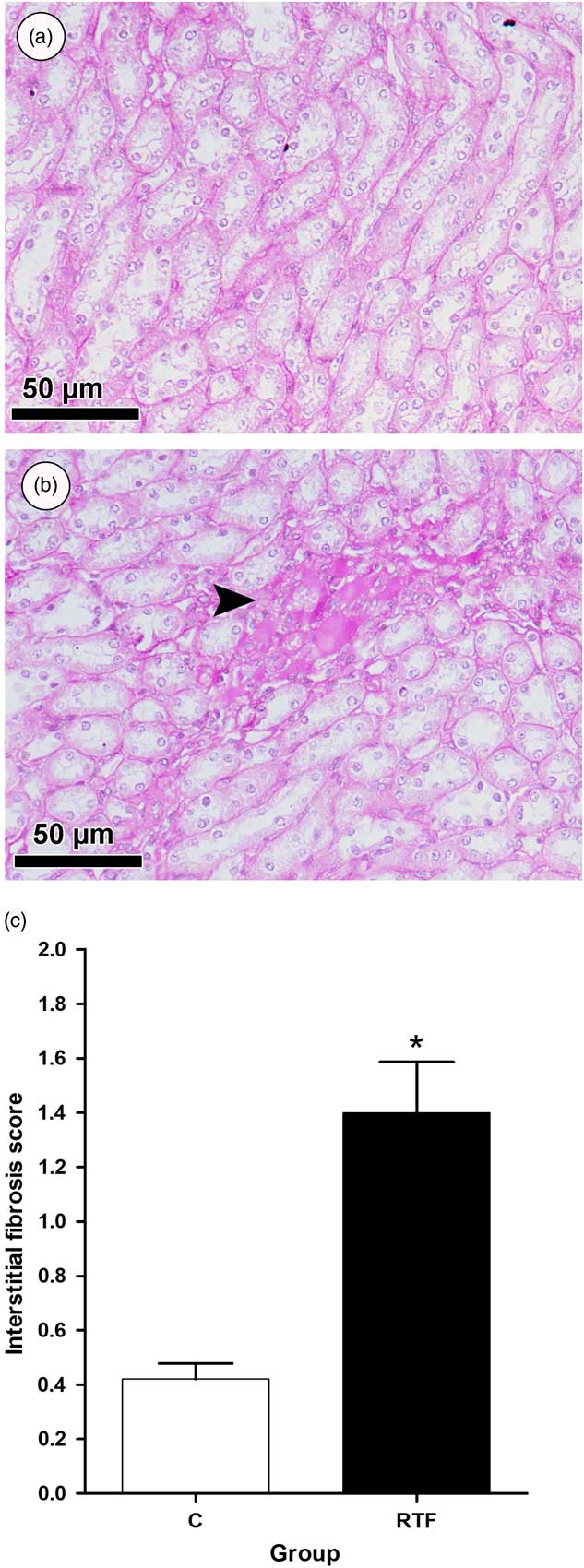
Glomerulsclerosis and interstitial fibrosis were assessed semi-quantitatively in PAS-stained sections. The data of glomerulosclerosis damage score exhibited mild glomerulosclerosis in RTF renal cortex compared with control renal cortex (Fig. 8a–8c). Although, the degree of interstitial fibrosis was significantly increased (P<0.05) in RTF renal medulla compared with control renal medulla (Fig. 9a–9c).
Periodic acid–Schiff-stained sections of the renal cortex from the control (a) and Ramadan-type fasting (RTF) (b) groups at day 112. The animals of RTF group show mild glomerulosclerosis (arrow). Glomerulosclerosis damage score (c) for six individual rats from each group showed an increase (P=0.056) of glomerulsclerosis in RTF kidney compared with control kidney, however, it did not reach a statistically significant difference.

Representative renal medulla sections stained by periodic acid–Schiff from the control (a) and Ramadan-type fasting (RTF) (b) groups at day 112 showed moderate interstitial fibrosis in RTF kidney (arrow). Accumulative data of interstitial fibrosis scoring for six individual rats from each group indicated a significant increase (P<0.05) of interstitial fibrosis in a RTF kidney compared with a control kidney (c). * indicates significant vs C group P at<0.05.

Discussion
Despite the well-documented deleterious effects of maternal malnutrition in the development of disease,Reference Paulino-Silva and Costa-Silva 31 , Reference Alzamendi, Zubiría and Moreno 32 limited data are available on the long-term effects of fasting exposure during gestation, especially on renal programming. Food intake and body weight gain were significantly lower in the fasting group compared with the control. This level of reduction in food intake is similar to the food restriction employed by othersReference Léonhardt, Lesage and Dufourny 33 , Reference Akitake, Katsuragi and Hosokawa 34 ; but differs in terms of the timing of food availability. This finding is also consistent with the well-established observation of fasting or food restriction in pregnant ratsReference Fleeman, Cappon, Chapin and Hurtt 35 and humansReference Awwad, Usta and Succar 36 when subjected to weight loss as a result of decreased energy intake.
The present study shows that exposure of pregnant rats to a 16-h fast significantly increased gestation length. Reduced maternal food intake in sheep also increased gestational length.Reference Cleal, Poore and Newman 37 In contrast, Almond and MazumderReference Almond and Mazumder 38 found that maternal fasting in humans resulted in a significant reduction of gestational length. Total protein was not affected by maternal fasting; however, blood glucose was significantly reduced in the maternal fasting group. A reduction in blood glucose can initiate a signaling cascade that leads to permanent physiological adaptations, which increase the risk of developing chronic disease.Reference Sharp 39 The results of our study showed that birth weight was significantly lower in RTF offspring, which may be due to a reduction in maternal food intake and overall weight gain. These findings are consistent with findings obtained from clinical studies in humans, in which in utero exposure to fasting during Ramadan has been associated with lower birth weight among Michigan Arab mothersReference Almond and Mazumder 40 and Saudi Arabian mothers.Reference Alwasel, Abotalib and Aljarallah 1
In the present study, exposure to fasting during pregnancy had no effect on litter size. Similar observations were made by Woodall et al.Reference Woodall, Johnston, Breier and Gluckman 41 who reported that 30% nutritional restriction did not affect litter size. Both low birth weight and early catch-up growth are considered developmental markers for later adult disease.Reference Osmond, Barker, Winter, Fall and Simmonds 42 An interesting finding in the current study is that RTF delayed nephrogenesis. Kidney sections of RTF displayed less well-differentiated glomeruli, more connective tissue, fewer medullary rays and significant increase in area of nephrogenic zone. In addition, sections showed a significant increase in apoptosis in RTF rat kidneys at birth when compared with controls. These findings are in accordance with a study conducted by Tafti et al.Reference Tafti, Nast and Desai 43 which showed that maternal undernutrition upregulated most of genes involved in both the extrinsic and intrinsic pathways of apoptosis as well as the downstream caspase cascade in offspring nephrogenesis. Previous studies have shown that protein restriction in pregnancy was associated with decline in the final number of glomeruli with aberrant nephrogenesis induced by increased apoptosis of mesenchymal cells at the start of rat metanephrogenesis.Reference Welham, Wade and Woolf 44
Another major finding of this study was that offspring exposed to maternal RTF exhibited a ~31% reduction in nephron number compared with the control, suggesting that maternal RTF may have slowed or halted nephrogenesis. Evidence from animal studies suggests that maternal protein or global nutrient restriction, uterine artery ligation, hyperglycemia, and exposure to various agents, such as glucocorticoids or alcohol, lead to the production of offspring with fewer nephrons.Reference Singh, Moritz, Bertram and Cullen-McEwen 45 Studies have suggested different mechanism(s) leading to reduced nephron number; for example, the depletion of stem cells by increased apoptosis,Reference Pham, MacLennan and Chiu 46 , Reference Hokke, Armitage and Puelles 47 inhibition of ureteric branchingReference Awazu and Hida 48 and early cessation of nephrogenesis.Reference Vaccari, Mesquita, Gontijo and Boer 49 The second finding of this study was that maternal RTF had no effect on kidney volume or total glomerular volume. Consistent with the present findings, offspring exposed to 50% food restriction in utero had a similar kidney volume to the controls.Reference Benz and Amann 50 Accordingly, mean glomerular volume of RTF offspring was significantly greater than that of the control animals, probably due to a decrease in nephron number. Additionally, some studies investigating nutritional programming have revealed an inverse relationship between nephron number and blood pressure.Reference Bagby 6 , Reference Woods, Weeks and Rasch 51 The results of the current study show that maternal RTF during pregnancy resulted in retarded nephrogenesis. Similar observations have been made in previous studiesReference Vaccari, Mesquita, Gontijo and Boer 49 using different models of nutritional programming. Delayed nephrogenesis may occur due to increased apoptosis, as observed in the present study. Pathological structural observations in adult kidneys may be the result of low nephron number. Similar observations have shown that 50% protein restriction during pregnancy induces renal injury with enhanced tubular dilatation, tubular atrophy and interstitial fibrosis.Reference Hokke, Armitage and Puelles 47
A limitation of our study is the duration over which fasting was imposed upon the pregnant rats. Pregnant women observing Ramadan fast for 1 month out of their 9 months of pregnancy, whereas in our rat model the animals were subject to daily fasting from conception to term. We chose this approach in order to illicit the maximum response, as in principle Ramadan could fall at any stage of human pregnancy, By fasting our rats throughout gestation we were able to impose the greatest challenge on the fetus; however we recognize that the magnitude of the impact on the developing kidneys may be less if the rats were fasted for shorter periods of time.
In conclusion, we have developed a rat model of maternal Ramadan fasting, in which the kidneys of offspring exhibit retarded nephrogenesis and low nephron number, partly due to increased apoptosis.
Financial Support
The authors would like to extend their sincere appreciation to the Deanship of Scientific Research at King Saud University for funding this research RG-164. The Center for Stochastic Geometry and Advanced Bioimaging is supported by the Villum Foundation (Denmark).
Conflicts of Interest
None.
Ethical Standards
The authors assert that all procedures contributing to this work comply with the ethical standards of the relevant national guides on the care and use of laboratory animals.
Acknowledgments
The authors gratefully acknowledge the excellent technical assistance provided by Maj-Britt Lundorf during the stereological investigation of this study at Stereological Research and Electron Microscopy Laboratory, University of Aarhus, Aarhus, Denmark.