Introduction
Capuchin monkeys are neotropical primates that, under captivity, are commonly used as experimental animals for other primate species including human (Philips et al., Reference Philips, Bales, Capitanio, Conley, Czoty, t’ Hart, Hopkins, Hu, Miller, Nader, Nathanielsz, Rogers, Shively and Voytko2014). Regarding female reproductive studies, the morphology and morphometry of the reproductive tract (Domingues et al., Reference Domingues, Caldas-Bussiere, Martins and Carvalho2007; Lima et al., Reference Lima, Guimaraes, Branco, Giese, Muniz, Pereira, da Cunha, Ricci and Miglino2015), the protein profile in oocyte−cumulus–oocyte according the follicular phase (Domingues et al., Reference Domingues, Caldas-Bussiere, Petretski, Ohashi, Lima, Santos, Cordeiro and Gomer de Castro2010), fetal development by ultrasonographic imaging (Miranda et al., Reference Miranda, Leao, Oliveira, Sodre and Domingues2018), and the population of preantral follicles (Domingues et al., Reference Domingues, Ferreira, Muniz, Lima, Ohashi, Figueiredo and Silva2003, Reference Domingues, Diniz, Furtado, Ohashi, Rondina and Silva2004) have been described previously. Furthermore, advances have been observed in the in vitro production of S. apella embryos from parthenogenetically activated or in vitro matured and fertilized oocytes (Lima et al., Reference Lima, Leao, Sampaio, Brito, Santos, Miranda, Ohashi and Domingues2013), the in vitro development of preantral follicles enclosed in the ovarian tissue from S. apella (Brito et al., Reference Brito, Santos, van den Hurk, Lima, Miranda, Ohashi and Domingues2013), and the cryopreservation of S. apella ovarian tissue (Brito et al., Reference Brito, Brito, Scalercio, Percario, Miranda, Rocha, Diniz, Oskam, van den Hurk, Paris, Domigues and Santos2014). Domingues et al. (Reference Domingues, Diniz, Furtado, Ohashi, Rondina and Silva2004) studied the morphometry and morphology of preantral and antral follicles from Sapajus apella (previously called Cebus apella). However, as have other researchers, they measured the mean diameters of the follicular compartments using a micrometric eye lens coupled to an optical microscope, instead of measuring the follicular and oocyte area. In this study, we proposed to accurately measure the follicular dimensions with the help of software. It is well known that preantral follicles represent the gametes reserve from females, and a proper knowledge on their morphology will support the analysis of techniques developed for these reproductive cells. Furthermore, it is of importance to detect the expression of the main proteins related to oocyte and granulosa cells quality and development. Besides the importance of growth factors (Scalercio et al., Reference Scalercio, Brito, Domingues, Santos and Amorim2015), follicular development is also characterized by its dependence on angiogenic factors (Torres-Ortiz et al., Reference Torres-Ortiz, Gutierrez-Ospina, Gomez-Chavarin, Murcia, Alonso-Morales and Perera-Marin2017). Therefore, the immunolocalization of related proteins, e.g. vascular endothelial growth factor (VEGF) and cluster of differentiation 31 (CD31) in the ovarian tissue and preantral follicles at different developmental stages will help to standardize analysis and will provide extra tools to evaluate follicular and ovarian tissue quality after bioprocessing. Therefore, in the present study, we aimed to characterize the morphology, morphometry and the expression of VEGF and CD31 in preantral follicles, and ovarian tissue, from S. apella.
Material and methods
Chemicals
Unless mentioned otherwise, the chemicals used in the present study were purchased from Sigma Chemical Co. (St Louis, USA).
Animals
Five healthy and sexually mature capuchin monkey females (7–9 years old, weight range: 1.7–2.6 kg) born in captivity at National Primate Center, Pará, Brazil, were selected for our study. The females were kept indoors under a natural photoperiod and housed individually in cages (80 × 90 × 80 cm) at the National Primate Center. Their daily diet consisted of fresh fruits, vegetables and commercial monkey pellets (MEGAZOO® P18, Betim, Brazil). Tap water was available ad libitum.
Ovarian tissue collection
All procedures were performed under general anaesthesia. Each animal was anesthetized with tiletamine hydrochloride and zolazepam hydrochloride (Zoletil®100; 4 mg/kg; IM; Virbac do Brazil Indústria e Comércio Ltda, São Paulo, Brazil) associated with tramadol hydrochloride (Tramal; 0.05 mg/kg; IM; Grünenthal GmbH; Aachen; Alemanha). Anaesthesia was maintained with 2% isoflurane (Tanohalo; Cristália, São Paulo, Brazil). The females were placed in dorsal recumbence and ovarian biopsies were collected by exploratory laparotomy. For this, a ventral midline skin incision was made to reach the ovaries, as described by Domingues et al. (Reference Domingues, Caldas-Bussiere, Martins and Carvalho2007). Ovarian biopsies were recovered by a ‘trap door’ technique as described by Santana et al. (Reference Santana, Brito, Brito, Lima, Domingues and Santos2013). In brief, using a no. 15 blade (Paramount Surgimed Ltda, New Delhi, Delhi, India) a 1 mm thickness circular incision in the surface of the ovary, and ovarian cortical envelope flap was removed. Suture was unnecessary, and bleeding was repressed by gently pressing the sampled area with cold (15°C) 0.9% saline-soaked gauze. After surgery, antibiotic (Chemitril; Chemitec, São Paulo, Brazil), and anti-inflammatory (Ketofen; Merial Saúde Animal Ltda, São Paulo, Brazil) drugs were administered to the animals for 1 week and 2 days, respectively. The removed tissue was kept in pre-warmed (37°C) HEPES-buffered minimum essential medium (MEM) supplemented with 10 mg/ml bovine serum albumin (BSA) and subjected to fragmentation to obtain cortical pieces of 1 × 1 × 2 mm.
Morphological and morphometric analysis
After fixation for 24 h in Davidson solution at room temperature, the ovarian fragments were dehydrated in ethanol, clarified in xylene and embedded in paraffin wax. Serial sections (5 µm) of ovarian tissue were cut, every tenth section was mounted on glass slides, and stained with haematoxylin-eosin (Merck, Darmstadt, Germany). Preantral follicles with an oocyte surrounded by one layer of flattened granulosa cells were classified as primordial, while were denominated transition follicles if the oocyte was surrounded by one layer of flattened and cuboidal granulosa cells. Primary follicles presented one layer of cuboidal granulosa cells surrounding the oocyte, and secondary follicles presented two or more layers of granulosa cells surrounding the oocyte (Almeida et al., Reference Almeida, Santos, Scalercio, Leao, Haritova, Oskam and Domingues2012). Just follicles with a visible nucleus were considered to avoid counting a follicle more than once.
Preantral follicles were classified as morphologically normal or atretic as described by Domingues et al. (Reference Domingues, Ferreira, Muniz, Lima, Ohashi, Figueiredo and Silva2003). All histological sections were examined by using a light microscope (DME; Leica Microsystems B.V., Amsterdam, The Netherlands) at a magnification of × 400. Images were captured with a digital camera (Sony DSC-W730, Sony Corporation, Tokyo, Japan) coupled in a light microscope (DME, Leica), and measurements were performed with the aid of morphometric analysis software (ImageJ 1.47), and at least 30 follicles per animal were evaluated and measured. Granulosa cell counts, and the area of follicles, oocytes, and oocyte nuclei were measured. To compare the present data with other studies that applied diameter measurements instead of area, we assumed the area of the oocyte and follicle as a spherical shape. Thus, the area is considered as A = πr 2 . Based on this, we calculated the theoretical diameter as obtained r × 2.
Immunohistochemistry
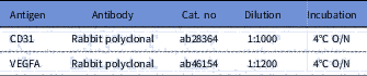
After overnight fixation in 4% paraformaldehyde (pH 7.2) at 4ºC, the ovarian fragments were dehydrated in ethanol, clarified in xylene and embedded in paraffin wax. Serial sections (5 µm) of ovarian tissue were cut and every tenth section was mounted on glass slides. Paraffin sections were deparaffinized with xylene (xylene, Sigma-Aldrich, USA) and rehydrated in alcohol series. After blocking endogenous peroxidase activity with 3% H2O2 diluted in methanol, a demasking step was performed for 75 min at 98ºC with citrate buffer in Tween 20 before the sections were subjected to an antigen retrieval step. Antigen retrieval steps, antibody dilutions and incubation conditions were performed as described in Table 1. Diaminobenzidine was used as a chromogen (Vector Laboratories, Peterborough, UK). The slides were then counterstained with haematoxylin and mounted. Negative controls consisted of the dilution solution without any primary antibody. All positive controls were processed in the same way as the tested tissues. For quantitative analysis of VEGFA and CD31 protein, expression in the ovarian tissue, as well as in primordial, transition, primary and secondary follicles, was evaluated in all animals.
Antibodies dilutions used in the present study

O/N, overnight.
Statistical analysis
Statistical analysis was performed applying the GraphPad Prism 7.0 program for Windows (GraphPad Software, La Jolla California, USA). Comparisons between the number of follicles per follicular class, number of granulosa cells surrounding the oocyte per follicular class, and morphometric data were compared among different follicular class using analysis of variance (ANOVA) and Tukey as a post-hoc test. A linear regression was applied for the correlation of follicular growth using the nucleus and oocyte diameter as predicted variable. Within each follicular class (primordial, transition, primary, and secondary), nucleus diameter was correlated with the follicle and oocyte diameter, as well as thickness and number of granulosa cell layers surrounding the respective follicles. Results were expressed as mean±standard error of the mean (SEM) and differences were considered significant when P-values were <0.05.
Results
Morphological analysis
In total, 213 preantral follicles were examined (33–45 follicles per female) by classical histology for morphometry and morphological classification. From this total, 20 (9.4%) were follicles that enclosed two or more oocytes, i.e. multi-oocyte follicles (MOFs) (Fig. 1), from which 90% were morphologically normal and enclosed in primordial 35%, transition 35%, primary 20%, and secondary 10% follicles.
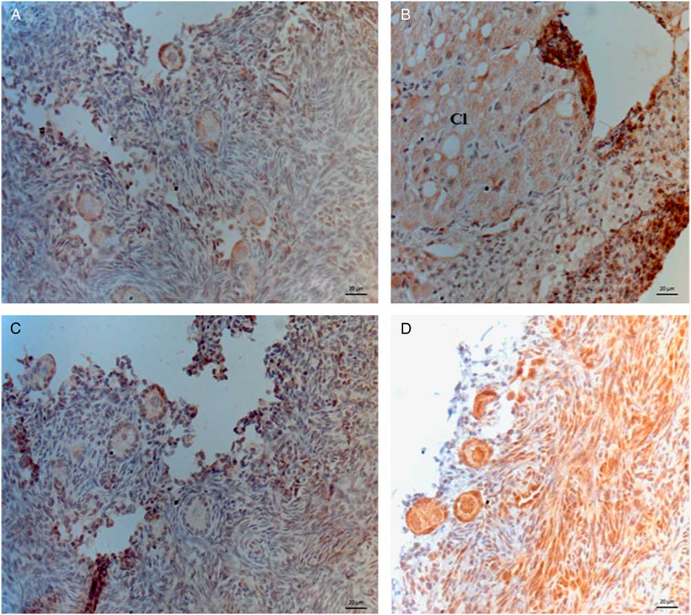
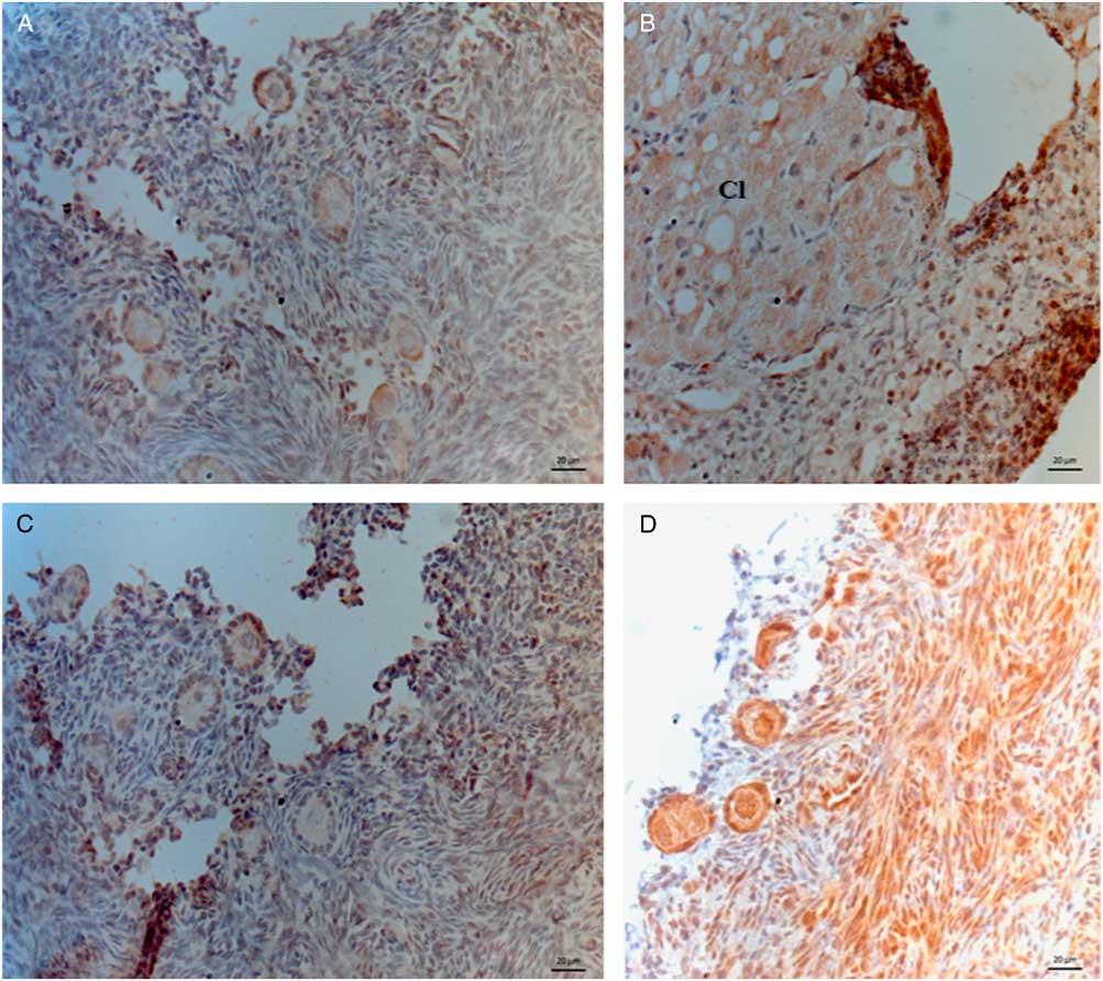
Sections of ovarian tissue from Sapajus apella showing the expression of CD31 (A–C) and VEGF (D). Corpus luteum is indicated by Cl. Bars indicate 20 µm.
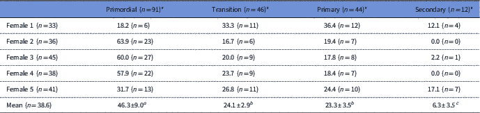
From the 193 follicles enclosing a single oocyte, most of these was classified as primordial (91; 46.3%), and the smallest population was that from secondary follicles (12; 6.3%). Table 2 lists mean values and the number of total follicles evaluated per female.
Number of preantral follicles evaluated per female capuchin monkey (in brackets), and mean percentages (± SEM) of total preantral follicles from each follicular class
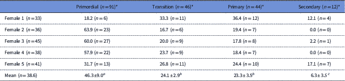
a–c Different superscripts indicate significant differences within the same row (P < 0.05).
* MOFs were not considered in this calculation.
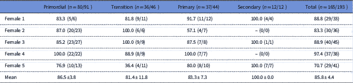
The majority of the total preantral follicles was morphologically normal (>85%), and the distribution of normal follicles enclosing a single oocyte, per class, is shown in Table 3. All the follicular classes presented similar percentages of morphologically normal follicles.
Mean (± SEM) percentages of morphologically normal preantral follicles from each follicular class per female capuchin monkey

No significant differences were observed when comparing means of morphologically normal follicles per category, i.e. primordial, transition, primary, and secondary.
Follicular morphometry and granulosa cells counting
From the above evaluated follicles, only those morphologically normal and enclosing a single oocyte (n = 165) were submitted to morphometric analysis. Mean morphometric data, as well as the mean number of granulosa cells surrounding each follicle, are presented in Table 4. The mean number of granulosa cells surrounding primordial, transition, primary, and secondary follicles significantly differed among all follicular classes, with the lowest number being found in primordial follicles (c. nine cells surrounding an oocyte) and the highest number in secondary ones (c. 45 cells surrounding an oocyte). Morphometric evaluation resulted in similar results when considering follicular diameter. Oocyte diameter started to increase significantly only in primary and secondary follicles, and oocyte nucleus increased significantly in diameter only when follicles were at the secondary stage.
Mean (± SEM) number of granulosa cells (♯GC) per follicle, and area of the follicle, oocyte, and oocyte nucleus per follicular class

a–d Different superscripts indicate significant differences within the same column (P < 0.05).
Immunohistochemistry
The expression of CD31 was strong in vessels, corpus luteum, and also present in normal preantral follicles (oocytes and granulosa cells) at all developmental stages. VEGFA expression was also observed in vessels and preantral follicles (granulosa cells, oocyte and oocyte nucleus) (Fig. 1).
Discussion
In the present study we characterized the morphology and morphometry of normal and atretic preantral follicles in ovaries from Sapajus apella. Furthermore, we determined the localization of the proteins CD31 and VEGF in the ovarian tissue, as well as in the oocyte and granulosa cells from S. apella preantral follicles.
In a previous study, Domingues et al. (Reference Domingues, Ferreira, Muniz, Lima, Ohashi, Figueiredo and Silva2003) characterized the preantral follicle population of Cebus apella (approximately 48,000 preantral follicles per ovary), now recognized as Sapajus apella. The change in nomenclature occurred after a detailed morphological study from Silva Jr (Reference Silva2002), in which the subgenus Sapajus was then recognized as a genus independent from the Cebus.
In the study from Domingues et al. (Reference Domingues, Ferreira, Muniz, Lima, Ohashi, Figueiredo and Silva2003), the rates of primordial, primary and secondary follicles were 77.3, 17.1 and 5.6%, respectively. In the present study, we observed rates of 46.3, 24.1, 23.3 and 5.6% for primordial, transition, primary and secondary follicles, respectively. As Domingues et al. (Reference Domingues, Ferreira, Muniz, Lima, Ohashi, Figueiredo and Silva2003) characterized transition follicles as primordial ones, both studies give similar results per follicular class. These percentages were also similar to those described for Saimiri sciureus (73.3, 18.6 and 8.1% for primordial, primary and secondary follicles, respectively; Almeida et al., Reference Almeida, Santos, Scalercio, Leao, Haritova, Oskam and Domingues2012) and human (81.7, 15.3 and <3% for primordial, primary and secondary follicles, respectively; Gougeon & Chainy, Reference Gougeon and Chainy1987).
In the present study we measured the area of the follicle, oocyte and oocyte nucleus. However, most morphometric studies from other groups measured the mean diameter. To allow a comparison with other studies, we calculated the theoretical diameter of the evaluated parameters. We observed that follicular, oocyte and oocyte nucleus dimensions obtained in the present study were similar to those observed for Cebus apella (Domingues et al., Reference Domingues, Diniz, Furtado, Ohashi, Rondina and Silva2004), and for Saimiri sciureus (Almeida et al., Reference Almeida, Santos, Scalercio, Leao, Haritova, Oskam and Domingues2012) except for the size of the oocyte nucleus, which was twice as big as that in S. apella. It is important to note that the secondary follicles evaluated in the present study were much smaller than those measured from Almeida et al. (Reference Almeida, Santos, Scalercio, Leao, Haritova, Oskam and Domingues2012). This difference can be explained by the fact that we evaluated early secondary follicles (with up to three layers of granulosa cells), and the former authors measured more advanced ones. Primordial and primary follicles from Alouatta caraya (Lopes et al., Reference Lopes, Lucci, Garcia, de Azevedo and Báo2006) were slightly bigger than those from S. apella. Interesting, both species presented secondary follicles and oocytes with similar dimensions. Dimensions of primordial, transition and primary follicles were also similar to those of Saimiri cassiquiarensis, S. macrodon and S. vanzolinii (Lopes et al., Reference Lopes, Santos, Almeida, Brito, Queiroz and Domingues2017), except for the oocyte nucleus, which was larger in S. apella. Moreover, preantral follicles from P. anubis (Amorim et al., Reference Amorim, Moyal, Donnez and Dolmans2016) were twice as big as those from S. apella.
The number of granulosa cells surrounding the primordial follicles (6–11) did not differ from that observed in human (Gougeon & Chainy, Reference Gougeon and Chainy1987), S. sciureus (Almeida et al., Reference Almeida, Santos, Scalercio, Leao, Haritova, Oskam and Domingues2012) and Alouatta caraya (Lopes et al., Reference Lopes, Lucci, Garcia, de Azevedo and Báo2006), but it was much lower than the number of granulosa cells that surrounded the primordial follicles from Papio anubis, c. 25 cells (Amorim et al., Reference Amorim, Moyal, Donnez and Dolmans2016). No other authors evaluated the morphometry of transition follicles from primates. In the present study, transition follicles presented 10–15 granulosa cells surrounding the oocyte. These data still cannot be put in parallel with those from baboon (Amorim et al., Reference Amorim, Moyal, Donnez and Dolmans2016) because these authors considered transition as primary follicles. For primary follicles, the number of granulosa cells that surrounded the oocyte was very variable (10–30 cells), being again similar to observation in human (Gougeon & Chainy, Reference Gougeon and Chainy1987), S. sciureus (Almeida et al., Reference Almeida, Santos, Scalercio, Leao, Haritova, Oskam and Domingues2012), Alouatta caraya (Lopes et al., Reference Lopes, Lucci, Garcia, de Azevedo and Báo2006), and Papio anubis (Amorim et al., Reference Amorim, Moyal, Donnez and Dolmans2016). As expected, the number of granulosa cells that surrounded oocytes in secondary follicles was variable (28–67 cells), similar to Alouatta caraya (Lopes et al., Reference Lopes, Lucci, Garcia, de Azevedo and Báo2006), but much lower than found in secondary follicles from S. sciureus (Almeida et al., Reference Almeida, Santos, Scalercio, Leao, Haritova, Oskam and Domingues2012) and Papio anubis (Amorim et al., Reference Amorim, Moyal, Donnez and Dolmans2016). Considering that secondary follicles are surrounded by two or more layers of granulosa cells, it is very probable that, in the present study, we measured the secondary follicles with less layers than those measured by Almeida et al. (Reference Almeida, Santos, Scalercio, Leao, Haritova, Oskam and Domingues2012) & Amorim et al. (Reference Amorim, Moyal, Donnez and Dolmans2016). Therefore, it is more difficult to compare the data within this follicle class. The number of granulosa cells surrounding an oocyte is a result of their mitotic index, and this index will be obviously higher in those follicles selected for growth and ovulation (Gougeon, Reference Gougeon1996). In the present study, we observed a huge range in the numbers of granulosa cells surrounding the oocytes per follicular category. This range was caused by individual variation once all females were similarly housed, were fed the same diets that complied with the nutritional requirements and did not present any reproductive disease. Furthermore, it was shown that even a previously low energy diet would not affect the mitotic index of granulosa cells from preantral follicles; only from large antral ones (Rondina et al., Reference Rondina, Freitas, Bruno, Celestino and Santos2017).
With advance in the development of preservation protocols for gametes from endangered species, it is expected that cryobanked tissues will be able to revascularize after transplantation or in vitro culture (Santos et al., Reference Santos, Amorim, Cecconi, Fassbender, Imhof, Lornage, Paris, von Schonveldt and Martinez-Madrid2010). Among the angiogenic factors, VEGF expression is often investigated in ovarian transplanted tissue, because its function can be affected by cryopreservation (Choi et al., Reference Choi, Seok, Choi, Park, Shin, Lee, Paik and Lee2015) or simply by graft revascularization (Scalercio et al., Reference Scalercio, Amorim, Brito, Percario, Oskam, Domingues and Santos2016); special attention was paid to VEGFA, which promotes endothelial cells migration (Trau et al., Reference Trau, Brannstrom, Curry and Duffy2016). Another marker for angiogenesis is CD31, a marker for angiogenesis that is found on the endothelial cell surface (Kaufman et al., Reference Kaufman, Lewis, Hanson, Auerbach, Plendl and Thomson2004; Scalercio et al., Reference Scalercio, Amorim, Brito, Percario, Oskam, Domingues and Santos2016). Localization of these factors in ovarian tissue will support identification of procedures that may affect further follicles development due to negative effects on angiogenic factors.
The determination of morphometric characteristics evaluated here would be useful for future studies on S. apella, as information cannot always be extrapolated from one species to another. Our results support the analysis of follicular quality. Furthermore, determination of angiogenic factor expression in the ovarian tissue and preantral follicles will be valuable when applying reproductive technologies for ovarian function.
Financial support
This study was supported by CNPq-Brazil (project no. 483439/2009-6). ABB is a recipient of a grant from CNPq (Brazil). The authors thank Aline Amaral Imbeloni and Osvaldo Leal dos Santos Filho for the technical support.
Conflicts of interest
The authors declare that there is no potential conflict of interest that can be perceived as prejudicing the impartiality of the research reported.
Ethical standards
Our study was approved by the local Ethical Committee on Animal Research from the Evandro Chagas Institute (IEC/SVS/MS no. 022/2014) and by the Brazilian Institute for Wildlife and Environment (SISBIO/IBAMA no. 32632-147602-1/2015) and followed the guidelines of the National Council of Control Animal Experimentation of Brazil. Procedures followed biosecurity guidelines, and the involved professionals used adequate and sterile utensils for tissue collection. All procedures were supervised and controlled by a veterinarian.