Williams syndrome is caused by a microdeletion within q11.23 region of chromosome 7.Reference Collins1 Elastin, coded by the ELN gene, is a connective tissue protein critical to the myocardium and systemic vasculature and one of the genes deleted in Williams syndrome.Reference Collins1 This absence of elastin can predispose to CHD and a systemic vasculopathy; however, venous anomalies have not been previously described in children or infants with Williams syndrome.Reference Collins1,Reference Pober2
Case
A 3-year-old girl with Williams syndrome was followed for progressive supravalvar aortic stenosis since birth. The diagnosis was confirmed via chromosomal micro-array with identified deletion of 7q11.23. On physical exam, her weight was 12.4 kg (8th percentile CDC 2000), height was 0.89 m (2nd percentile CDC 2000), systolic blood pressure in right arm of 104 mmHg, systolic blood pressure of 98 mmHg in left arm, systolic blood pressure in right leg of 118 mmHg, pulse of 118 beats per minute, respiratory rate of 28 breaths per minute, and pulse oximetry on right hand of 100% on room air. She had a harsh grade 2 systolic ejection murmur at the upper right sternal border that radiated to the carotid arteries, with no thrills or gallops. Her upper and lower extremity pulses were full and brisk, with a capillary refill of <3 seconds. No oedema was noted in the upper or lower extremities, and no dilation of the superficial veins was noted. An electrocardiogram demonstrated normal sinus rhythm with a rate of 106 beats per minute, QRS axis of 95°, QRS duration of 95 ms, and a corrected QT interval of 412 ms. An echocardiogram demonstrated trivial peripheral pulmonary stenosis with right pulmonary artery hypoplasia, normal right ventricular pressure less than 30 mmHg above right atrial pressure, normal mitral valve without evidence of mitral valve prolapse or regurgitation, trileaflet aortic valve without stenosis or regurgitation, moderate to severe supravalvar aortic stenosis with peak gradient of 69 mmHg and mean gradient 32 mmHg, mild aortic arch hypoplasia without evidence of coarctation, and mild concentric left ventricular hypertrophy with normal systolic function (ejection fraction 66%). Due to the severity of the supravalvar aortic stenosis and potential need for surgical intervention, a surveillance MRI of the head, neck, chest, and abdomen was obtained with general anaesthesia to fully assess the systemic vasculature prior to any invasive procedures. She was allowed clear fluids ad lib up to 2 hours prior to the MRI to minimise hypotension from hypovolaemia. In addition, oral premedication with midazolam was given 20 minutes before the induction of anaesthesia and an intravenous line placed facilitated by 70% nitrous oxide with 30% oxygen. We induce with etomidate intravenously followed by rocuronium to facilitate muscle relaxation, intubation, and breath holding sequences when indicated.Reference Latham, Ross and Eissess3 General anaesthesia was maintained with isoflurane at 0.5% to 1 MAC and BP monitored by cuff every 3 minutes along with continuous EKG, SPO2, and ETCO2 monitoring. No hypotension occurred. The study was performed on a Phillips 1.5 Tesla MRI scanner (Ingenia, Philips Healthcare, Best, the Netherlands). The study demonstrated the following findings:
1. Moderate supravalvar aortic stenosis
2. Trivial right pulmonary artery hypoplasia
3. Very mild transverse and descending aortic hypoplasia
4. Multifocal venous ectasia with a beaded configuration in bilateral subclavian, internal jugular, axillary, and basilic veins.
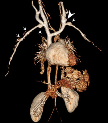
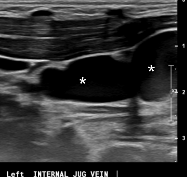
The multifocal ectasia noted in multiple upper extremity veins was the unexpected finding, seen in Figure 1. To our knowledge, this is the first report of systemic venous abnormalities related to Williams syndrome in a child. This venous abnormality was followed for the past 2 years, and a repeat vascular ultrasound of upper and lower extremities was demonstrated: multifocal venous ectasia with beaded configuration of bilateral central internal jugular veins, central subclavian veins, left greater than right, consistent with findings on the prior MRI. An example of the abnormal ultrasound findings is seen in Figure 2. Ultrasound of the lower extremities was normal.
3D-volume rendered magnetic resonance angiography image shows variable degrees of segmental dilation of the central portion of the left internal jugular vein (arrowhead) and bilateral subclavian veins (arrows) resulting in a beaded appearance.

Ultrasound image of the left internal jugular vein reveals similar findings (*) seen in magnetic resonance angiography.

Discussion
Venous anomalies in Williams syndrome
Our case reflects the first reported account of diffuse venous ectasia in a patient with Williams syndrome. This venous anomaly is likely related to the deficiency of elastin seen in patients with Williams syndrome and potentially similar to the pathology of varicose veins. Varicose veins are related to a deficiency in elastin, which allows for deformation of the vein.Reference Bujan, Gimeno and Jimenez4 Varicose veins are deficient in elastin due to increased elastase enzyme and decreased elastin synthesis, similar to the haploinsuffiency of the ELN gene leading to deficiency in elastin as seen in Williams syndrome.Reference Collins1,Reference Bujan, Gimeno and Jimenez4 Clinically, venous abnormalities are better tolerated than arterial abnormalities but do have important implications. Patients with Williams syndrome frequently require central venous access during procedures, catheterizations, and surgeries for vessel rehabilitation. Additionally, the abnormalities can lead to abnormal venous drainage, resulting in oedema, pain, and potentially venous thromboses.
Benefits of MRI in Williams syndrome
Arterial stenoses in Williams syndrome are commonly detected in the great arteries by echocardiography and the renal arteries by ultrasound, though they have a limited role in the assessment of the distal vasculature of the lung bed and distal arterial tree. Advanced imaging studies are helpful to assess the entire vascular tree and in detecting subclinical arterial stenosis in more distal vessels.Reference Hills, Zarate and Danylchuk5
At our institution, we have a comprehensive Williams syndrome clinic and routinely perform MRI of the entire arterial system if there is evidence of significant arterial stenosis or systemic hypertension. This allows us to assess the area of interest and potentially detect subclinical arterial stenosis within the distal vasculature. Screening MRI may be beneficial in Williams syndrome patients to detect subclinical arterial and venous anomalies.
Conclusion
Williams syndrome is associated with a systemic vasculopathy that commonly affects the arterial system and potentially the venous system. This case report highlights the first reported child with Williams syndrome and diffuses venous ectasia. MRI is useful to fully assess the vasculature and serially follow disease progression before and after interventions. MRI screening in Williams syndrome can detect subclinical cardiovascular disease that may improve long-term morbidity and mortality.
Acknowledgements
None.
Financial Support
This research received no specific grant from any funding agency, or not-for-profit sectors.
Conflicts of Interest
None.
Ethical Standards
NA, the report reviews the patient’s case.