Introduction
The aging process can be defined as a progressive and general decline of the organism's functions that leads to a low ability to adaptively react to the constant changes which an individual faces and thus, preserve homeostasis. This dynamic balance, so-called homeostasis, is necessary for the maintenance of health in all biological responses to molecular, physiological, and social stimuli. Because of this balance deteriorates with age, there is an age-related increase in the risk of morbidity and mortality (Refs Reference De la Fuente and Miquel1, Reference De la Fuente, Fernández-Ballesteros, Benetos and Robie2). Although aging is not an illness (it would not be understood that a disease affects 100% of people) the risk of suffering diseases increases with age. Indeed, a growing percentage of the world´s population is over 60 years old, which is translated to a high number of older people, with the social and economic repercussions that this entails for any country. In fact, the denominated demographic pyramid is becoming increasingly regressive and will be inverted in the coming years, representing more older people than children and young people. In addition, the aging process, which starts at adult age (in humans in their twenties), is the longest period of our life. The aging process ends with the death of the individual, and therefore determines his/her life expectancy, which we can also term mean longevity. This longevity, which can be defined as the mean length of time that members of a population, who were born on the same date, live, has increased globally, almost doubling in Western countries since the last decades of the last century. However, this increase is not necessarily associated with an extended health span, which describes both the length of healthy life and the fraction of the total lifespan free from disease.
Collectively, these reasons explain that aging research is entering a new era that has unique biological, medical, commercial, and social implications, demonstrating the need to ensure the best possible conditions to carry out this process. To achieve this, it is necessary to be able to maintain health, which is determined by homeostatic systems' functionality. Accordingly, the nervous, endocrine, and immune systems must function properly as well as the communication that exists among them. This maintenance depends, in each individual, on his/her genes, but to a greater extent on the environment and lifestyle. Both factors (genes and environment) will affect each subject, from the foetal life, the subsequent postnatal development, and growth, conditioning how adulthood is reached, and how the long process of aging is carried out (Ref. Reference De la Fuente, Fernández-Ballesteros, Benetos and Robie2). Thus, the changes that occur during the development of an individual are crucial for the correct development of his/her homeostatic systems, affecting his/her morbidity and, therefore, his/her survival (Ref. Reference Torday3). Depending on how a subject has his/her homeostatic systems in the adult age, so will carry out his/her aging.
Based on the above, it can be understood that the development and function of the homeostatic systems are different among subjects. The same happens with the multidirectional communication that these systems establish among them, generating the denominated neuroimmunoendocrine system. As a consequence, it can be considered that each individual has his/her neuroimmunobiography (Ref. Reference De la Fuente4). This concept reflects one of the most important characteristics of aging, its heterogeneity. The aging rate varies for different individuals of the same chronological age (CA). Thus, the term biological age (BA), indicative of this rate of aging, was established. A younger BA than CA is associated with better health, a slower rate of aging, and, consequently, a longer health span. Nonetheless, it is still difficult to find a way to accurately quantify this BA, and in this context, several clocks have been proposed in the last years, such as the epigenetic clock and the telomere clock, among others. In this sense, our research group has established the ‘Immunity clock’ (Ref. Reference Martínez de Toda5), which will be discussed later.
Achieving successful aging requires maintaining health and this depends on adequate homeostatic systems' functionality. The assessment of the function of the nervous system can be done easily, but indirectly, through the analysis of the behavioural response. Similarly, the functional state of the endocrine system is indirectly assessed by the amounts of circulating hormones. Nevertheless, the immune system is the only one of the three systems in which it is possible to directly analyse the cell function state. This is because leucocytes recirculate through the body, and they can be easily obtained in a minimally invasive way, i.e., in blood samples. For this reason and because of the relevant role of immune cells in our body, the functional state of these cells was proposed as the best markers of health that we could have (Refs Reference De la Fuente, Fernández-Ballesteros, Benetos and Robie2, Reference Martínez de Toda5, Reference Martínez de Toda6).
With aging, the three homeostatic systems are affected as well as the neuroimmunoendocrine communication. If we focus on the immune system, it is an evident fact that with aging there is decreased resistance to infections and increased autoimmune processes and cancers, as well as a low response to most vaccines, which indicate the presence of a less competent immune system. The age-related alterations in this system, a process known as immunosenescence, exert a great influence on the increase in morbidity and mortality associated with aging. This immunosenescence is characterised by the dynamic remodelling of the immune system with a decrease in many functions in both innate and adaptive immune responses (Ref. Reference Bauer and De la Fuente7). In this regard, we have described that several functions of immune cells such as chemotaxis of phagocytes and lymphocytes, phagocytosis, Natural Killer (NK) cytotoxic activity, and proliferative response to mitogens of lymphocytes, can be used in the determination of BA through the Immunity Clock (Refs Reference Martínez de Toda5, Reference Martínez de Toda6, Reference Martínez de Toda8), and they can work as predictors of lifespan in mice (Ref. Reference Martínez de Toda9).
Oxi-inflamm-aging
In the oxidative-inflammatory theory of aging (Ref. Reference De la Fuente and Miquel1), it is proposed that the basis of aging is the chronic oxidative and inflammatory stress, two very interrelated processes (Ref. Reference De la Fuente, Fulop, Franceschi, Hirokawa and Pawelec10), as a consequence of the imbalance due to excess of oxidant and inflammatory compounds versus antioxidants and anti-inflammatory defences generated with the advance of age. These age-associated oxidative and inflammatory stresses affect all cells of the organism, but in a more relevant way those of the homeostatic systems, the nervous, endocrine, and immune systems, affecting their functions and thereby increasing morbidity and mortality. Moreover, this theory proposed the involvement of immune cells, which need to generate oxidant and inflammatory compounds to carry out their functions, in ‘oxi-inflamm-aging’ and, consequently, in the rate of aging of each individual (Refs Reference De la Fuente and Miquel1, Reference De la Fuente11).
Changes in the aging rate through the modulation of the neuroimmunoendocrine system
Altogether, it is understandable that many studies have focused on investigating how environmental and lifestyle factors may be involved in the rate of aging, and more specifically how these can influence the immunosenescence process. Indeed, the goal is not only to increase lifespan but also to increase health span. Within the framework of neuroimmunoendocrine communication, on one hand, everything that involves an inadequate response to stress (negative emotionality, anxiety state, among others) will generate an acceleration of immunosenescence. On the other hand, interventions such as diet, and physical and mental exercise, among others, are effective in preventing the decline in function of immune cells (Refs Reference De la Fuente, Fulop, Franceschi, Hirokawa and Pawelec10, Reference De la Fuente11). Although the need to improve immune health and resistance to infectious diseases in older adults has been brought into sharp focus by the COVID-19 pandemic, it must be considered that because the homeostatic systems are interconnected, any intervention that affects one of these systems such as the immune system, will also affect the others. Humans did not evolve to be alone, for this reason, the social environment has been studied as a characteristic of lifestyle very relevant to the maintenance of health and as a modulator of the rate of aging and longevity. Its impact on lifespan has been analysed using rodents (as rats and especially mice), which similarly to humans are social species. In this context, understanding the mechanisms by which social environment, especially social positive support, promotes resilience to stress and increases mental and physical health during aging will allow new preventive strategies in humans.
In this review, to understand the role that the social environment can have on immunosenescence and therefore on the aging rate of each individual as well as several concepts such as stress, hormesis, and resilience in this context will be discussed. Afterwards, and in the frame of the role of stress in neuroimmunoendocrine communication, an update on the effects that a negative social environment or a positive one has on the homeostatic systems, especially on the immune system will be commented on. Finally, a series of possible mechanisms involved in such effects will be suggested.
Stress response, hormesis, and resilience
Depending on the circumstances, social interactions can be a source of stress or provide a buffer against stress. For these reasons, our body constantly has to adapt to different social situations. On one hand, exposure to a social stressor can induce physiological responses that are protective against subsequent stress exposures in a way that the body is anticipating more stress. On the other hand, constant high-level exposure can increase tolerance to avoid overstressing the system. Such adaptations are essential because the environmental changes can be unpredictable, and the body needs to adjust the trade-off among different functions such as fecundity versus longevity, or energy conservation versus expenditure. In this sense, nervous, endocrine, and immune systems are clearly affected, sensing the environmental changes and communicating the perceived change to the rest of the body. As a consequence, when a stimulus appears a stress response is triggered.
The stress response can be defined as the adaptive physiological modifications that occur as a consequence of any internal or external changes or threats (stressors). Thus, an individual reacts to a physical or mental stressor that is potentially health-threatening by activating interconnected neuroimmunoendocrine circuits. In this regard, the Sympathetic-Adreno-Medullary (SAM) and Hypothalamic-Pituitary-Adrenal (HPA) axes act as the main pathways involved in the stress response, which work through the release of catecholamines (CA) and glucocorticoids (GC), respectively. Nevertheless, in a stressful situation, in addition to the CA and GC mentioned, other neuroendocrine pathways are also activated and other modulators are released such as opioids, dehydroepiandrosterone (DHEA), growth hormone (GH), or prolactin, among others.
Nonetheless, the activation of these neuroendocrine axes depends on various factors, such as how the individual perceives and responds to that stressor. In this sense, it has been proposed that the SAM axis is predominantly activated when the individual facing a stressful situation can maintain control of the situation, whilst the HPA axis seems to be more activated in a stressful situation that causes a feeling of loss of control or even helplessness in the individual (Ref. Reference Henry and Wang12). Nevertheless, the stress response is a pro-survival response that allows the body to face and deal with the challenge, re-establishing the homeostatic equilibrium and promoting health. However, when individuals perceive a noxious stimulus as too intense, or its duration as too long, they may fail to cope with it, maladaptation occurs and neuroimmunoendocrine parameters remain altered, which will accelerate the rate of aging and the appearance of age-related diseases (Refs Reference De la Fuente, Fernández-Ballesteros, Benetos and Robie2, Reference Sayed13, Reference Mariotti14). In this sense, several years ago we demonstrated that women who have moderate and high-perceived stress exhibit worse immune functionality and higher oxidative and inflammatory stress compared to women with low-stress perception. In addition, a positive correlation was found between how they perceived the stress situation and their biological age (Ref. Reference Martínez de Toda15).
This ‘buffering capacity’ that an individual has to respond to and adapt to different stressors has been also referred to as the homeodynamic space and also as ‘survival ability’ (Ref. Reference Rattan16). Nonetheless, as we age the ability to quickly and completely recover after deviation from a normal physiological state or damage caused by a stressor or an adverse health event decline (Refs Reference Kirkland, Stout and Sierra17–Reference Arbeev21). This fact is translated into that an older person will recover less efficiently and have lower chances of survival following the same adverse health event compared to his/her younger self. For instance, at age 85 an average individual has a higher chance of dying following an acute hip fracture than s/he would have had in the 20s (Refs Reference Schnell22, Reference Lin23). The decline in this ability to respond to and adapt to different stressors can contribute to the increased mortality risk associated with age that eventually limits longevity even in people without chronic diseases (through lower survival chances at older ages after acute events, such as fractures, flu, bleeding, wounds, etc.). Among the oldest old individuals, this capacity can also influence their chances of becoming centenarians (Ref. Reference Ukraintseva24).
In the context of the stress response, the dose and the intensity of the stressor are important factors. Accordingly, exposure to mild stresses relates to better health, whereas severe stress is associated with impaired health. Indeed, exposure to mild stress over time can have a beneficial impact on the health of the organism, triggering counteracting mechanisms that over time translates into a strengthened resistance capacity, a concept that was referred to as ‘hormesis’ (Refs Reference Rattan25, Reference Calabrese26). The molecular mechanisms associated with this hormetic effect of strengthening relate to the triggering of different stress response pathways (oxidative, heat-shock, DNA damage stress responses, among others) which activate particular transcription factors that increase the expression of antioxidants, chaperones, or repair proteins which not only resolve the damage caused by the disturbance but can also help repair other damages present or prevent future damages. In this sense, exposure to a short-term and mild variety of stressors has been proposed as an efficient intervention to promote health (Refs Reference De la Fuente11, Reference Rattan25, Reference Dhabhar27).
Importantly, stress responses have been reported to be very different not only depending on the type, duration, or intensity of stressors but also on the characteristics of each individual (personality, life experiences, social situation, controllability, and perception of the situation, among others). All these, cause a very high inter-individual variability in stress responses, which can have as a result a successful or unsuccessful adaptation, and consequently, a maintained or impaired health (Ref. Reference De la Fuente, Fernández-Ballesteros, Benetos and Robie2). It is known that when two individuals are exposed to an adverse event, one can efficiently recover from the situation whereas the other one can be tremendously affected forever. This fact made the term resilience emerge. Resilience is a complex and dynamic process, broadly defined as the ability to adapt successfully to adversity, stressful life events, significant threat, or trauma. It is best conceptualised as a continuum, with potential for change across the life span of an individual (Ref. Reference Southwick28). Studies have repeatedly shown that severity and frequency of exposure to stress matter. Resilient outcomes are less likely in the face of severe adversity. This is especially true of trauma or neglect during childhood when biological stress response systems are under development (Refs Reference Gee and Casey29, Reference VanTieghem and Tottenham30), and of chronic adversity (Ref. Reference Hobfoll31). Additionally, risk factors commonly co-occur and accumulate over time, and higher cumulative risk generally predicts worse outcomes (Refs Reference Burns32–Reference Green34). Yet even at the most severe exposure levels, it is often possible to identify individuals who recover from stress, manage to lead meaningful lives, or find ‘relative resilience’ to severe adversity (Ref. Reference Feder35).
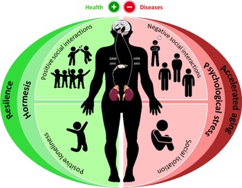
Resilience is thought to operate by attenuating the appraisal of the threat posed by a stressor, by providing effective means of coping with stressors that are appraised as threatening. Also, it acts by reducing affective (e.g., anxiety, depression, anger), behavioural (e.g., smoking, alcohol consumption, loss of sleep, poor diet), and in turn physiological (e.g., activation of SAM and HPA axes) responses to stressors that put people at risk for immune dysfunction and consequently for infections and inflammatory diseases (Ref. Reference Cohen, Gianaros and Manuck36). Although the mechanisms underlying resilience are not clear yet, ranges of psychosocial factors that contribute to resilience have been identified (Ref. Reference Liu37). The factors include active coping (Refs Reference Snow-Turek, Norris and Tan38, Reference Hanton, Neil and Evans39), optimism (Ref. Reference Warner40), cognitive reappraisal (Refs Reference Maren41–Reference Troy43), prosocial behaviour (Ref. Reference Staub and Vollhardt44), social support and others (Refs Reference Ozbay45, Reference Cai46). Concretely, social support is one of the main protective elements that influence family well-being, parenting quality, and child resilience (Ref. Reference Armstrong, Birnie-Lefcovitch and Ungar47). A 10-year longitudinal study found that social support from partners promoted resilience in response to economic stress (Ref. Reference Conger and Conger48). In contrast, poor social support enhances inappropriate stress responses, leading to elevated heart rate (Ref. Reference Stansfeld49), depression (Ref. Reference Oxman and Hull50), increased susceptibility to post-traumatic stress disorder (Ref. Reference Johnson51), and immune alterations (Ref. Reference Cruces52) (Fig. 1).
Dual role of social experiences on health. Depending on the circumstances, social relationships can be a source of stress or a buffer against it. In this regard, negative social interactions, as well as loneliness and social isolation, are associated with the overactivation of the neuroendocrine SAM and HPA axes, releasing CA and GC. If these social-psychological stresses are maintained over time, alterations in all physiological systems are triggered, promoting the acceleration of aging, and increasing the risk of diseases. However, when these social environments are positive such as positive social interactions or positive loneliness (when the individual wants to be alone), they buffer against stress. These positive social experiences as well as low doses and short times of exposure to social threats (perceived as a mild stress situation) can help to develop mechanisms improving the subsequent stress responses and the general function of the organism, process so-called hormesis. Moreover, depending on the individual context (personality, …), the development of hormesis can promote a high resilience capacity, improving all physiological responses and achieving better maintenance of health. CA, catecholamines; GC, glucocorticoids; HPA, Hypothalamic-Pituitary-Adrenal; SAM, Sympathetic-Adreno-Medullary.

Interestingly, not only resilience can modulate immune system response, but also the immune system is capable to change resilient responses (reviewed in Ref. Reference Dantzer53). Accordingly, the inflammatory state of an individual has been described as a modulator of resilient responses, although experimental research demonstrating this relation is still limited. In this sense, only two studies have examined this important relationship. In the first (Ref. Reference Wright54), administration of S. Typhi vaccine, but not placebo, was associated with increased IL-6 concentrations and the subsequent increased negative mood and decreased positive mood. In the second (Ref. Reference Janicki-Deverts55), healthy adults exposed to two different cold viruses (influenza and flu virus) exhibited lower positive affect, a behavioural change associated with daily production of IL-6, but not IL-1beta or TNF-alpha. These findings support a causal association (cytokines can predict the change of mood). Even so, future research trying to shed light on these relations should be undertaken.
Social contexts as modulators of immunosenescence
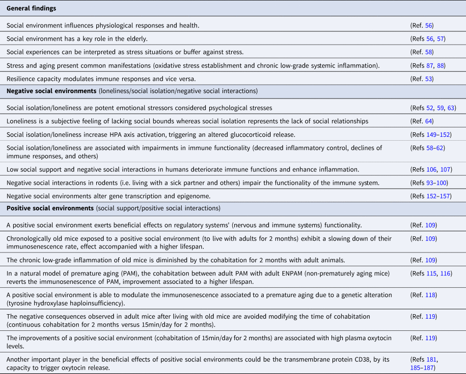
Relations between environmental conditions and health have been long described in humans and rodents. An individual's social environment includes his/her society and all the surroundings influenced in some way by humans. It comprises all relationships, institutions, culture, and physical structures. Although the social context is important throughout life, it is in the elderly when it seems to have a key role, due to physical limitations characteristic of this age (Refs Reference Seeman and Crimmins56, Reference Wahl and Weisman57). Social relationships are a ubiquitous part of life, which affect social, psychological, physiological, and behavioural functions. Depending on the source and nature of the relationship, social interactions can promote negative effects on regulatory systems due to stress or buffering these detrimental consequences, acting as a positive stimulus against stress situations. Nonetheless, to date, most empirical studies of social influences on immunity have focused on the negative aspects of social interactions. Even so, considering this dual role of social contexts, this review will summarise the most important findings related to how loneliness in humans and social isolation in rodents as well as different social interactions in both species can affect immunosenescence (Table 1).
Overview of the most important findings about effects of social environments on immunosenescence and health

Loneliness and social isolation
Social species, such as humans and rodents, need to establish and develop quality social relationships, which ensure an adequate neuroimmunoendocrine system function, preserving homeostasis and hence health. In addition, the maintenance of these relationships enhances the survival and reproductive success of social species by providing protection from environmental threats. By contrast, the disruption of social interactions produces significant detrimental effects on health, affecting all regulatory systems and ultimately, leading to the loss of homeostasis and promoting the development of a wide range of pathologies. Accordingly, negative social environments such as social isolation and loneliness have been associated with increased vascular resistance, elevated blood pressure, increased activation of the HPA axis, and glucocorticoid receptor (GR) dysregulation. As a consequence, alterations in immune functionality, such as decreased inflammatory control, impairment of immune responses, and others have been reported (Refs Reference Cruces52, Reference Avitsur58–Reference Budiu61). Therefore, social isolation and loneliness can be defined as potent emotional stressors, which have been considered a type of psychological stress (Refs Reference Cruces52, Reference Cacioppo62, Reference Menec63). For this reason, facing these situations entails adaptive changes that alter the homeostatic balance. However, if this situation is maintained, the changes become chronic, and maladaptation appears. Indeed, although loneliness and social isolation are important at any age, they are more common in older people either due to the loss of close ones or the difficulty of establishing or maintaining quality relationships. Even so, it is worth noticing that social isolation and loneliness are different concepts. While social isolation represents the lack of significant social relationships, loneliness is related to the subjective feeling of lacking these social bonds (Ref. Reference Hsu64). These differences affect the way both pathological situations can be investigated, given that whereas social isolation can be easily performed in animal models, generating the feeling of loneliness in animals is complicated.
Considering that social isolation and loneliness are both psychological stresses, it is necessary to define what a psychological stress is and how it is able to affect the immune system. Psychological stress can be described as an environmental threat that does not necessarily induce the typical physical ‘fight-or-flight’ response but rather, provokes typical stress-related psychological and physiological alterations. In the context of the immune system, several authors have described that psychological stress induces both quantitative and qualitative changes in innate immunity components. In general, while acute stress increases the number and activity of NK cells (Refs Reference Naliboff65, Reference Segerstrom and Miller66), chronic stress decreases the activity of this immune component (Refs Reference Segerstrom and Miller66–Reference Cruces68). Moreover, the cytokine response of NK cells is also affected by this chronic stress (Refs Reference Gouin, Hantsoo and Kiecolt-Glaser67, Reference Cruces68). However, in the case of phagocytes, a great controversy exists regarding the effects of psychological stress on this immune cell type. Some authors have described that mice exposed to repeated social stress increase the number of splenic monocytes/macrophages, increasing, as a consequence, their pro-inflammatory cytokine production and Toll-like receptor (TLR) 2 and 4 expression, potentiating their antimicrobial activity (Ref. Reference Bailey69). Similar results have been reported in peripheral blood neutrophils from individuals exposed to psychological acute stress (Ref. Reference Khanfer70). Paradoxically, other authors indicated that the released Norephinefrine (NE) after acute psychological stress can diminish the activation of M1 macrophages, which are essential in the healing process (Ref. Reference Kuebler71). Although this latter finding suggests that this acute stress could produce an immunosuppression process, it is widely accepted that CA activates the immune responses. Among the reasons behind this discrepancy, differences in experimental designs and methodologies used could explain this fact.
In the case of acquired immunity, and specifically B-lymphocytes, several studies have described that psychological stress induces changes in their capacity to produce antibodies. In fact, chronic stress is associated with a deficient response of antibodies against vaccines such as influenza virus or hepatitis B (Refs Reference Cohen, Tyrrel and Smith72–Reference Glaser75). Among the molecular mechanisms of these alterations, GC, (Refs Reference Cohen, Tyrrel and Smith72–Reference Glaser75) as well as other stress hormones, such as NE, Ephinefrine (E), prolactin, and growth hormone (GH) have been proposed as important contributors (Ref. Reference Wu76). In addition, psychological stress has been related to worse control of viral infections, which are in a latent state in physiological conditions, such as the herpes virus (Refs Reference Glaser77, Reference Glaser and Kiecolt-Glaser78). This loss of control could be due to the alteration of the antigenic presentation process from B cells to T lymphocytes by psychological chronic stress, promoting a decrease of specific T memory cells against this type of virus (Refs Reference Glaser77, Reference Glaser and Kiecolt-Glaser78). Moreover, this inadequate control against latent viruses together with alterations in antibody production against vaccines could increase the susceptibility to suffering a variety of pathologies, which could be fatal in the elderly. This feature has been described in individuals exposed to psychological chronic stress (Refs Reference Cohen, Tyrrel and Smith72, Reference Glaser79, Reference Dimsdale80).
T lymphocytes, in a similar way to B lymphocytes, are greatly affected by psychological stress, which produces a general impairment in the functions of these cells. In fact, psychological stress promotes a low lymphoproliferative response to several antigenic stimuli (Refs Reference Cruces52, Reference Bartrop81, Reference Delahanty82). Moreover, psychological chronic stress is also able to induce changes in the balance of Th1/Th2 cytokine response, increasing Th2 responses and promoting a decrease in the levels of Th1 cytokines, such as interferon (IFN), IL-2, and IL-12. These changes could potentiate and diminish the immune response at the same time. Accordingly, the decrease of Th1 cellular response could increase the vulnerability to suffering infections and cancer, whereas an increase in Th2 humoral response might increase the probability to suffer allergy and autoimmune diseases. This unbalance would explain the immune changes that appear in individuals exposed to psychological chronic stress with diseases related to this stressful situation. As a consequence, these individuals present an inefficient immune response due to the high activation of the immune system (Ref. Reference Marshall83).
Altogether, social isolation and loneliness, both processes considered a type of psychological stress, are able to produce a general impairment of immune function, which might potentiate the loss of homeostasis and hence health. Moreover, since in the elderly the immune system suffers a lot of changes that affect almost every immune component, the absence of social bonds could be critical in the establishment of the immunosenescence process and therefore in the maintenance of health. In fact, aged individuals exposed to social isolation or loneliness could suffer the consequences of immunosenescence before, affecting regulatory systems' functionality and promoting the loss of homeostasis and hence health. Moreover, these individuals would be more susceptible to suffering diseases, exhibiting high morbidity and mortality (Refs Reference House, Landis and Umberson84–Reference Pantell86). Therefore, although stress situations affect individuals of all ages, the older can be more affected. In fact, with aging, alterations in the HPA and SAM axes appear and they are responsible for decreasing stress adaptability and increasing physiological systems' impairments. Thus, there is an overlap between the alterations that appear with aging and inadequate stress responses in physiological systems. This is clearly observed in the consequences that the immune system suffers with both processes. Aging is associated with impairments of immune function and low responses against immune challenges, alterations that also are observed in individuals exposed to chronic stress. Moreover, in both stress and aging, a chronic oxidative stress situation and a chronic low-grade systemic inflammation develop (Refs Reference Chovatiya and Mdezhitov87, Reference Garrido88). These oxidative and inflammatory states, as mentioned previously, are associated with accelerated biological aging and thus, with an increased risk of age-related diseases and premature mortality.
Finally, it is worth noticing that not all people that suffer social isolation/loneliness are affected in the same way. Concepts such as resilience or people's attitude have a particular influence on how we are affected by these psychological stresses. One example is how an individual responds to such stress. Experimental models have shown that rats and mice that respond to isolation with higher anxiety levels have a more significant immune impairment (Ref. Reference Cruces68), leading to lower longevity (unpublished data). Another example is the attitude; a positive attitude focused on positive emotions could improve the cognitive and immune impairment caused by loneliness and isolation (Ref. Reference Kearns and Creaven89). In addition, during the last two years, social isolation and its adverse effects on health have become very important due to the confinement caused by the COVID-19 pandemic, particularly among the elderly (Refs Reference Pietrabissa and Simpson90, Reference Holt-Lunstad and Perissinotto91), who have suffered the most important consequences of this isolation and whose consequences are yet to be determined. Future research will demonstrate the consequences of this epidemiological decision on people from different age groups.
Effects of negative social interactions
Social stress is often a recurring aspect of the environment in humans and other animal species. As previously commented, these stressful social situations can lead to a dysregulation of the HPA axis, which results in systemic changes affecting all physiological systems (Ref. Reference Smith and Wang92). Accordingly, several previous studies have reported how negative social interactions, such as living with a sick partner, can modify the functionality of these physiological systems. In fact, the cohabitation of healthy mice with sick individuals deteriorates the immune system of the former. Accordingly, in female mice, cohabitation with a sick cage-mate decreases the levels of NE and increases the turnover rate of hypothalamic NE (Ref. Reference Alves93), decreases neutrophil and macrophage oxidative burst in response to a stimulus as well as the percentage of neutrophil and macrophage phagocytosis (Refs Reference Alves93–Reference Palermo-Neto and Alves97), modifies dendritic cells phenotype (Ref. Reference Tomiyoshi98), and increases the number and concentration of tumour cells in the ascetic fluid (Ref. Reference Machado99). In males, cohabitation with a sick cage-mate induces an increase in the number of eosinophils and neutrophils, and the levels of IL-4 and IL-5, as well as a decrease in bone marrow cell count and the levels of IL-10 and IFN-gamma, indicating an exacerbation of the pro-inflammatory response (Ref. Reference Hamasato100).
In the case of humans, negative social interactions are the most frequent daily stressors, which are more highly associated with poor well-being than non-interpersonal stressors (Ref. Reference Almeida101). Relative to aged people, it has been reported that they have fewer negative interactions and are more likely to avoid them (Ref. Reference Birditt, Fingerman and Almeida102). However, when they experience these negative situations, their well-being is often profoundly affected (Ref. Reference Birditt103) because their HPA axis is less flexible, recovering slower from stress (Ref. Reference Giordano104) and showing less ability to maintain homeostasis (Ref. Reference Majnaric105). Besides, there is some evidence showing an association between low social support and shorter leucocyte telomere length (Ref. Reference Carroll106). Therefore, it is to be expected that low social support or negative social interactions deteriorates immune functions and enhances pro-inflammatory activity (Refs Reference Carroll106–Reference Cole108), increasing the risk of disease and premature death by accelerating the aging process (Ref. Reference Martínez de Toda8).
Effects of positive social interactions
The beneficial effects that social environment can exert on regulatory systems' functionality in aged individuals, and specifically, on immunosenescence have been hardly studied. The first experimental approximation in this sense was reported in (Ref. Reference Garrido109). In that work, chronologically old female mice (20 months of age) cohabited continuously (for two months) with adult counterparts (9 months of age) in a ratio of 2:5 per cage. In parallel, one group of chronologically old mice and adult mice lived with counterparts of the same chronological age. After that social experience, several immune functions were assessed in peritoneal leucocytes from all experimental groups. Paradoxically, while a general impairment of immune functions was observed in the chronologically old control group, peritoneal leucocytes from old mice exposed to a predominantly adult environment exhibited broad functional immune improvements. In fact, significantly higher phagocytosis, chemotaxis, NK activity, and lymphoproliferative capacity in response to LPS were reported compared with their control aged-counterparts. Thus, these results evidence the positive effects that the social environment can have on immune functionality in aged individuals, slowing down the impairments associated with immunosenescence. Moreover, the improvements of that cohabitation were also observed in the control of chronic low-grade inflammation. This sterile inflammation is a typical phenomenon of aging and it forms part of the immune signature of immunosenescence (Ref. Reference Barth110). In fact, peritoneal leucocytes from these aged mice after living with adults exhibited a recovered inflammatory balance, releasing lower pro-inflammatory cytokines (IL-1beta and IL-6) and higher anti-inflammatory cytokines (such as IL-10) in absence of any antigenic stimulus compared to those aged animals that did not cohabit with adults. Another important finding of that work is that the values obtained in almost all immune parameters analysed in aged individuals that lived with adult mice were similar to those obtained in control adults, which highlights the enormous extent that this social strategy can have on immunosenescence. Further, after this analysis all animals were maintained until their natural death, trying to decipher whether these improvements could positively affect their lifespan. Importantly, these improvements were translated into a longer lifespan in chronologically aged mice subjected to this cohabitation. Therefore, this cohabitation between aged mice and adult counterparts entails a positive social experience for aged mice, which is translated into a slower establishment of immunosenescence, and a higher lifespan.
Afterwards, the same experimental design was carried out in a natural model of premature aging in mice based on the stress response that chronologically adult mice exhibit when exposed to a new environment (like a T-maze). These prematurely aging mice (PAM) exhibit an inadequate stress response in that environment, being characterised by a premature immunosenescence establishment together with a higher oxidative-inflammatory stress in their leucocytes compared to their chronologically non-prematurely adult counterparts, termed exceptional non-prematurely aging mice (ENPAM). Indeed, these PAM exhibit a shorter lifespan compared to ENPAM (Refs Reference Garrido88, Reference Martínez de Toda111–Reference Viveros114). However, when these PAM lived in a predominantly ENPAM environment for 2 months, a general improvement in immune functions of their spleen and thymus leucocytes was observed, showing higher chemotaxis, Natural Killer activity and lymphoproliferative responses to Concanavalin A (ConA) and Lipopolysaccharide A (LPS) compared to control PAM (PAM that cohabited with PAM) (Ref. Reference Garrido115). Similarly, this improvement was also observed in their peritoneal leucocytes, exhibiting higher chemotaxis, phagocytosis, NK activity, and lymphoproliferation in response to ConA and LPS compared to their control PAM (Ref. Reference Garrido116). These results resemble the previous one obtained in chronological aging, indicating, therefore, the importance of an adequate social environment in the establishment of immunosenescence.
Considering the positive results obtained in this natural model of premature aging based on the inadequate stress response, the authors then sought to know whether the beneficial effects of social environment could be enough to revert or at least ameliorate immunosenescence in a genetic model of premature aging, based on the deficiency of the gene encoding for tyrosine hydroxylase (Th), the enzyme that governs the production of CA. This genetic alteration had been previously described as a model of premature aging, since the Th haploinsufficient (hemizygotic: HZ) mice (TH-HZ) exhibit at the adult age a premature immunosenescence, negatively affecting their lifespan, which is shorter than in wild-type mice (Ref. Reference Garrido117). In fact, after chronologically adult TH-HZ mice cohabited for two months in a predominantly wild-type environment (ratio 2:5) they improved several immune functions of their peritoneal leucocytes, exhibiting higher chemotaxis of macrophages and lymphocytes, phagocytosis, NK activity, and LPS-stimulated lymphoproliferation compared to their control TH-HZ counterparts that did not live in this social environment (Ref. Reference Garrido118). Therefore, these results point out that this social environment not only can slow down the immunosenescence establishment in a natural model of premature aging (PAM) but is also able to modulate the effects of premature aging due to a genetic alteration (TH-HZ mice), demonstrating the importance and also the potential of social context beneficial effects on immunosenescence.
However, this social context was not beneficial for all the animals included in that cohabitation. In fact, the function of peritoneal leucocytes from adult animals that lived with old individuals exhibited an impairment in almost all the immune functions compared to their control adult counterparts, although these negative effects did not affect their lifespan (Ref. Reference Garrido109). Similarly, the immune functions of ENPAM were also compromised after the cohabitation with PAM, although in a similar way to chronologically adult animals, the lifespan was not affected (Ref. Reference Garrido115). In addition, adult wild-type mice that cohabited with Th-Hz mice presented a deterioration of their immune functions (Ref. Reference Garrido118). Therefore, although this continuous social environment positively affects the immune function of aged individuals, it has negative consequences for non-aged individuals. Trying to avoid the negative effects on non-aged individuals, the conditions of cohabitation were then changed. When the cohabitation time is reduced to 15 min/day for 2 months, the previously commented improvement in immune functionality as well as in lifespan in aged mice is maintained, avoiding not only the deleterious effect previously described in adult animals but also improving their immune response. Moreover, these adults that cohabited with old mice maintained better immune activity until reaching old age, showing a slowdown of the immunosenescence process (Ref. Reference Díaz-Del Cerro119). Similarly, when the cohabitation between PAM and E-NPAM is shortened to 15 min/day for 2 months, it is possible to maintain the previously observed positive effects on immune functions of PAM, avoiding the immune deteriorations above commented in E-NPAM (Ref. Reference Carr120).
Finally, although in the case of humans these kinds of experimental studies are too complicated to carry out, several previous reports have described how the social context can affect their immune system response. In fact, it has been observed that a shared environment (socio-economic status, diet, smoking, alcohol intake, exercise levels…) acts in some way to bring immune profiles towards a convergent equilibrium (Ref. Reference Díaz Del Cerro, Félix and De La Fuente121). Therefore, a positive social environment or social support from a committed partner or best friend can ameliorate stress, reducing the risk of diseases and improving well-being (Refs Reference Heinrichs122–Reference Karelina and DeVries124). It has also been observed that social support optimises surgery recovery in older people (Ref. Reference Zhang and Tumin125). In a study about the links between relationships and immunity, it was found that people with strong social networks were less likely to get sick when exposed to a cold virus (Ref. Reference Cohen126). According to a work carried out by Harvard Medical Center, those with stronger social relationships improved their life expectancy by 50 per cent (Ref. Reference Holt-Lunstad, Smith and Layton85). Another study found that participants who had multiple social conflicts in their lives were at greater risk of developing an infection when subsequently exposed to a common cold virus than those with fewer or no conflicts (Ref. Reference Cohen126). In addition, those participants with greater perceived support were protected from stress-associated susceptibility to infection. In fact, social support can also alleviate the impact of stress by providing a solution to the problem, reducing the perceived importance of the problem, or providing a distraction from the problem. It might facilitate healthful behaviours such as exercise, personal hygiene, proper nutrition, and rest (Refs Reference House, Landis and Umberson84, Reference Cohen127). In this sense, previous work with primates has shown that those that spend more of their time engaged in affiliative behaviours (e.g., touch, close proximity) showed less suppression of mitogen-stimulated lymphocyte proliferation following continuous exposure to a social stressor (living in an unstable as opposed to stable social group) than those spending less time affiliating with other counterparts (Ref. Reference Cohen128). Interestingly, humans who reported being regularly hugged by others showed a similar resilience to the potential immune effects of conflicts with others. In a viral-challenge study, those with conflict and few days being hugged were at greater risk of infection in response to exposure to a common cold virus, but those who received many days of hugging were protected from the role of conflict in increasing risk (Ref. Reference Cohen126).
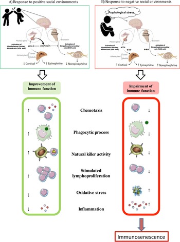
Positive social environments have been closely related to an increase in plasmatic oxytocin (OXT) levels. Considering that this neuropeptide can enhance immunity (Ref. Reference Smith and Wang92), the beneficial effects on health and especially on the immune system function in aging promoted by positive social environments and/or social support may occur via an OXT-mediated mechanism (Fig. 2). A detailed explication of the possible role of OXT on the beneficial effects of this kind of environments will be summarised in the next section.
Effects of positive and negative social environments on immune function. Exposure to positive social environments triggers the release of OXT, which produces the blockage of the neuroendocrine SAM and HPA axes. As a consequence of this blockage and the immunomodulatory capacities of Oxytocin, several immune functions are improved. In fact, leucocytes from individuals exposed to positive social environments exhibit high chemotaxis and phagocytic process, both processes closely linked with innate immunity. Also, the natural killer activity of these individuals is potentiated. With regard to acquired immunity, a high lymphoproliferation in response to stimuli has been also reported. These immune improvements are associated with low oxidative stress and low sterile inflammation establishment. However, if an individual is exposed to negative social environments, the overactivation of both SAM and HPA axes is promoted, triggering the increase of its neuroendocrine mediators, such as catecholamines (epinephrine and norepinephrine) and glucocorticoids (cortisol), respectively. If this psychological stress situation is maintained over time, an impairment in leucocyte functions is triggered. Among them, low chemotaxis, phagocytosis, and natural killer activity together with low stimulated lymphoproliferation appear. Moreover, oxidative and inflammatory stresses are established. Therefore, these negative social environments accelerate the appearance of immunosenescence. HPA, Hypothalamic-Pituitary-Adrenal; SAM, Sympathetic-Adreno-Medullary.

The key role of physical contact in the beneficial effects of social environment
Although the importance of positive and strong social networks has been previously commented on, the physiological mechanisms underlying these effects are mainly unknown. Several physiological mechanisms could be related to the improvements described by the social environment. Thus, visual (Ref. Reference Mesik129), olfactory (Ref. Reference Patel and Larson130), and auditory (Ref. Reference Knutson, Burgdorf and Panksepp131) mechanisms could be involved in the observed effects, but it is known that the sense of touch is of great importance in social interactions as it is by the skin that we feel and interact with other individuals (Ref. Reference McGlone, Wessberg and Olausson132). In fact, touch is the main way of interindividual communication and evolutionarily precedes oral communication (Ref. Reference Field133), which suggests that touch plays an important role in social behaviour and human communication. In addition, some authors postulate that physical contact produces a biological stimulation that is needed throughout life (Ref. Reference McGlone, Wessberg and Olausson132).
Before birth, the foetus receives tactile stimulation through the mother's abdominal walls (Ref. Reference Lagercrantz and Changeux134). After birth, the newborn continues receiving touch stimulation by being cuddled breastfed, and held skin-to-skin, which is essential for growth and development and also calms infants in pain and discomfort (Refs Reference Bellieni135–Reference Cooijmans137). The importance of physical contact in the early stages of development is that touch deprivation in infants is related to later cognitive and neurodevelopmental delays (Refs Reference MacLean138, Reference Nelson139). This is important because what one individual experiences, from before birth, through youth and adulthood, will influence his or her aging process (Ref. Reference De la Fuente and Miquel1). In this context, many studies have demonstrated that physical contact, in addition to providing information, emotions, or social support (Ref. Reference Field133), is also able to impact people's health (Ref. Reference McGlone, Wessberg and Olausson132) improving cardiovascular responses, decreasing blood pressure and heart rate (Refs Reference Christenfeld and Gerin140, Reference Grewen141). Nevertheless, the number of touches matters (Ref. Reference Vaidis and Halimi-Falkowicz142). It has been observed that touch therapy is useful to treat pain, cancer, dementia, and multiple sclerosis among others (Refs Reference Finch143, Reference Celebioglu144). In the context of aging, physical contact has been demonstrated to be useful to reduce anxiety, physiological stress, and pain and improve sleep and respiratory rate in older people (Ref. Reference Papathanassoglou and Mpouzika145).
These effects of physical contact are not only observed in therapy conditions, but also during cohabitation. Recently, our research group observed that during cohabitation, in female CD1 mice, physical contact is essential to obtain the social environment effects commented before (unpublished data). This study demonstrated that skin-to-skin contact between old and adult female CD1 mice, and between PAM and E-NPAM, old mice and PAM resulted in an improvement of their behavioural responses, immune function, redox state, and greater longevity with respect to those that cohabited but without physical contact. In addition, this skin-to-skin contact strategy did not have deleterious effects on adults and E-NPAM and was able to increase the lifespan of adult mice.
For all the above, physical contact has been suggested as a stress buffer, playing a regulatory role in the body's responses to stress (Ref. Reference Morrison146). This is possible thanks to the skin being considered a neuroimmunoendocrine organ (Ref. Reference Roosterman147) since it contains immune and neural cells influencing such systems through neurotransmitters or hormones such as CA or OXT (Ref. Reference Roosterman147). OXT is the principal hormone that is released during physical contact (Ref. Reference Morrison146). OXT modulates the HPA axis and can antagonise corticotropin-releasing hormone (CRH) through noradrenergic signalling, thus decreasing cortisol production and, therefore, improving stress responses. This fact would lead to an improvement in the behavioural and immune response since GC exerts a suppressive effect on them. In addition, OXT also exhibits anti-inflammatory and antioxidant effects (Ref. Reference Morrison146). For this, physical contact has been suggested to be anti-inflammatory, improving immune function, which is a great marker and predictor of longevity (Refs Reference De la Fuente and Miquel1, Reference Martínez de Toda6, Reference Martínez de Toda8, Reference Martínez de Toda9).
Therefore, the importance of the observed effects in positive social networks lies in the physical contact, and although new online social interactions are being explored (Ref. Reference Panzarasa148) they may not be as beneficial as a relationship that includes skin-to-skin contact.
Molecular mechanisms of social environment
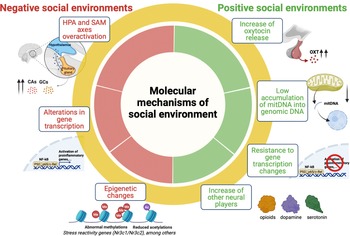
As was previously indicated, poor social support and/or negative social interactions in aged individuals accelerate the immunosenescence process, contributing to a higher rate of aging and an increased risk of disease. Conversely, social support and/or the existence of positive social relationships are an essential component of healthy aging and are associated with better immunity, overall health, and a longer and healthier lifespan. Here, the molecular mechanisms underlying these negative and positive effects of the social environment on the immune system and especially on immunosenescence will be summarised (Fig. 3).
Several molecular mechanisms of social environment effects. The mechanisms underlying the effects that the social environment has on the organism may depend on the type (positive or negative environments). Thus, on one hand, in individuals exposed to negative environments (loneliness, social isolation, or negative social interactions) the overactivation of neuroendocrine stress axes (HPA and SAM axes) appears, triggering the continuous release of glucocorticoids (GC) and catecholamines (CAs). Indeed, alterations in gene transcription have been reported. Among them, the activation of pro-inflammatory genes which will increase the inflammatory state of the individual is one the most known. Finally, changes in epigenetic mechanisms such as abnormal methylations and reduced acetylations are produced. On the other hand, when an individual is exposed to positive environments, they show an increase in oxytocin (OXT) release, mainly produced by the posterior pituitary. Also, mechanisms generating resistance to changes in gene transcription have been suggested. Similarly, the increase of other neural players, such as opioids, serotonin, and/or dopamine has been described. Finally, a decrease of mitochondrial DNA (mitDNA) insertions into the genomic DNA, a process closely related to immunosenescence, could be occurring in leucocytes from these individuals. BDNF, Brain-Derived Neurotrophic Factor; CA, catecholamines; GC, glucocorticoids; HPA, Hypothalamic-Pituitary-Adrenal; OXT, oxytocin; SAM, Sympathetic-Adreno-Medullary; mitDNA, Mitochondrial DNA.

Molecular mechanisms of loneliness/social isolation
It is well accepted that detrimental effects of social isolation/loneliness are produced by HPA and SAM axis overactivation, which are translated into an abrupt increase in plasmatic levels of GC and CA (Refs Reference Cruces52, Reference Steptoe149, Reference Stansfeld49). Among these molecular players, the effects of GC on regulatory systems have been extensively studied. These hormones have been proposed as regulators of a wide variety of physiological processes related to social isolation/loneliness via nuclear hormone receptors-mediated control of gene expression (Ref. Reference Cole108).
In response to an acute stress, cortisol activation of the GC receptor (GR) exerts broad anti-inflammatory effects by inhibiting nuclear factor (NF)-kB/Rel transcription factors and other pro-inflammatory signalling pathways (i.e., the Janus kinase signal transducer and activator of transcription (JAK/STAT) and interferon response factor (IRF) signalling) (Ref. Reference Xavier151). Given the broad anti-inflammatory effects of GC, it is normal to consider that chronically lonely people with elevated cortisol levels should be relatively protected from inflammation-mediated processes rather than having the increased risk empirically observed (Refs Reference Cruces52, Reference Cole108, Reference Xavier151). This contradictory effect involves the functional desensitisation of the GR pathway that mediates the transcriptional response to GC. In this sense, several molecular mechanisms have been shown to render cells insensitive to the anti-inflammatory effects of GC in vitro, including decreased expression of the NR3C1 gene (encoding for GR), post-translational modifications of GR protein, increased expression of GR antagonists, and decreased activity of GR transcription co-factors (Ref. Reference Ferland and Schrader150). Accordingly, in both humans and rodents, prolonged stress has been linked to reduced cellular expression of NR3C1/Nr3c1 and increased cellular resistance to GC inhibition of pro-inflammatory cytokine responses (Refs Reference Ferland and Schrader150, Reference Xavier151). Therefore, the pro-inflammatory signalling seems to persist in socially isolated individuals with high cortisol levels because of the impaired GR-mediated signal transduction, which prevents the cellular genome from effectively ‘hearing’ the anti-inflammatory signal sent by circulating GC.
Indeed, social experiences can themselves alter gene transcription and have consequences for health. In particular, social isolation/loneliness can modulate gene expression across tissues (Refs Reference Xavier151, Reference Kent and Agrawal152) and especially in immune cells. Blood leucocytes from isolated people present genetic expression changes, exhibiting a high expression of pro-inflammatory mediator genes, such as those related to the NF-kB signalling pathway compared to non-isolated individuals. Indeed, these isolated subjects show in their leucocytes low expression levels of genes related to anti-viral responses and antibody production genes (Ref. Reference Cole108), promoting the inadequate immune response against future infections. Thus, these transcription alterations contribute to the immune impairments and the establishment of the inflammatory stress observed in isolated individuals, at least in part.
Finally, classical epigenetic mechanisms for the control of gene expression are also implicated in the effects of prolonged social isolation (Refs Reference Weaver153–Reference Wang156). Intermittent social isolation in rodents can modulate DNA methylation and histone post-translational modifications at regulatory elements of genes involved in stress reactivity, including the GR gene (Nr3c1) (Ref. Reference Weaver153) and the mineralocorticoid receptor (MR) gene (Nr3c2) (Ref. Reference Gapp157). This alteration has been associated with a rewiring of stress responses and behavioural adaptations. Indeed, chronic social isolation can also alter the epigenome throughout life. In this sense, mice at postnatal day (PND) 21 subjected to social isolation for 2 months show a global increase in the level of the repressive histone post-translational modification H3K9me2 in neurons, an effect correlated with increased transcription of the H3K9me2 histone methyltransferase (HMT) G9a and GLP in the hippocampus (Ref. Reference Wang156). These genetic alterations have been also demonstrated in adulthood. In fact, chronic social isolation for 3 months in adult mice produced a significant global increase in DNA methylation, H3K4 di-methylation, and trimethylation, as well as a trend towards an increase in the global levels for H3K9ac, in the midbrain (Ref. Reference Siuda155). In all cases, the increase in epigenetic modifications was associated with an increase in the catalytic processes leading to such epigenetic modifications. For instance, H3K4 HMT activity was significantly enhanced in the midbrain of socially isolated male mice, which could suggest increased transcription of genes encoding for H3K4 HMT (Ref. Reference Siuda155). Prolonged social isolation also increased the transcription of genes encoding for histone deacetylases (HDAC), such as Hdac1 and Hdac3, which correlated with decreased CpG methylation at their promoter regions (Ref. Reference Siuda155). This supports the hypothesis that chronic isolation can affect gene regulatory networks by altering epigenetic modifiers. In contrast, the transcription of the gene encoding for the serotonin transporter Slc6a4 is markedly reduced by social isolation and this correlates with increased DNA methylation at its promoter region (Ref. Reference Siuda155). In this sense, although these molecular signatures of social isolation/loneliness have not been studied in immune cells yet, it is probably that similar molecular changes occur in this regulatory system. Moreover, considering the communication that exists between the nervous and immune systems, the molecular alterations previously reported in the nervous system could be affecting immune function. In sum, all these genetic alterations contribute to impairments related to social isolation and loneliness, including the inflammatory profile exhibited by individuals who suffer these social threats.
Molecular mechanisms of positive social environments
Compared to social isolation/loneliness, the underlying mechanisms by which positive social environments and social support promote healthy aging have been poorly studied. Even so, several molecular candidates, such as OXT, CD38, and other neural players have arisen as possible main actors in these beneficial effects.
Traditionally, OXT has been considered for its role in lactation and parturition. However, growing evidence in the last years has pointed out diverse roles related to pro-social functions. Investigations across species have shown that this neuropeptide plays a key role in encoding information relevant to social interactions and is critically involved in the regulation of complex social cognition and behaviour, including attachment, social recognition, social exploration as well as anxiety and fear-related behaviours (reviewed in Ref. Reference Gapp157). Apart from these pro-social functions, social support and positive social interactions have also been closely related to an increased OXT release. Both men and women had higher plasma OXT levels after having supportive partners (Ref. Reference Grewen159). Similarly, couples engaged in a programme of affiliative touch over 4 weeks resulted in higher post-treatment salivary OXT concentrations than couples in the non-intervention group (Ref. Reference Holt-Lunstad, Birmingham and Light160). After these studies, other authors have also reported similar OXT-related effects in different positive social environments (Refs Reference Scheneiderman161, Reference Plasencia162). Therefore, these data evidence the correlation between OXT and social support/positive social environments.
In addition, the OXT receptor (OXTR) is expressed in numerous tissues throughout the body, which demonstrates the capacity of OXT to regulate diverse functions by endocrine and paracrine bias. Among them, the modulation of the immune system by this neuropeptide has been widely characterised (reviewed in Ref. Reference Plasencia162). In fact, OXTR is expressed in the thymus (Ref. Reference Hansenne164), monocytes as well as macrophages (Ref. Reference Yamaguchi165), and this hormone can act on the immune system by activating OXTR directly and/or through sympathetic outflow (Ref. Reference Yang, Wang and Ju166). OXT promotes the formation of human hematopoietic stem cells (Ref. Reference Elabd167) and rat bone marrow mesenchymal stem cell migration (Ref. Reference Kim168). Furthermore, OXT also has important regulatory roles in immunogenic defence and homeostasis. Accordingly, OXT significantly increases peripheral blood mononuclear cell responses to phytohemagglutinin (Ref. Reference Macciò169) and decreases both superoxide production and release of pro-inflammatory cytokines from OXTR-bearing monocytes and macrophages, promoting an adequate immune response (Ref. Reference Szeto170). Moreover, peripheral OXT administration inhibits adipose tissue inflammation by reducing IL-6 production (Ref. Reference Nation171) and continuous OXT delivery reduces inflammation and cellular apoptosis (Ref. Reference Jankowski172). OXT also has important regulatory roles in the recovery of immune homeostasis in adverse conditions. For instance, septic shock results from an excessively defensive and inflammatory response by releasing cytokines into the circulation from activated immune cells. Interestingly, in the early phase of sepsis (4–6 h), there is an increase of OXT in plasma, which decreases nitrite, TNF-alpha, and IL-1 beta levels in macrophages (Ref. Reference Oliveira-Pelegrin173), an effect demonstrated in both humans and animals and proposed as a molecular mechanism to minimise immunologic damage (Ref. Reference Ross, McDonald-Jones and Miller174). Indeed, growing evidence demonstrates that OXT also acts as an anti-inflammatory protective molecule for the adult gastrointestinal system (reviewed in Ref. Reference Ross, McDonald-Jones and Miller174). In adult rodents, stress-induced colitis increases intestinal damage, oxidative stress, neutrophil infiltration, the expression of inflammatory pathway genes, and anxiety-like behaviour. OXT treatment dampens the severity of intestinal damage and alleviates anxiety, whereas co-administration of an OXTR antagonist prevents OXT´s protective effects (Refs Reference Cetinel176, Reference Welch177). These anti-inflammatory effects are related to OXT-attenuated oxidative stress and inflammatory cytokine release, as has been previously reported in several experimental conditions (Refs Reference Szeto170, Reference Nation171, Reference Iseri178, Reference Kiyikli179). Similarly, OXT also preserves mitochondrial function, reducing, therefore, oxidative stress (Ref. Reference Kaneko180). In sum, the modulatory effects of OXT on immune cells and the increased expression levels of this neuropeptide in individuals exposed to social support and/or positive social relationships reinforce the role of this neuropeptide as a mediator of the beneficial effects exerted by positive environments. Accordingly, we have recently reported how the intermittent cohabitation (15 min/day for 2 months) of chronologically aged mice with adult animals produces an improvement of several immune functions as well as slows down the immunosenescence process establishment in peritoneal leucocytes from aged animals, which is associated with higher plasmatic concentrations of OXT (Ref. Reference Díaz-Del Cerro119). These same effects were then observed in prematurely aging mice (unpublished data). Even so, further research trying to define how OXT improves these immune functions in the context of immunosenescence as well as if other molecular mechanisms related to OXT are also involved should be addressed.
In addition, another recently described player related to OXT release is the multifunctional transmembrane protein CD38, which is widely expressed in immune cells. Since its discovery, growing evidence suggests that CD38 contributes to cell signalling modulating antigen-mediated T-cell responses (Ref. Reference Glaria and Valledor181). Indeed, this protein plays multiple roles derived from intrinsic enzymatic activities as CD38 converts nicotinamide adenine dinucleotide (NAD) phosphate (NADP) to generate nicotinic acid adenine dinucleotide phosphate (NAADP) (Ref. Reference Kar, Mehrotra and Chatterjee182). As a consequence, the enzymatic products generated in these reactions, which are calcium-mobilising second messengers, exert relevant signalling functions in diverse cellular contexts. In the immune response, they are important for the recruitment of different leucocytes toward several chemotactic signals produced at the site of infection (Refs Reference Glaria and Valledor181–Reference Piedra-Quintero183). Also, CD38 regulates leucocyte infiltration to infected/inflamed tissues and exhibits an important role in phagocytosis (Ref. Reference Lischke184). Thus, CD38 acts as an important modulator of immune function. Similarly, in the context of OXT secretion, the role of CD38 has already been established (Refs Reference Tolomeo185, Reference Makhanova186). This protein through its enzymatic function mediates ADP-ribose (ADPR) and cyclic ADPR (cADPR) production, extracellular signal-regulated kinase (ERK1/2) activation, and Ca2+ mobilisation, processes triggering OXT release (Ref. Reference Jin187). Additionally, it also promotes OXT release by activating molecular cascades of OXT autoregulation (Refs Reference Tolomeo185, Reference Makhanova186). Therefore, considering the importance of this protein in the release of OXT and the role it plays in the control of immune responses it is conceivable that CD38 could be involved in the beneficial effects previously commented in individuals with social support/positive social environments. Nonetheless, these hypothetical mechanisms have not been studied yet.
Another important finding regarding the improvements due to this positive social environment is the recovery of the inflammatory balance that is exhibited by old and prematurely aging mice after living in a younger environment. In fact, peritoneal leucocytes from chronologically and prematurely aging mice after living with adult animals exhibit high anti-inflammatory together with low pro-inflammatory cytokine release (unpublished data). The abolition of this chronic low-grade inflammation might be related to a lower accumulation of mitochondrial DNA fragments inside the nuclear genome of T-lymphocytes. In fact, previous reports have described that the constant generation of ROS in the mitochondria (mitROS) together with impairments in the DNA-repair systems produces single and double-strand breaks in mitochondrial DNA (mitDNA), giving rise to highly oxidised mitDNA (Ref. Reference Suter and Richter188–Reference Singh, Choudhury and Tiwari190). These oxidised fragments can migrate into the nucleus, increasing the genomic instability as well as eventually affecting proper chromosome segregation (Refs Reference Patrushev191–Reference Caro193), both processes contributing to cellular T-cell senescence. As a consequence, the functions carried out by these immune cells, i.e., the lymphoproliferative responses to mitogens are impaired (Refs Reference Martínez de Toda5, Reference Martínez de Toda6, Reference Martínez de Toda8). Interestingly, this recently proposed molecular mechanism of immunosenescence is reverted when aged or prematurely aging mice live in a predominantly adult environment, showing in their peritoneal T-lymphocytes lower amounts of oxidised mtDNA and exhibiting an improvement in the lymphoproliferative responses to antigens (unpublished data).
Although alterations in gene transcription due to social support/positive social environments is still not well described, it has been indicated how these positive environments can be protective against these molecular alterations. In this sense, rats exposed to a chronic unpredictable stress protocol for 18 days followed and living in pairs for 1 month maintained HDAC5 expression compared to isolated counterparts. As a consequence, the maintenance of HDAC5 expression ensured adequate levels of Brain-Derived Neurotrophic Factor (BNDF) as well as enhanced neural functions essential for appropriate behavioural responses to stress, highlighting the buffering capacity that these positive environments give to individuals exposed to stressful situations (Ref. Reference Borges194). In this regard, there is increasing evidence to support the hypothesis that the balance of MRs to GRs can be crucial for effective regulation of the stress response and the development of resilience capacity (Ref. Reference Oitzl195). Social support modulates the stress-related gene expression in various brain regions as well as ensures the balance between both receptors, which generates a better response to stress (Ref. Reference Kanitz196). Considering the communication that exists between the nervous and immune systems, this adequate control of stress responses will be translated into an appropriate immune function and in the case of old people, a decreased immunosenescence rate and, therefore, a long health span.
Finally, other neural actors have been also related to the positive effects of social support/positive social environments, such as serotoninergic, dopaminergic as well as opioid neural pathways. In this line, it has been indicated that a polymorphism in the serotonin transporter gene (5-HTTLPR) is critically involved in setting sensitivity to social experiences. Accordingly, individuals with the short allele of this gene in a positive environment, and particularly the short/short genotype, function better psychologically than those with the long/long genotype. However, when they are exposed to adverse environments or in the absence of social support, those with the short allele are at higher risk for a variety of negative health outcomes. This serotoninergic duality in social sensitivity seems to occur in concert with other neurochemical systems, such as the opioid system (Refs Reference Way and Taylor197, Reference Pearson198). Similarly, other studies have pointed out the importance of dopaminergic pathways in social support/positive social environments, which state that the level of dopaminergic system activity could influence social support via the capacity of cognitive function (Refs Reference Lin199, Reference Sotoyama200). Nonetheless, further research trying to decipher the role of each neural pathway on the effects of social support/positive social environments, especially in the context of immunosenescence, should be addressed.
Conclusions
Immunosenescence leads to high susceptibility to suffering a broad variety of infections and inflammatory diseases, promoting premature death. However, the establishment of these immune alterations in aged individuals does not appear at the same rate. Thus, while some old individuals exhibit immune impairments at early ages, others experience these alterations later on. This difference in the rate of immune function decline indicates the existence of several factors that modulate this age-related impairment. Accordingly, the social environment, as it has been summarised in this review, is one crucial factor able to modulate the immunosenescence rate, accelerating it or slowing it down. Depending on the type of social environment, it can delay the loss of function of immune cells and therefore the aging rate, as occurs in individuals exposed to positive social environments or vice versa in the case of the exposure to negative social environments such as loneliness in humans and social isolation in rodents.
In addition, depending on the circumstances, social interactions can be a source of stress or a buffer against it. Accordingly, our body constantly has to adapt to different social situations, triggering the activation of neuroendocrine stress axes and generating a wide range of physiological effects. However, how individuals face these social situations is decisive for the outcome. In this regard, the characteristics of each individual (personality, life experiences, social situation, controllability, and perception of the stressful situation, among others) can modulate the individual responses to this social stress, promoting or not impairments in the immune system and accelerating or slowing down immunosenescence. Indeed, low doses and short times of exposure to a social threat (perceived as a stress situation) can help to develop mechanisms improving the subsequent stress responses, a process known as hormesis. However, if the social stress is maintained over time or is too high, detrimental effects on immunity and hence on health appear. If this chronic stress exposure is suffered by old individuals, the consequences can be fatal. This higher susceptibility of old people is due to their lower flexibility in their neuroendocrine stress axes, an impairment that provokes a slower recovery from stress and a lower ability to restore homeostasis. As a consequence, it accelerates the aging of the immune system, the loss of health, and promotes premature death. Another important consideration in this stress framework is the resilience capacity. It is known that when two individuals are exposed to a stress threat, one can efficiently respond whereas the other one can be tremendously affected. These different responses are due to their different resilient capacities. Accordingly, resilient people can cope with stressful situations better, responding more adequately and faster than non-resilient people.
Finally, several molecular players have been suggested in this review as mediators of the effects generated by social environment. On one hand, the chronic release of GC triggers molecular changes, such as the functional desensitisation of GR, alterations in epigenetic modifiers, and transcription expression levels among others, which can be responsible for the negative effects reported in negative social contexts. On the other hand, OXT seems to be one of the most important neuropeptides mediating the beneficial effects of positive social environments. This neurohormone acts at different levels (immune, nervous, and endocrine cells) promoting beneficial responses in all of them (increases immune control; modulates stress responses; decreases GC release, among others). Nevertheless, further research is needed to investigate other mediators as well as their regulation of molecular pathways implicated in the observed beneficial and negative effects of different social contexts.
Considering the high percentage of older people around the world and the social and economic repercussions that this entails for any country it is important to find new strategies to palliate the consequences of immunosenescence, and therefore the aging process. In this sense, the capacity of the social environment to modulate the immunosenescence rate should be considered. Interestingly, social modifications promoting positive social environments (such as the cohabitation of aged people with adult ones or potentiating the existence of social bonds as well as providing social support to aged individuals) could be effective lifestyle strategies, which might reduce the economic and social impact of aging in our societies. Therefore, they can be an excellent way to slow down the aging rate and consequently increase the life span and more importantly the health span.
Acknowledgements
The figures were created using Biorender.
Financial support
This work was supported by the grants of the Research Group of Complutense University of Madrid (UCM) (910379).
Conflict of interest
Authors declare no conflict of interest.
Ethical standards
Not applicable.