INTRODUCTION
Babesia organisms are obligate intraerythrocytic protozoa capable of invading a wide range of vertebrate hosts subsequently leading to serious economic problems of the livestock industry throughout tropical and subtropical regions of the world (Kuttler, Reference Kuttler and Ristic1988). These protozoan parasites, taxonomically classified in the phylum Apicomplexa, class Piroplasmea, order Piroplasmida and family Babasidae, initiate infection in mammalian hosts as sporozoites, transmitted through bites of infected ticks, that invade and replicate within erythrocytes until leading eventually to acute babesiosis. Fatal infections are due to the ability of parasites to sequester in microcapillaries of kidneys, lungs, and brain with consequent organ failure and systemic shock (Kuttler, Reference Kuttler and Ristic1988; Wright, Reference Wright1991; Homer et al. Reference Homer, Aguilar-Delfin, Telford, Krause and Persing2000). Babesia gibsoni is a causative agent of canine babesiosis known to be prevalent in many areas throughout the world. Signs of acute infection include lethargy, pallor with haemolytic anaemia, fever, hepatomegaly, splenomegaly, and haemoglobinuria occasionally leading to hypotensive shock and death (Macintire et al. Reference Macintire, Boudreaux, West, Bourne, Wright and Conrad2002; Muhlnickel et al. Reference Muhlnickel, Jefferies, Ryan and Irwin2002; Boozer and Macintire, Reference Boozer and Macintire2003).
The invasion of erythrocytes by merozoites is a multistep process that includes attachment, reorientation of the apical end of merozoite in order to contact the erythrocyte surface, junction formation and, finally, entry into host cells. To carry out this process, rhoptries, micronemes, and dense granules (spherical bodies in Babesia) are specialized organelles of apicomplexan parasites that are believed to play important roles in the attachment and invasion into the host cell (Preiser et al. Reference Preiser, Kaviratne, Khan, Bannister and Jarra2000; Yokoyama et al. Reference Yokoyama, Suthisak, Hirata, Matsuo, Inoue, Sugimoto and Igarashi2002; Kaneko, Reference Kaneko2007); a better understanding of the role of these organelles might provide important knowledge about the basic biology of intracellular parasites and serve as potential therapeutic targets. Rhoptries of intra-erythrocytic protozoan parasites are known to be pear-shaped organelles localized to the apical pole of apicomplexan parasites and contain highly immunogenic proteins that are released during invasion and are involved in the process of attachment to erythrocytes (Sam-Yellowe, Reference Sam-Yellowe1996; Preiser et al. Reference Preiser, Kaviratne, Khan, Bannister and Jarra2000; Blackman and Bannister, Reference Blackman and Bannister2001).
Consistent with a role for rhoptries in erythrocyte attachment, immunization of cattle with purified B. bigemina rhoptries elicits protection upon challenge with the majority of antibodies in immunized animals directed to the rhoptry-associated protein-1 (Machado et al. Reference Machado, McElwain, Pancracio, Freschi and Palmer1999). The rhoptry associated protein-1 (RAP-1) occurs in all Babesia species, has significant sequence homology with those from other apicomplexan parasites, and includes neutralization sensitive B-cell and T-cell epitopes detected on the merozoite surface of each species (McElwain et al. Reference McElwain, Perryman, Davis and McGuire1987; Suarez et al. Reference Suarez, Palmer, Jasmer, Hines, Perryman and McElwain1991a, Reference Suarez, McElwain, Stephens, Mishra and Palmerb, Reference Suarez, McElwain, Echaide, Torioni de Echaide and Palmer1994). Importantly, cattle immunized with purified native B. bigemina RAP-1 and recombinant B. bovis RAP-1 resulted in promising protection upon challenge infection (McElwain et al. Reference McElwain, Perryman, Musoke and McGuire1991; Wright et al. Reference Wright, Casu, Commins, Dalrymple, Gale, Goodger, Riddles, Waltisbukl, Abetz, Berrie, Bowles, Dimmock, Hayes, Kalnins, Leatch, McCrae, Montague, Nisbet, Parrodi, Peters, Scheiwe, Smith, Rode-Bramanis and White1992; Machado et al. Reference Machado, Palmer and McElwain1997; Fish et al. Reference Fish, Leibovich, Krigel, McElwain and Shkap2008). Furthermore, closer examination into putative roles for these proteins using monoclonal antibodies directed against B. bovis RAP-1 and B. bigemina RAP-1 showed inhibition of invasion of sporozoites and merozoites (Mosqueda et al. Reference Mosqueda, McElwain, Stiller and Palmer2002; Yokoyama et al. Reference Yokoyama, Suthisak, Hirata, Matsuo, Inoue, Sugimoto and Igarashi2002). In contrast, Norimine et al. (Reference Norimine, Mosqueda, Suarez, Palmer, McElwain, Mbassa and Brown2003) noted that immunization with recombinant B. bovis RAP-1 elicited specifically strong Th1 cell and IgG responses that were insufficient to protect calves against virulent B. bovis challenge. In terms of other related parasites, experimental immunization of Saimiri monkeys with purified complexes of P. falciparum RAP-1 and PfRAP-2 have been shown to confer partial protection against infection and similarly PfRAP-1 MAbs reduced the replication of the parasite in vitro (Schofield, Reference Schofield, Bushell, Cooper, Saul, Upcroft and Kidson1986; Ridley et al. Reference Ridley, Takacs, Etlinger and Scaife1990; Howard et al. Reference Howard, Jacobson, Rickel and Thurman1998). Taken together, these earlier findings point to biological and immunological characteristics of RAP-1s and demonstrate their potential as subunit vaccines against apicomplexan parasite infection.
With this in mind, we present a molecular characterization of 3 rhoptry-associated proteins expressed in the merozoite stage of B. gibsoni including their cellular localization, binding affinity to canine erythrocytes and the antigenicity of each BgRAP-1 with experimentally and naturally infected sera.
MATERIALS AND METHODS
Parasites and experimental animals
The B. gibsoni NRCPD strain isolated from a hunting dog in Hyogo Prefecture, Japan (Ishimine et al. Reference Ishimine, Makimura, Kitazawa, Tamura and Suzuki1978) was maintained in 1-year-old splenoctomized beagle dogs by intravenous serial passage of infected erythrocytes (Fukumoto et al. Reference Fukumoto, Xuan, Nishikawa, Inoue, Igarashi, Nagasawa, Fujisaki and Mikami2001). The B. gibsoni Oita strain was cultured in fresh canine erythrocytes in vitro as described previously (Sunaga et al. Reference Sunaga, Namikawa and Kanno2002). Beagle dogs (Nihon Nosan, Japan) and ddY (SLC, Japan) mice were used for infection and immunization experiments. All animal experiments described in this article were conducted in accordance with the Guiding Principles for the Care and Use of Research Animals Promulgated by Obihiro University of Agriculture and Veterinary Medicine.
The cDNA library and gene sequencing
The full-length cDNA library was made using the vector capping method (Kato et al. Reference Kato, Ohtoko, Ohtake and Kimura2005). Total RNA was prepared using infected erythrocytes collected from a splenectomized beagle dog. cDNA was synthesized with 5 μg total RNA by the G-Capping method, ligated into the plasmid vector pGCAP1 before resulting plasmids were transformed into the electrocompetent DH12S cells (Invitrogen, USA). A total of 10 000 recombinant transformants from the library were randomly selected for plasmid DNA purification and sequencing. The nucleotide sequences were determined using an automated sequencer (ABI PRISM 310 Genetic Analyzer, USA). Complete nucleotide sequences of identical cDNAs were analysed using basic local alignment search tool (BLAST) accessed through the National Center for Biotechnology Information NCBI (http://www.ncbi.nlm.nih.gov). The cDNA clones encoding BgRAP-1s were selected for further analysis. The presence and location of a putative N-terminal signal peptide in the identical sequences were predicted using the SignalP server (http://www.cbs.dtu.dk/services/SignalP/). The presence of functional motifs and domains was predicted using ExPASy proteomics server (http://myhits.isb-sib.ch/cgi-bin/motif_scan). The amino acids sequences of RAP-1s were aligned for phylogenetic analysis using the MegAlign program (DNASTAR software; NetWell Corporation, Tokyo, Japan) by the CLUSTAL W method.
Recombinant protein expression and polyclonal antibody production
Three different cDNA clones encoding RAP-1 homologues were identified from the EST database of B. gibsoni. The full length genes were obtained after sequencing each clone with universal and internal DNA primers by using an automated sequencer (ABI PRISM 310 Genetic Analyzer, USA). The entire fragment encoding BgRAP-1a, BgRAP-1b and BgRAP-1c (lacking signal peptides) were amplified using primers shown in Table 1. The PCR products were subcloned into the pGEX-4T-3 vector (Amersham Pharmacia Biotech, USA) using appropriate enzyme sites. All were expressed as GST-fusion proteins in E. coli BL21 strain. The proteins were purified from the soluble fraction using glutathione-Sepharose 4B beads (Amersham Biosciences, USA). Thereafter, the sequences encoding C-terminal BgRAP-1a/CT (329-474 aa), C-terminal BgRAP-1b/CT (334-484 aa), and C-terminal BgRAP-1c/CT (337-437 aa) were cloned into the pGEX-4T-3 vector using appropriate enzyme sites (Table 1), expressed and purified using the same procedure described above. To prepare antiserum for each recombinant protein, 6-week-old ddY mice (n=5) were immunized intraperitoneally with 100 μg of the purified BgRAP-1a, BgRAP-1b and BgRAP-1c emulsified in Freund's complete adjuvant (Sigma, USA). Two boosters were given intraperitoneally using 50 μg of the same proteins emulsified in Freund's incomplete adjuvant (Sigma, USA) at 14-day intervals. Sera were collected 2 weeks after the last booster and checked for specific antibodies by an indirect immunofluorescence assay (IFAT).
The primers used to amplify the BgRAP-1a, BgRAP-1a/CT, BgRAP-1b, RAP-1b/CT, RAP-1c and RAP-1c/CT fragment for cloning with enzyme sites presented underlined.

Reverse transcription-polymerase chain reaction (RT-PCR)
Total RNA was extracted from the B. gibsoni-infected canine erythrocytes at the peak of parasitaemia using Trizol (Sigma, USA). Total RNA (0·25 μg) was used as template in the RT-PCR reaction with gene-specific primers. In parallel, the actin gene (internal control) was amplified using gene-specific primers (Zhou et al. Reference Zhou, Huang, Suzuki, Fujisaki, Igarashi and Xuan2006a). All were subjected to one-step RT-RNA kit according to the manufacturer's instruction (Takara, Japan).
SDS-PAGE and Western blot analysis
Protein expressions were verified by sodium dodecyl sulfate–polyacrylamide gel electrophoresis (SDS-PAGE) stained with Coomassie blue and their antigenicities were confirmed by Western blot analysis. B. gibsoni-infected erythrocytes were obtained from infected dogs at the peak of parasitaemia, and then the mononuclear cells were removed by HISTOPAQUE®-1077 (Sigma, USA) according to the manufacturer's instructions, while the buffy coat was removed by repeating the washing with PBS and centrifuging 4 times at 400 g. The erythrocytes were later lysed with 0·25% saponin, the pellet was washed 4 times with cold PBS, and then the final pellet was resuspended in PBS, disrupted 3 times by a freeze-thaw cycle in liquid nitrogen, and then sonicated in ice slurry. The protein concentration of the lysates was determined by a BCA protein assay kit (Pierce, USA), and finally stored at −80°C until use. Proteins in the extract were size-separated by electrophoresis in 12% SDS–PAGE and then electroblotted onto a nitrocellulose membrane. The membrane was blocked with 0·05% phosphate-buffered saline Tween 20 (PBST) plus 5% skimmed milk, and probed with specific primary antibodies. After washing with PBST, secondary antibody, horseradish peroxidase-conjugated to anti-dog or mouse IgG (Bethyl, USA) was applied. Finally, bands were visualized by using ECL detection kit (Amersham, USA).
IFAT and confocal laser microscopic observation
B. gibsoni-parasitized erythrocytes were coated on IFAT slides, dried, and fixed in absolute acetone for 10 min. For confocal microscopic observation, thin blood smears of B. gibsoni-infected erythrocytes, obtained from either in vitro culture or in vivo infected dog, were fixed with absolute methanol at −20°C for 30 min. A standard protocol for immunofluorescence was carried out to obtain the cellular localization of the proteins (Fukumoto et al. Reference Fukumoto, Xuan, Nishikawa, Inoue, Igarashi, Nagasawa, Fujisaki and Mikami2001). Briefly, the antiserum against each rBgRAP-1 (1:200) was applied as the first antibody to the fixed smears and incubated for 1 h at 37°C in moist chambers. After washing with PBST 3 times, Alexa-Fluor® 488-conjugated to goat anti-mouse immunoglobin G (IgG) (Molecular Probes, USA) was applied as a secondary antibody (1:400), and then incubated for 30 min at 37°C. The slides were washed 3 times with PBST and incubated with 2·5 μg/ml propidium iodide (PI) (Molecular Probes, USA) containing 50 μg/ml RNase (Qiagen, Germany) for 10 min at 37°C. After washing with PBS twice, the glass slides were mounted by adding 10 μl of a 50% glycerol-PBS (v/v) solution and covered with a glass cover-slip. For co-localization assays, anti-rBgP50 rabbit serum was used as specific surface marker (Fukumoto et al. Reference Fukumoto, Xuan, Nishikawa, Inoue, Igarashi, Nagasawa, Fujisaki and Mikami2001) that stained with Alexa-Fluor® 594-conjugated goat anti-rabbit immunoglobin G (IgG) (Molecular Probes, USA). Imaging was performed using a confocal laser scanning microscope (TCS NT, Leica, Germany). The overall percentage fluorescence due to the reaction anti-BgRAP-1s sera with parasite antigens was determined in the blood smears in 3 independent experiments.
Erythrocyte binding assay
The assay was carried out as described previously (Kumar et al. Reference Kumar, Yokoyama, Kim, Huang, Inoue, Xuan, Igarashi and Sugimoto2004). Briefly, erythrocytes were separated from the blood collected from a healthy dog and then the mononuclear cells were removed by HISTOPAQUE®-1077 (Sigma, USA) according to the manufacturer's instructions. Thereafter, the buffy coat was removed by washing 3 times with RPMI medium and once with Tris–saline (50 mm Tris–HCl, pH 8·0, 150 mm NaCl). The erythrocyte pellet was incubated for 30 min at 4°C with (1:10) TNET buffer [(50 mm Tris–HCl, pH 8·0, 150 mm NaCl, 5 mm EDTA, 0·05% (v/v) Triton X-100)] containing protease inhibitors (Roche Diagnostic GmbH, Germany). Triton X-100-insoluble material was pelleted by centrifugation at 25 000 g for 10 min at 4°C. The pellet was washed 3 times at 4°C and finally suspended in equal volumes of Tris–saline. Recombinant BgRAP-1s (20 μg) or protein-lysate (100 μg) of B. gibsoni (Yokoyama et al. Reference Yokoyama, Suthisak, Hirata, Matsuo, Inoue, Sugimoto and Igarashi2002) were mixed with the insoluble erythrocyte suspension up to a final volume of 500 μl, then incubated on a slow speed roller at 4°C for 1 h. The suspension was centrifuged and the pellet was washed 5 times with 10X vol. of Tris–saline. The final pellet was suspended in 100 μl of SDS sample buffer (62·5 mm Tris–HCl, pH 6·8, 10% glycerol, 5% 2-mercaptoethanol, 2% SDS, 0·01% bromophenol blue) and heated at 100°C for 5 min, and then subjected to 12% SDS–PAGE and Western blot analysis. The membranes were probed with 1:500 anti-GST serum or anti-BgRAP-1s as a primary antibody. After appropriate washing with PBST, secondary antibody horseradish peroxidase-conjugated (Bethyl, USA) anti-mouse IgG was applied. Finally, bands were visualized using an ECL detection kit (Amersham, USA). The efficacy of anti-BgRAP-1s sera to inhibit the binding of rBgRAP-1s with canine erythrocytes was also assessed in a similar manner. Briefly, aliquots of recombinant protein (20 μg) were incubated at room temperature for 1 h with different amounts (25, 50, 100 μl) of each antiserum. After incubation these recombinant protein samples were used for erythrocyte binding assays (Wickramarachchi et al. Reference Wickramarachchi, Yengkhom, Asif and Virander2008).
ELISA
The standard ELISA was performed as described previously (Terkawi et al. Reference Terkawi, Jia, Zhou, Lee, Igarashi, Fujisaki, Nishikawa and Xuan2007). Briefly, 96-well plates (Nunc, Denmark) were coated overnight at 4°C with 1 μg/ml of each above recombinant proteins and GST emulsified in a coating buffer (0·05 m bicarbonate buffer, pH 9·6). The plates were washed thrice with 0·05% PBST and blocked with 5% skimmed milk in PBS for 1 h at 37°C. The antigen-coated wells were sequentially incubated with the test sera (1:100) and horseradish peroxidase-conjugated to anti-dog (1:6000) as a secondary antibody. The enzyme reaction was developed with ABTS [2,2′-azinobis (3-ethylbenzthiazolinesulfonic acid)] (Sigma, USA) at room temperature. Optical density (OD) was measured by MTP-500 microplate reader (Corona Electric, Tokyo, Japan) at 415 nm. The positive cut-off value was calculated as the mean OD value of the 25 serum samples from free B. gibsoni-infected dogs plus 3-fold of their standard deviation, for BgRAP-1a/CT (0·16) and BgRAP-1c/CT (0·13).
For detection the BgRAP-1s in the supernatant of B. gibsoni culture, the cultures were harvested at the peak-parasitaemia, centrifuged at 2000 g for 5 min at 4°C, then the supernatant was again centrifuged at 25 000 g for 1 h, emulsified in a coating buffer (v:v) and lastly 100 μl was coated on 96-well plates (Nunc, Denmark) overnight at 4°C. The coated wells were sequentially incubated with each anti-BgRAP-1 serum, anti-GST serum and pre-immune serum (1:500) and horseradish peroxidase-conjugated to anti-mouse (1:6000) as the secondary antibody. The colour development and the reaction detection were performed as described above.
Sera and blood samples
Canine serum samples used to evaluate the BgRAP-1s as a diagnostic reagent by ELISA. Sequential serum samples (from day 0 to 541 days post-infection) were obtained from a dog experimentally infected with B. gibsoni NRCPD strain. The 10 sera from dogs experimentally infected with B. gibsoni; 25 sera from specific pathogen-free (SPF) dogs (Nihon Nosan, Japan); 3 serum samples from dogs experimentally infected with B. canis canis; 2 sera from dogs experimentally infected with B. canis vogeli; 3 sera from dogs experimentally infected with B. canis rossi; 3 sera from dogs experimentally infected with Neospora caninum. In total, 107 samples (blood and serum) were collected from domestic dogs, comprising males and females of different breeds and ages, admitted to 8 different veterinary hospitals in Japan between April and October 2006.
PCR
DNA was extracted from canine whole-blood samples using the QIAamp DNA Blood Mini-Kit according to the manufacturer's instructions. Parasite DNA was detected by semi-nested PCR as described previously (Goo et al. Reference Goo, Jia, Aboge, Terkawi, Kuriki, Nakamura, Kumagai, Zhou, Lee, Nishikawa, Igarashi, Fujisaki and Xuan2008).
Nucleotide sequence Accession numbers
The complete gene sequences of BgRAP-1b and BgRAP-1c have been submitted to the GenBank with the Accession numbers; AB480715 and AB480716, respectively.
RESULTS
Molecular characterization of the genes encoding BgRAP-1a, BgRAP-1b and BgRAP-1c
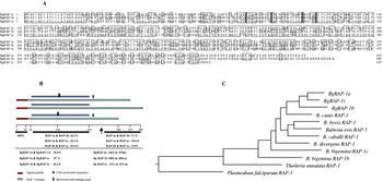
Due to the promising results using RAP-1s from other apicomplexan parasites, we decided to explore the use of RAP-1 homologues from B. gibsoni as potential therapeutic agents. Full length BgRAP-1a was reported previously (Zhou et al. Reference Zhou, Jia, Nishikawa, Fujisaki and Xuan2007) (Accession number DQ386864) to contain a single open reading frame of 1425 nucleotides encoding a polypeptide of 474 amino acids corresponding to a 51 kDa predicted mature protein. BgRAP-1b and BgRAP-1c, each showing significant homology to BgRAP-1a, were identified from a B. gibsoni cDNA expression library. BgRAP-1b contained 33·7% identity with BgRAP-1a and an ORF of 1452 nucleotides encoding a polypeptide of 484 aa weighing 53 kDa while BgRAP-1c had 57% identity with BgRAP-1a and a 1311 nucleotide ORF encoding a polypeptide of 437 aa with a molecular weight of 47 kDa. Within this similarity both these proteins shared structural characteristics of the Babesia-RAP-1 family including the presence of a signal peptide (1 to 21aa), the absence of transmembrane regions, a serine-rich N-terminal region, RAP-1 homology sequence (40 to 293 aa), and positionally conserved cysteine residues (Fig. 1A, B). Furthermore, cell-attachment motifs (RGD) at aa 156 and a conserved sequence (KKNV) at aa 313 reported to possess human erythrocyte binding activity (Calvo et al. Reference Calvo, Guzmán, Perez, Segura, Molano and Patarroyo1991) were found in both BgRAP-1a and BgRAP-1c but not BgRAP-1b (Fig. 1A, B). The phylogenetic tree demonstrated the evolutionary relationships of BgRAP-1s amino acids with other RAP-1s from other apicomplexan parasites (Fig. 1C) and indicated that BgRAP-1c was more closely related to BgRAP-1a than BgRAP-1b, which seemed to be divergent but more related to RAP-1 proteins identified in other species, such as B. canis and B. bovis.
(A) Multiple sequence alignments of the Babesia gibsoni rhoptry-associated proteins BgRAP-1a, BgRAP-1b and BgRAP-1c. (B) Schematic representation of the structures, including domains and motif, and identity percentages of BgRAP-1s. The arrow indicting the C-terminal truncated BgRAP-1s. (C) Phylogenetic tree of B. gibsoni rhoptry proteins with those from other apicomplexan parasites.

Characterization of BgRAP-1s
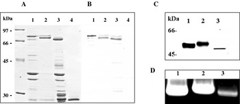
In order to understand a potential role for the BgRAP-1s genes, BgRAP-1a (22–474 aa), BgRAP-1b (22–484 aa) and BgRAP-1c (22–437 aa), were PCR amplified and cloned into the pGEX-4T-3 plasmid. The recombinant BgRAP-1a, rBgRAP-1b and rBgRAP-1c were expressed in E. coli with molecular masses estimated by SDS-PAGE to be 77 kDa, 79 kDa and 74 kDa, including an additional 26 kDa from the GST tag, respectively. Sera from dogs experimentally infected with B. gibsoni specifically reacted with the recombinant BgRAP-1a, BgRAP-1b and BgRAP-1c but not with GST protein by Western blotting (Fig. 2B), suggesting antigenicity of these proteins during infection. We therefore used the recombinant proteins as antigens for antibody production in mice. Western blot analysis of B. gibsoni lysate probing with these antibodies revealed a 51 kDa protein using anti-rBgRAP-1a antiserum, a 53 kDa protein with anti-rBgRAP-1b and a 47 kDa protein with anti-rBgRAP-1c, all consistent with expected molecular weights for each mature protein. Furthermore, this analysis revealed that all BgRAP-1s were expressed in the merozoite stage to varying degrees; and BgRAP-1a seemed to be the most abundant of the 3 proteins followed by BgRAP-1b and BgRAP-1c (Fig. 2C). Despite immunizing mice with identical and equal doses of the proteins simultaneously, there is a possibility of these antibodies not having an equal affinity to their relative proteins. Therefore, transcriptional analysis of merozoite RNA was performed and the results suggested that BgRAP-1a is the highest expressed gene among the 3 BgRAP-1s genes (Fig. 2D).
(A) 12% SDS-polyacrylamide gel (SDS-PAGE) stained with Coomassie blue: recombinant BgRAP-1a (lane 1), BgRAP-1b (lane 2), BgRAP-1c (lane 3) and GST (lane 4). (B) Western blot analysis of rBgRAP-1s (corresponding to SDS-PAGE); the blot was incubated with acutely Babesia gibsoni-infected dog serum. (C) Western blot analysis of native BgRAP-1a (lane 1), BgRAP-1b (lane 2) and BgRAP-1c (lane 3). (D) Transcriptional analysis of B. gibsoni BgRAP-1s expression in B. gibsoni merozoites by RT-PCR. Lane 1, BgRAP-1a; 2, BgRAP-1b; 3, BgRAP-1c.

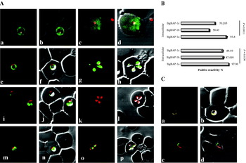
Localization of proteins can often reveal clues to their functions and therefore the antibodies were used for IFAT analysis. Confocal microscopy of both intracellular and extracellular parasites demonstrated the localization of the BgRAP-1s within the cytoplasm of the various developmental stages of merozoites. Interestingly, BgRAP-1a was found discharged into the cytoplasm of erythrocytes in a stage-dependent manner as evidenced by the specific fluorescent staining seen within the cytoplasm of infected erythrocytes collected from both a dog during peak-parasitaemia (in 31·7±10·4%) and from infected in vitro cultures (in 2·2±1·8%) (Fig. 3A; a–d). Notably, this secretion of BgRAP-1a appeared to occur only during the early phase of single merozoites and by the time of the ring-form stage of merozoites, fluorescence appeared restricted to within the merozoite (Fig. 3A; e, f). At the cell division stage, localization of BgRAP-1a appeared diffuse with more intensity within the cytoplasm of parasites (Fig. 3A; g, h). This is in contrast to both BgRAP-1b and BgRAP-1c whose localization appeared unchanged and restricted to within the parasite over the various developmental stages of B. gibsoni (Fig. 3A; i–p).
Localization of the BgRAP-1s. (A) Confocal laser microscopic observation of BgRAP-1s in various intraerythrocytic stages of Babesia gibsoni. anti-rBgRAP-1s serum (green) and nuclear staining (red). Anti-BgRAP-1a serum during the early stage (a, b, c, d), ring stage (e, f) and with sequential dividing form of B. gibsoni merozoite (g, h). Anti-BgRAP-1b serum during the ring stage of merozoites (i, j) and with the sequential dividing form of B. gibsoni (k, l). Anti-BgRAP-1c serum during the ring stage of merozoites (m, n) and with sequential dividing form of B. gibsoni (o, p). (a, e, i, m, c, g, k, o) Overlaid image of BgRAP-1s (green) and red nuclear staining, (b, d, f, h, j, l, n, p) overlaid image of BgRAP-1s (green) and red nuclear staining on phase-contrast images of the parasites. (B) The overall positivity of each anti-BgRAP-1 serum with extracellular and intracellular parasites. (C) Co-immunostaining study with surface resident protein BgP50 (red) and BgRAP-1a (green) during the intracellular ring stage (a, b) and extracellular stage (c, d).

Interestingly, all 3 proteins appeared to localize to the apical end of these extracellular merozoites near the site of invasion. Altogether, significant differences in the overall reactivity of the antisera with the parasites were apparent (Fig. 3B) and consistent with reactivity seen in Western blots with anti-BgRAP-1a serum reacting significantly higher than anti-BgRAP-1b or anti-BgRAP-1c serum. To further define the localization of each BgRAP-1s in the merozoites, co-localization studies were carried out with BgP50, a known surface protein. Again, differences were seen in the localization of BgRAP-1a, which, unlike BgRAP-1b or BgRAP-1c, co-localized with BgP50 in the ring stage of merozoites. As above, all 3 BgRAP-1s were seen to localize in a bright punctate pattern at the apical end of the free parasite near the site of invasion independent of BgP50 (Fig. 3C and data not shown). Taken together, all BgRAP-1s were expressed in different manners in the merozoite stage with localization primarily in the matrix of the merozoites near the apex. Interestingly, a difference was seen since BgRAP-1a was also detected on the surface of the parasite as well as secreted into the cytoplasm of infected erythrocytes.
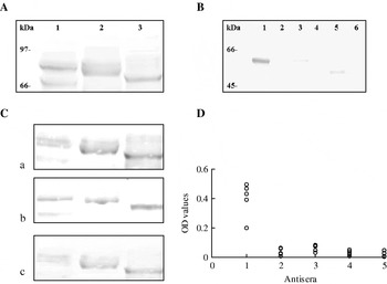
Due to the apical localization of all 3 BgRAP-1s during the extracellular stage of B. gibsoni infection we suspected that these proteins might be involved in binding erythrocytes. Therefore an in vitro erythrocyte-binding assay was carried out to determine the ability of BgRAP-1s to bind canine-erythrocytes. Recombinant BgRAP-1s were incubated with the insoluble fraction of the erythrocytes before examination by Western blotting. Indeed, all rBgRAP-1s (Fig. 4A, lane 1, BgRAP-1a; lane 2, BgRAP-1b; lane 3, BgRAP-1c) but not control GST (data not shown), showed affinity to canine erythrocytes. Moreover, canine erythrocyte precipitated all native BgRAP-1s from the merozoite lysate in (Fig. 4B). As further confirmation of the binding ability of these proteins to erythrocytes, polyclonal antibodies against each rBgRAP-1 were tested for an inhibitory effect on this binding. The antisera raised against each BgRAP-1 could prevent noticeably the interaction between the recombinant proteins with canine erythrocytes in a dose-dependent manner (Fig. 4C panels A to C correspond to increasing antibody concentration). Taken together, including the apical localization of the proteins, it appears the BgRAP-1s play a role in parasite adherence to erythrocytes. Furthermore, the combination of apical localization in extracellular merozoites, the apparent secretion of BgRAP-1a into the erythrocytes, and the high affinity of BgRAP-1s to bind canine erythrocytes encouraged us to explore whether those proteins can be secreted into the culture supernatant. Indeed, the presence of BgRAP-1a, but neither BgRAP-1b nor BgRAP-1c, could be detected in the supernatant of B. gibsoni culture by ELISA (Fig. 4D), consistent with the observations made during localization studies.
(A–C) In vitro erythrocyte binding affinity of rBgRAP-1s. (A) Western blot with anti-GST serum of recombinant proteins incubated with insoluble canine erythrocyte fraction. Lane 1, BgRAP-1a; lane 2, BgRAP-1b; lane 3, BgRAP-1c. (B) Western blot using anti-BgRAP-1s of precipitated proteins from merozoite extracts incubated with insoluble canine erythrocyte suspension in the presence of 50 mm CaCl2: Lane 1, BgRAP-1a; 3, BgRAP-1b, 5, BgRAP-1c; and lanes 2, 4, 6 are canine erythrocyte incubated with medium. (C) Inhibition of BgRAP-1s erythrocyte binding with different concentrations of specific antisera. Each recombinant was pre-incubated with its corresponding antiserum: 25 μl (a), 50 μl (b) and 100 μl (c) Lanes 1, 2, and 3 correspond to BgRAP-1a, BgRAP-1b, and BgRAP-1c respectively. (D) ELISA detection of BgRAP-1s in the supernatant of Babesia gibsoni culture. Lane 1, supernatant of culture incubated with anti-BgRAP-1a serum; 2, anti-BgRAP-1b serum; 3, anti-BgRAP-1c serum; 4, anti-GST serum and 5, pre-immune serum.

Development of rBgRAP-1s for diagnostics
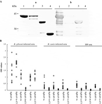
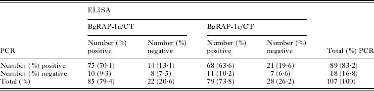
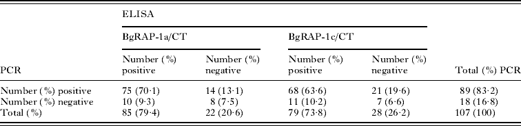
The antigenicity of the rBgRAP-1s was evaluated and compared using ELISA on variously infected serum samples. Although high reactions were observed with rBgRAP-1s when tested with B. gibsoni-infected sera, non-specific backgrounds were observed with sera from healthy dogs (SPF) and dogs infected with closely related parasites. In an attempt to improve their specificity, the highly conserved N-terminal regions of the BgRAP-1s were truncated leaving the C-terminal sequences to be cloned into the pGEX-4T-3 vector (C-terminal BgRAP-1a/CT (329–474 aa), C-terminal BgRAP-1b/CT (334–484 aa), and C-terminal BgRAP-1c/CT (337–437 aa)). The recombinant proteins had molecular weights of 40, 43, and 37 kDa, for BgRAP-1a/CT, BgRAP-1b/CT, and BgRAP-1c/CT, respectively (Fig. 5). Notably, the reactivity of rBgRAP-1s/CT were much higher than those of the non-truncated rBgRAP-1s, likely due to the hydrophobic nature of the BgRAP-1s' N-termini that would lead to significant amounts of insoluble forms. Unexpectedly, rBgRAP-1b/CT was not antigenic with B. gibsoni-infected sera by either Western blotting (Fig. 5A) or ELISA (Fig. 5B). On the other hand, both BgRAP-1a/CT and BgRAP-1c/CT showed higher sensitivity and specificity than their original recombinant proteins by ELISA (Fig. 5C). Next, the antigenicity of rBgRAP-1a/CT and rBgRAP-1c/CT was compared by testing sera obtained sequentially after infection of a dog with B. gibsoni. Although the antibody responses to rBgRAP-1a/CT were detectable 2 days earlier than the antibody to rBgRAP-1c/CT, no significant differences in the OD values were observed during acute and chronic stages (data not shown). Moreover, the same antigens were tested using 107 field samples collected from dogs clinically diagnosed as having B. gibsoni infections and checked previously by PCR (Goo et al. Reference Goo, Jia, Aboge, Terkawi, Kuriki, Nakamura, Kumagai, Zhou, Lee, Nishikawa, Igarashi, Fujisaki and Xuan2008). As seen in Table 2, 85 (79·4%) and 79 (73·8%) samples tested positive by ELISA using rBgRAP-1a/CT and rBgRAP-1c/CT, respectively, while 89 (83·2%) were positive by PCR. On the other hand, 75 (70·1%) and 68 (63·6%) samples were positive for both PCR and ELISA of each individual antigen; rBgRAP-1a/CT and rBgRAP-1a/CT, respectively. Ten (9·3%) and 8 (10·2%) samples were negative to PCR but positive to rBgRAP-1a/CT and rBgRAP-1a/CT, respectively. The discrepancies in percentage of positive PCR and ELISA can be explained potentially by differences in the timing of the presence of parasites and antibody responses in infected animals as well as stage of infection. Lastly, the ODs of rBgRAP-1a/CT were higher than those of rBgRAP-1c/CT in all positive samples tested, again consistent with initial Western blot and RT-PCR analysis that revealed higher expression for BgRAP-1a. These results revealed that BgRAP-1a has higher antigenicity than the other BgRAP-1s and its C-terminal region could be promising for further sero-epidemiological investigations.
(A) Western blot analysis of C-terminal proteins. (a) Stained 15% SDS-polyacrylamide gel with Coomassie blue of recombinant BgRAP-1a/CT (lane 1), BgRAP-1b/CT (lane 2), BgRAP-1c/CT (lane 3) and GST (lane 4). (b) Western blot analysis of rBgRAP-1s (corresponding to SDS-PAGE) probed with B. gibsoni-infected dog serum. (B) The reactivity of dog sera with recombinant BgRAP-1a, BgRAP-1b, BgRAP-1c and truncated C-terminal proteins by ELISA.

The summary of ELISA results using BgRAP-1a/CT and BgRAP-1c/CT with field serum samples
DISCUSSION
Babesia gibsoni is an obligate intracellular protozoan parasite that depends on the successful invasion of appropriate host cells for its survival. Several attempts over the past decade have been undertaken to develop an anti-Babesia vaccine, however, no vaccine has yet proven to be completely effective in preventing the infection (Brown and Palmer, Reference Brown and Palmer1999). During the past 2 decades, vaccine development efforts have been directed toward the use of antigenically defined immunogens (Wright, Reference Wright1991; Wright et al. Reference Wright, Casu, Commins, Dalrymple, Gale, Goodger, Riddles, Waltisbukl, Abetz, Berrie, Bowles, Dimmock, Hayes, Kalnins, Leatch, McCrae, Montague, Nisbet, Parrodi, Peters, Scheiwe, Smith, Rode-Bramanis and White1992; Dalrymple, Reference Dalrymple1993) with proteins derived from apical complex organelles believed to be ideal candidates for recombinant vaccines for both babesial and related malarial parasites (Perkins, Reference Perkins1992; Machado et al. Reference Machado, McElwain, Pancracio, Freschi and Palmer1999; Preiser et al. Reference Preiser, Kaviratne, Khan, Bannister and Jarra2000). Therefore, as a target for Babesia prevention more understanding of the biogenesis and functions of proteins from one such organelle, the rhoptry, is vital.
With this in mind, we have hereby identified and characterized 3 B. gibsoni cDNA-genes encoding BgRAP-1s. Indeed, one member of this family was identified previously by Zhou et al. (Reference Zhou, Jia, Nishikawa, Fujisaki and Xuan2007) via immuno-screening for immuno-dominant antigens during B. gibsoni infection. The phylogenetic analysis indicated high conservation between members of the BgRAP-1s ranging between 33 and 57%. This remarkable conservation stems from specific domains found in the N-terminal domains of these proteins including a signal peptide, a rhoptry-associated protein-1 sequence and oligopeptide motifs. In contrast, the C-terminal regions were distinguishable between the proteins. This family of genes shares similarities and is found in other parasites including 2 polymorphic linked genes in B. bovis, 4 genes in B. bigemina, 5 in B. ovis, 3 in B. divergens and 1 gene in each B. canis, and B. caballi (Goff et al. Reference Goff, Davis, Palmer, McElwain, Johnson and Bailey1988; Suarez et al. Reference Suarez, Palmer, Jasmer, Hines, Perryman and McElwain1991a; McElwain et al. Reference McElwain, Perryman, Musoke and McGuire1991; Dalrymple et al. Reference Dalrymple, Casu, Peters, Dimmock, Gale, Boese and Wright1993; Skuce et al. Reference Skuce, Mallon and Taylor1996). These similarities in the encoded proteins include an amino terminal cysteine-rich region and conserved short oligopeptide motifs at the N-terminus but not at the C-terminus (Suarez et al. Reference Suarez, McElwain, Stephens, Mishra and Palmer1991b, Reference Suarez, McElwain, Echaide, Torioni de Echaide and Palmer1994). This high degree of conservation among the apicomplexan RAP-1s points to a potential essential role for these parasite proteins in parasite survival.
Further analysis showed that all BgRAP-1s were expressed during the merozoite stage of development to varying degrees. This is consistent with previous findings where Suarez et al. (Reference Suarez, Palmer, Florin-Christensen, Hines, Hötzel and McElwain2003) reported several RAP-1s expressed during the merozoite stage of B. bigemina in various amounts. Immunofluorescence of the BgRAP-1s revealed their localization within the matrix of the merozoites, in addition to their presence during different developmental stages of these merozoites. Divergence was seen between the 3 proteins, however, when it was observed that localization of both BgRAP-1b and BgRAP-1c was restricted to within the parasite whereas BgRAP-1a was found secreted into the cytoplasm of the infected erythrocyte as well as in the supernatant of B. gibsoni culture. This putative secretion only appeared to occur during the early stages of merozoite development and by the ring stages BgRAP-1a was again found within the confines of the parasite, along the surface of the parasite before being uniformly distributed throughout the cytoplasm upon division. Another interesting observation was made during the extracellular stages of the parasite life cycle when all BgRAP-1s could be seen at the apex of the extracellular parasites near the site of invasion. This is consistent with earlier findings characterizing the B. bovis RAP-1 that was also detected at the apical end of that parasite, along the surface of the merozoites, within the matrix of infected erythrocytes as well as in the culture supernatant (Goff et al. Reference Goff, Davis, Palmer, McElwain, Johnson and Bailey1988; Suarez et al. Reference Suarez, McElwain, Echaide, Torioni de Echaide and Palmer1994; Johnson et al. Reference Johnson, Perryman and Goff1997; Yokoyama et al. Reference Yokoyama, Suthisak, Hirata, Matsuo, Inoue, Sugimoto and Igarashi2002). Moreover, the cytoplasmic secretions of RAP-1 within the host erythrocytes have been also reported in B. bigemina and B. equi (Kumar et al. Reference Kumar, Yokoyama, Kim, Huang, Inoue, Xuan, Igarashi and Sugimoto2004; Boonchit et al. Reference Boonchit, Alhassan, Chan, Xuan, Yokoyama, Ooshiro, Goff, Waghela, Wagner and Igarashi2006). In Plasmodium parasites, a similar process may be occurring as rhoptry proteins can be seen to be discharged into the erythrocyte membrane during invasion and persist in the host membrane and cytosol for some time before disappearing at the ring stage (Sam-Yellowe et al. Reference Sam-Yellowe, Shio and Perkins1988). The question arises as to how babesial RAP-1s interact with the membrane and cytoskeleton of erythrocytes during the invasion process. One possibility is that these proteins can be secreted from attached merozoites and inserted into the membrane or cytoplasm of erythrocytes to aid in the entry of parasites. However, since the proteolytic activities of rhoptry proteins have not been reported in Babesia, (unlike Plasmodium and Toxoplasma gondii rhoptry proteins that function as lytic substances that may be involved in the penetration process during invasion) (Nichols et al. Reference Nichols, Chiappino and O'Connor1983; Sam-Yellowe et al. Reference Sam-Yellowe, Shio and Perkins1988; Preiser et al. Reference Preiser, Kaviratne, Khan, Bannister and Jarra2000), further study is required to understand the biochemical function of these proteins.
The sequence analysis of BgRAP-1s revealed the presence of motifs involved in cell attachment and in combination with localization of BgRAP-1s in the free merozoites, it seems possible that these proteins are involved in attachment of the parasite to the erythrocytes. Indeed, erythrocyte binding assays demonstrated high affinity of all recombinant BgRAP-1s with membranes of canine erythrocytes. Furthermore, membrane fractions of canine erythrocytes were capable of precipitating BgRAP-1s. Furthermore, membrane fractions of canine erythrocytes were capable of precipitating BgRAP-1s. The absence of any clear erythrocyte binding motifs (RGD and KKNV) in BgRAP-1b is puzzling considering its affinity to canine erythrocytes and suggests other motifs and/or domains present in BgRAP-1s might be involved in this binding function. Likewise, B. bovis RAP-1 showed high affinity to bind bovine and equine erythrocytes (Yokoyama et al. Reference Yokoyama, Suthisak, Hirata, Matsuo, Inoue, Sugimoto and Igarashi2002). It is not just the Babesia RAP-1s that are able to bind erythrocytes as P. falciparum RAP-1 and RAP-2 were also shown to contain high binding affinity to human erythrocytes (Curtidor et al. Reference Curtidor, Ocampo, Tovar, López, García, Valbuena, Vera, Suárez, Rodríguez, Puentes, Guzmán, Torres and Patarroyo2004; Lopez et al. Reference López, Valbuena, Curtidor, Puentes, Rodríguez, García, Suárez, Vera, Ocampo, Trujillo, Ramirez and Patarroyo2004). Work to determine the identification of such domains in addition to their receptors on the surface of erythrocytes will be important to elucidate the biological basis of BgRAP-1s and erythrocyte interaction for potential therapeutic exploitation. Babesia parasites are thought to utilize a number of proteins known to reside on the surface, in micronemes and in rhoptries of extracellular merozoites in order to effectively attach and invade the erythrocytes (Yokoyama et al. Reference Boonchit, Alhassan, Chan, Xuan, Yokoyama, Ooshiro, Goff, Waghela, Wagner and Igarashi2006). These proteins include variable merozoite surface antigens VMSA believed to play key roles in the initial attachment of merozoites and sporozoites during their invasion. Microneme and rhoptry proteins, including apical membrane antigen 1 (AMA-1), thrombospondin-related anonymous protein (TRAP) and RAP-1s seem to be involved in the later stages of re-orientation and formation of a tight junction between merozoites and erythrocytes and have been shown to physically interact with host membranes (Gaffar et al. Reference Gaffar, Yatsuda, Franssen and de Vries2004a, Reference Gaffar, Yatsuda, Franssen and de Vriesb; Zhou et al. Reference Zhou, Fukumoto, Jia, Yokoyama, Zhang, Fujisaki, Lin and Xuan2006b; Yokoyama et al. Reference Boonchit, Alhassan, Chan, Xuan, Yokoyama, Ooshiro, Goff, Waghela, Wagner and Igarashi2006). The secreted nature of these proteins coupled with their affinity to bind host cells suggest that they play an important role in invasion and consequent parasite survival.
Finally, the antigenicity of the rBgRAP-1s was evaluated using ELISA on sera from dogs experimentally and naturally infected with B. gibsoni, in order to explore the use of these proteins for diagnostics. Although all rBgRAP-1s showed strong antigenicity with infected sera the background was high and therefore steps were taken to correct this including removing the N-termini of these proteins. In a surprising finding, rBgRAP-1b/CT was not antigenic with infected dogs' sera, whereas rBgRAP-1a/CT and rBgRAP-1c/CT showed high reactivity when tested by Western blotting and ELISA. It seems that infected B. gibsoni dog sera could recognize B-cell epitopes found in both BgRAP-1a/CT and BgRAP-1c/CT but not in BgRAP-1b/CT. Consistent with this notion, BgRAP-1a/CT shared higher homology with BgRAP-1c/CT (36·6%) than with BgRAP-1b/CT (7·1%). In related studies, Boonchit et al. (Reference Boonchit, Xuan, Yokoyama, Goff, Wagner and Igarashi2002, Reference Boonchit, Xuan, Yokoyama, Goff, Waghela, Wagner and Igarashi2004, Reference Boonchit, Alhassan, Chan, Xuan, Yokoyama, Ooshiro, Goff, Waghela, Wagner and Igarashi2006) also noted that full-length B. bovis and B. bigemina RAP-1 were unsuitable as ELSIA antigens due to cross-reactivity and found similarly that N-terminal truncation resulted in higher specificity and sensitivity when used as sero-diagnostic antigens. Notably, in the present study rBgRAP-1a/CT seemed to be more antigenic than rBgRAP-1c/CT as evidenced by higher sensitivity during sequential B. gibsoni-infected sera probing as well as the higher proportion of positive field samples. The higher antigenicity seen with rBgRAP-1a might be due to its localization and constant expression during the developmental stages of B. gibsoni merozoites. The high specificity and sensitivity of ELISA-rBgRAP-1a/CT and ELISA-rBgRAP-1c/CT were promising enough to pursue development of a reliable sero-diagnostic reagent for detection of B. gibsoni in dogs. However, it is worth noting that use of the rBgRAPs as diagnostic agents were compared against previous antigens. Specifically, the same field samples used in the previous studies were retested using both rBgRAP-1a/CT and rBgRAP-1c/CT and compared to rBgTRAP, rBgP50, rBgSA1, and rBgP32 (Goo et al. Reference Goo, Jia, Aboge, Terkawi, Kuriki, Nakamura, Kumagai, Zhou, Lee, Nishikawa, Igarashi, Fujisaki and Xuan2008). While rBgTRAP was found to be more sensitive than rBgRAP-1a (89·7% versus 79·4% positive, respectively) the use of BgRAP-1a still appears promising as a diagnostic agent.
In summary, we have identified and characterized 3 BgRAP-1s expressed in B. gibsoni merozoite stages and found differences in their expression pattern, localization and antigenicity during infection. Although, the BgRAP-1s are highly homologous, contain similar protein structures and an affinity to bind canine erythrocytes, BgRAP-1a seemed to be a more promising vaccine candidate than other BgRAP-1s. These findings should hopefully provide a basis for future work that could exploit these proteins as drug targets for therapeutic use.
This work was supported by a grant from the 21st Century COE Program (A-1) and a Grant-in-Aid for Scientific Research, both from the Ministry of Education, Culture, Sports, Science and Technology, Japan, and a grant from the Bio-orientated Technology Research Advancement Institution (BRAIN).