Introduction
Patients suffering from depressive disorder frequently complain about cognitive disturbances. Recent studies support the independent association of cognitive deficit with depression (Austin et al., Reference Austin, Mitchell and Goodwin2001), and even mildly depressed patients are more impaired in cognitive function than healthy controls (Brown et al., Reference Brown, Scott, Bench and Dolan1994). Although depressed patients complain of subjective cognitive problems in memory and attention, objective impairments of cognitive function are found in these and other cognitive domains (Austin et al., Reference Austin, Ross, Murray, O'Carroll, Ebmeier and Goodwin1992; Purcell et al., Reference Purcell, Maruff, Kyrios and Pantelis1997; Porter et al., Reference Porter, Gallagher, Thompson and Young2003; Baudic et al., Reference Baudic, Tzortzis, Barba and Traykov2004).
Depressive disorders accompanying cognitive decline sometimes make it difficult to discriminate depressive disorders from dementia. In addition, reduced cognitive function may be an initial sign of dementia or an independent risk factor for dementia in late-life depression (Modrego and Ferrández, Reference Modrego and Ferrández2004; Panza et al., Reference Panza2010). Therefore, assessment of cognitive function in depressed patients, especially in elderly patients, could be important for differential diagnosis and the recognition of potential risk factors for dementia.
Research to reveal which cognitive domains might be affected by depression has shown conflicting findings (Austin et al., Reference Austin, Ross, Murray, O'Carroll, Ebmeier and Goodwin1992; Purcell et al., Reference Purcell, Maruff, Kyrios and Pantelis1997; Porter et al., Reference Porter, Gallagher, Thompson and Young2003; Baudic et al., Reference Baudic, Tzortzis, Barba and Traykov2004; Fischer et al., Reference Fischer2008). Recent research has reported the recovery of cognitive function after treatment with antidepressants in depressed patients (Koetsier et al., Reference Koetsier, Volkers, Tulen, Passchier, van den Broek and Bruijn2002; Levkovitz et al., Reference Levkovitz, Caftori, Avital and Richter-Levin2002; Vythilingam et al., Reference Vythilingam2004), but results were inconsistent regarding which cognitive functions were recovered. Narrative reviews have also described no consistent pattern of association between depression and cognitive decline (Marazziti et al., Reference Marazziti, Consoli, Picchetti, Carlini and Faravelli2010; Wilkins et al., Reference Wilkins, Kiosses and Ravdin2010; Millan et al., Reference Millan2012).
We therefore conducted a meta-analysis to find out which cognitive tests have been used in studies of depression and what specific subsets of cognitive tests could sensitively differentiate depressed patients from patients with other disorders or from healthy controls.
Methods
Source of data
Relevant articles were identified through a literature search from the PubMed and Cochrane Library databases for the period between January 1997 and May 2011 using the following key words: “depression” or “major depression” or “depressive illness” or “major depressive disorder,” or “depressed” and “cognitive function” or “cognition” or “cognitive” or “neuropsychological” or “neuropsychology,” or “memory.” We included only randomized controlled trial (RCT) studies, cohort studies, and case-control studies. We also cross-checked the reference lists of identified articles to discover other relevant clinical trial reports.
Inclusion/exclusion criteria
We included studies that compared cognitive function between patients with major depressive disorder (MDD) and healthy controls. The inclusion criteria were as follows: Studies included a sample of patients diagnosed with MDD according to the Diagnostic and Statistical Manual of Mental Disorders, Versions III Revised, IV, or DSM-IV-TR, and measured depression severity using the Hamilton Rating Scale for Depression (Hamilton, Reference Hamilton1967), Beck Depression Inventory (Beck et al., Reference Beck, Ward, Mendelson, Mock and Erbaugh1961), Montgomery–Asberg Depression Rating scale (Montgomery and Asberg, Reference Montgomery and Asberg1979), Yale Depression Inventory (Mazure et al., Reference Mazure, Nelson and Price1986; Mazure et al., Reference Mazure, Nelson and Jatlow1990), Center for Epidemiological Studies Depression scale (Radloff, Reference Radloff1977), or the Geriatric Depression scale (Yesavage et al., Reference Yesavage1983). Because an association between depression severity and cognitive performance has been reported (McDermott and Ebmeier, Reference McDermott and Ebmeier2009), we selected only studies that restricted the sample of depressed individuals to those with moderate or severe depression (according to the scale used in each study). We also selected studies in which the depression symptoms of depressed patients were significantly more severe than healthy controls. Selected studies were also required to report means and standard deviations of neuropsychological test scores or to report sufficient data for these to be derived. Selected studies excluded participants who suffered from delirium, any neurodegenerative disease, including dementia, active substance abuse, or an unstable medical disease, or participants who had recently received electroconvulsive therapy or who had a current or past diagnosis of a psychotic disorder that could influence cognitive function. Patients with bipolar disorder or other mood disorder except for MDD were also excluded. Finally, all studies were published in English in peer-reviewed journals.
Cognitive measures
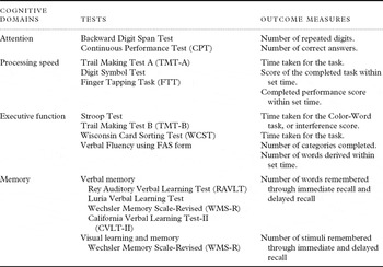
After reviewing available researches and literatures, cognitive measures of attention, processing speed, executive function, and memory were selected for study because these domains were most frequently reported to be impaired in depressive patients. After that, two psychiatrists and two clinical psychologists assigned each cognitive test to a particular cognitive domain according to traditional trends and clinical judgment, although such classification is ambiguous and has controversies for some cognitive tests. Table 1 presents the cognitive domains and the related individual neuropsychological tests and their outcome variables. For the Stroop Test, we used the times taken for the Color-Word test, in which participants should name the color which was incongruent with words. When the times data for the Color-Word test were unavailable, the interference score which was calculated as a time difference between the Color test and the Color-Word test was used. For the analysis of memory function, studies which employed the Rey Auditory Verbal Learning Test (RAVLT) (Rey, Reference Rey1964), the Luria Verbal Learning Test (Christensen, Reference Christensen1975), the California Verbal Learning Test, second edition (CVLT-II) (Delis et al., Reference Delis, Karmaer, Kaplan and Ober1987), and the original and revised version of the Wechsler Memory Scale (WMS/WMS-R) (Wechsler, Reference Wechsler1987; Delis et al., Reference Delis, Kramer, Kaplan and Ober2000) were selected. The data of original version of the WMS were assigned to the domain of immediate verbal memory since the test does not measure delayed recall and any visual memory. And in the test of WMS-R, measured values of logical memory subset and visual reproduction subset were extracted. As a dependent variable for our analysis, the number of words or stimuli remembered through immediate and delayed recall was used. Meta-analyses were conducted for individual cognitive tests in each of the separate cognitive domains.
Neuropsychological tests and outcome measures for each cognitive domain

Data extraction and analysis
Participants’ characteristics, including mean age, treatment, study procedures, diagnostic information (e.g., comorbid conditions), severity of depressive symptoms, and outcome measures, were collected. Literature selection and data extraction were initially performed by two psychologists and independently reassessed by two psychiatrists, all of whom extracted data independently from each study using a predetermined data extraction form. Meta-analysis and data extraction were performed with Review Manager 5.0 (Cochrane Collaboration, 2011). The mean difference for each measure of cognitive function between MDD patients and healthy controls was directly extracted from the cited studies or computed. Summary estimates of standardized mean differences (SMDs) and 95% confidence intervals (CI) were obtained by using a random-effects model in consideration of the standard error dispersion between the studies. A random-effects model was chosen because we could not assume a common effect size for studies conducted independently in which differences in participants and study settings may have had an impact on the results. Further, since the goal of this analysis was to generalize to a range of results, a random-effects model was regarded as more suitable than a fixed-effect model (Borenstein et al., Reference Borenstein, Hedges, Higgins and Rothstein2009). Statistical heterogeneity of effect size between studies was tested with the Q-test (significance threshold, p ≤ 0.1). A shortcoming of the Q-statistic is that it has poor power to detect true heterogeneity among studies when the meta-analysis includes a small number of study variance; therefore, a more liberal critical value of 0.10 was used for testing homogeneity (Colditz et al., Reference Colditz, Burdick and Mosteller1995; Higgins et al., Reference Higgins, Thompson, Deeks and Altman2003). The I 2 statistic was also examined to quantify the degree of heterogeneity, since the Q-test only informs us about the presence versus the absence of heterogeneity, and we considered an I 2 value of 50% or higher to indicate meaningful heterogeneity between the trials (Higgins et al., Reference Higgins, Thompson, Deeks and Altman2003).
When the heterogeneity between studies was significant, we performed post hoc subgroup analyses for age and medication status to determine sources of heterogeneity. For the subgroup analyses, we divided studies according to the mean age of participants (60 years or older versus younger than 60 years) and whether antidepressant medication was administered or not. Subgroup analyses were conducted by use of a mixed-effect model, and equal variance was assumed among subgroups.
Results
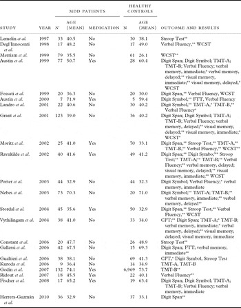
In total, 4,140 articles (3,308 articles from PubMed and 832 articles from the Cochrane Library) were identified via the search terms. From this list, 407 studies remained after excluding duplicates and trials that did not include depressive patients or for which the full-text version was inaccessible. Finally, 22 trials that met the inclusion criteria described above were selected for our meta-analysis (Lemelin et al., Reference Lemelin, Baruch, Vincent, Everett and Vincent1997; Degl'Innocenti et al., Reference Degl'Innocenti, Agren and Bäckman1998; Austin et al., Reference Austin1999; Fossati et al., Reference Fossati, Amar, Raoux, Ergis and Allilaire1999; Merriam et al., Reference Merriam, Thase, Haas, Keshavan and Sweeney1999; Austin et al., Reference Austin2000; Grant et al., Reference Grant, Thase and Sweeney2001; Landrø et al., Reference Landrø, Stiles and Sletvold2001; Moritz et al., Reference Moritz2002; Ravnkilde et al., Reference Ravnkilde, Videbech, Clemmensen, Egander, Rasmussen and Rosenberg2002; Nebes et al., Reference Nebes2003; Porter et al., Reference Porter, Gallagher, Thompson and Young2003; Stordal et al., Reference Stordal2004; Vythilingam et al., Reference Vythilingam2004; Constant et al., Reference Constant, Adam, Seron, Bruyer, Seghers and Daumerie2006; Gallassi et al., Reference Gallassi, Sarro, Morreale and Amore2006; Gualtieri et al., Reference Gualtieri, Johnson and Benedict2006; Kuroda et al., Reference Kuroda2006; Godin et al., Reference Godin2007; Ridout et al., Reference Ridout, O'Carroll, Dritschel, Christmas, Eljamel and Matthews2007; Fischer et al., Reference Fischer2008; Herrera-Guzmán et al., Reference Herrera-Guzmán2010). These trials included 955 MDD patients and 7,664 healthy controls. Nineteen trials were cross-sectional studies on cognitive function in patients with MDD and healthy controls, and in most trials, the age of MDD patients was matched with that of controls. The other trials were two RCT and one cohort study. Details of the 22 selected trials are summarized in Table 2. Six trials had samples (MDD patients or healthy controls, or both) with a mean age above 60 years (Austin et al., Reference Austin1999; Austin et al., Reference Austin2000; Nebes et al., Reference Nebes2003; Gallassi et al., Reference Gallassi, Sarro, Morreale and Amore2006; Godin et al., Reference Godin2007; Fischer et al., Reference Fischer2008). Participants of 14 trials were drug-free for a minimum of two weeks and a maximum of 18 weeks, whereas participants of eight trials were taking antidepressants or other psychotropic medications at the time of cognitive function measurement. The results of individual meta-analysis on tests within each cognitive domain are presented in Figures 1–4.
Summary of studies included in meta-analysis

MDD: Major Depressive Disorder; CPT: Continuous Performance Test; FTT: Finger Tapping Task; TMT-A: Trail Making Test A; TMT-B: Trail Making Test B; WCST: Wisconsin Card Sorting Test.
*p < 0.05, **p < 0.01.
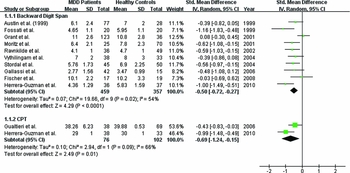
Meta-analysis of the attention domain in MDD patients compared with healthy controls. MDD: major depressive disorder; SMD: standardized mean difference; CI: confidence interval; SD: standard deviation; CPT: continuous performance test.

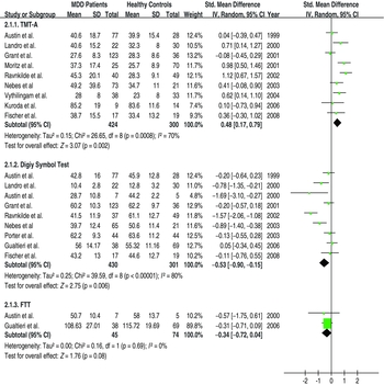
Meta-analysis of the processing speed domain in MDD patients compared with healthy controls. MDD: major depressive disorder; SMD: standardized mean difference; CI: confidence interval; SD: standard deviation; TMT-A: Trail Making Test A; FTT: finger tapping task.

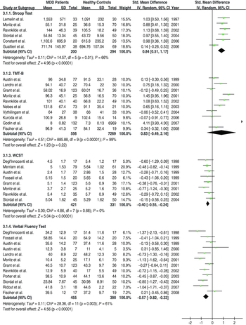
Meta-analysis of the executive function domain in MDD patients compared with healthy controls. MDD: major depressive disorder; SMD: standardized mean difference; CI: confidence interval; SD: standard deviation; TMT-B: Trail Making Test B; WCST: Wisconsin Card Sorting Test.

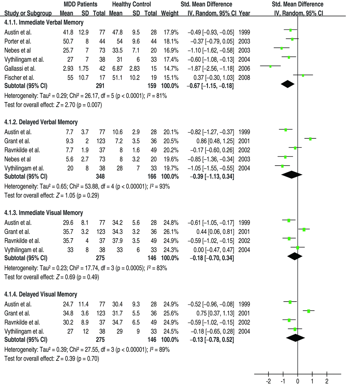
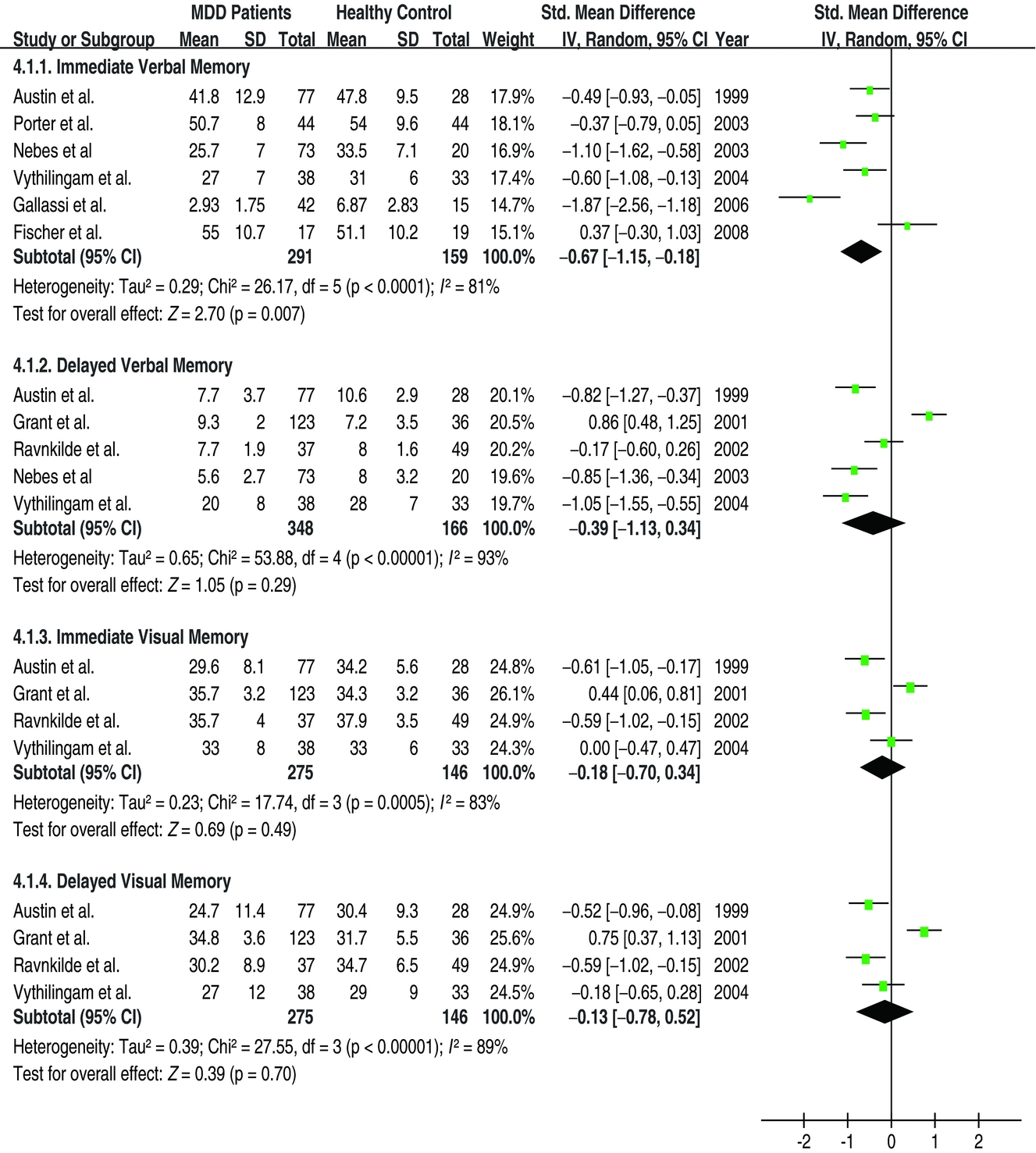
Meta-analysis of the memory domain in MDD patients compared with healthy controls. MDD: major depressive disorder; SMD: standardized mean difference; CI: confidence interval; SD: standard deviation.
Attention
The results for the Backward Digit Span Test and the Continuous Performance Test (CPT) were obtained from the data of ten and two trials, respectively (Figure 1). Overall performance on both tests was significantly impaired in MDD patients compared with healthy controls (Backward Digit Span Test: SMD = –0.50, 95% CI = –0.72~–0.27, p < 0.0001; CPT: SMD = –0.69, 95% CI = –1.24~–0.15, p = 0.01).
Heterogeneity was found between studies of the Digit Span Test (χ 2 = 19.66, df = 9 (p = 0.02), I 2 = 54%) and CPT (χ 2 = 2.94, df = 1 (p = 0.09), I 2 = 66%).
Processing speed
Nine trials were included in the analysis of the Trail Making Test A (TMT-A) and the Digit Symbol Test (Figure 2). Time taken for the task of TMT-A was significantly prolonged in MDD patients compared with healthy controls (SMD = 0.48, 95% CI = 0.17~0.79, p = 0.002), and performance on the Digit Symbol Test was also significantly impaired in MDD patients (SMD = –0.53, 95% CI = –0.90~–0.15, p = 0.006).
The meta-analysis for the Finger Tapping Task (FTT) was conducted using data from two papers (Figure 2), revealing an overall SMD of –0.34 (95% CI = –0.72~0.04, p = 0.08) and indicating that the performance difference was not significant between MDD patients and healthy controls. The two original studies also failed to show significant differences between the two groups on the FTT.
Between-study heterogeneity was significant in the trials included in the analysis of TMT-A (χ 2 = 26.65, df = 8 (p = 0.0008), I 2 = 70%) and the Digit Symbol Test (χ 2 = 39.59, df = 8 (p < 0.00001), I 2 = 80%). There was no heterogeneity between studies included in the analysis of FTT (χ 2 = 0.16, df = 1 (p = 0.69), I 2 = 0%).
Executive function
Data from six studies were extracted for the meta-analysis of the Stroop Test, ten studies for the Trail Making Test B (TMT-B), eight studies for the Wisconsin Card Sorting Test (WCST), and 12 studies for Verbal Fluency (Figure 3). Patients with MDD showed significantly reduced performance in the Stroop Test (SMD = 0.84, 95% CI = 0.51~1.17, p < 0.0001), WCST (SMD = –0.40, 95% CI = –0.55~–0.24, p < 0.00001), and Verbal Fluency (SMD = –0.57, 95% CI = –0.82~–0.33, p < 0.00001) compared with healthy controls. For the TMT-B, the overall SMD was 0.83 (95% CI = –0.49~2.16, p = 0.22), which indicated that performance on the TMT-B did not differ between patients with MDD and healthy controls.
Between-study heterogeneity was significant in the studies included in the meta-analysis of the Stroop Test (χ 2 = 14.57, df = 5 (p = 0.01), I 2 = 66%), TMT-B (χ 2 = 885.88, df = 9 (p < 0.00001), I 2 = 99%), and Verbal Fluency (χ 2 = 28.36, df = 11 (p = 0.003), I 2 = 61%), whereas it was not significant in the studies included in the meta-analysis of WCST (χ 2 = 4.86, df = 7 (p = 0.68), I 2 = 0%).
Memory
Meta-analysis using six studies showed significantly impaired performance in immediate verbal memory in patients with MDD compared with healthy controls (SMD = –0.67, 95% CI = –1.15~–0.18, p = 0.007) (Figure 4). The overall SMDs of the analyses for delayed verbal and immediate and delayed visual memory were not significant between the two groups (delayed verbal: SMD = –0.39, 95% CI = –1.13~0.34, p = 0.29; immediate visual: SMD = –0.18, 95% CI = –0.70~0.34, p = 0.49; delayed visual: SMD = –0.13, 95% CI = –0.78~0.52, p = 0.70). The analysis for verbal delayed memory used the data from five studies, and the analysis for visual memory immediate and delayed used the same four studies.
Studies included in the meta-analyses of cognitive tests for the memory domain were significantly heterogeneous (immediate verbal memory: χ 2 = 26.17, df = 5 (p < 0.0001), I 2 = 81%; delayed verbal memory: χ 2 = 53.88, df = 4 (p < 0.00001), I 2 = 93%; immediate visual memory: χ 2 = 17.74, df = 3 (p = 0.0005), I 2 = 83%; delayed visual memory: χ 2 = 27.55, df = 3 (p < 0.00001), I 2 = 89%).
Subgroup analysis
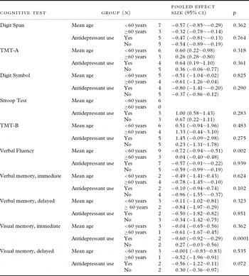
Subgroup analyses based on age of study participants and antidepressant use were performed to investigate the sources of heterogeneity. The results are shown in Table 3. Subgroup analysis for FTT and WCST was not done because heterogeneity was absent within the individual studies for these tests. The CPT was also excluded from subgroup analysis because the mean age of participants in both studies was below 60 years and no patient was taking antidepressant medication at the time of testing.
Subgroup analyses for cognitive impairment of MDD patients

MDD: Major Depressive Disorder; CPT: Continuous Performance Test; TMT-A: Trail Making Test A; TMT-B: Trail Making Test B.
The results show that performance of Verbal Fluency was significantly impaired in younger depressed patients (<60 years) and that immediate visual memory was significantly reduced in depressed patients using antidepressants compared with unmedicated patients.
Discussion
There has been controversy about whether neurocognition in depression is impaired in a global or specific way (Fischer et al., Reference Fischer2008), and even the relationship between cognitive function and depression has sometimes been denied (Grant et al., Reference Grant, Thase and Sweeney2001; Fischer et al., Reference Fischer2008). Our findings suggest that depression is related to reductions in a wide range of cognitive abilities, including attention, processing speed, executive function, and memory. Specifically, depressive patients showed significantly reduced performance across all of the above domains when compared with healthy controls. A number of studies have also shown that depressed patients often have reduced function in many cognitive domains (Ravnkilde et al., Reference Ravnkilde, Videbech, Clemmensen, Egander, Rasmussen and Rosenberg2002; Dotson et al., Reference Dotson, Resnick and Zonderman2008). The idea that depressed patients suffer from a global cognitive disturbance could be supported by existing evidence of neurological deterioration of specific brain regions. In studies investigating the brain structures responsible for various cognitive functions (including attention, executive function, memory, psychomotor speed, and others; Baxter et al., Reference Baxter1989; Bench et al., Reference Bench, Friston, Brown, Scott, Frackowiak and Dolan1992; Drevets et al., Reference Drevets, Videen, Price, Preskorn, Carmichael and Raichle1992; Dolan et al., Reference Dolan1993; Rezai et al., Reference Rezai, Andreasen, Alliger, Cohen, Swayze and O'Leary1993; Mayberg et al., Reference Mayberg, Lewis, Regenold and Wagner1994; Videbech et al., Reference Videbech2002), changes in cerebral blood flow and glucose metabolism in the frontal cortex, limbic system, thalamus, and striatum have been demonstrated in MDD patients.
Significantly impaired cognitive function in depressive patients was found in the attention domain via the Digit Span Test and CPT; in the processing speed domain via the TMT-A and the Digit Symbol Test; in the executive function domain via the Stroop test, WCST, and Verbal Fluency; and in the memory domain via tests of immediate verbal memory. The FTT in the processing domain, TMT-B in the executive function domain, and delayed verbal memory and immediate and delayed visual memory in the memory domain failed to separate MDD patients from healthy controls.
Cognitive deficits affecting depressive patients are sometimes considered a secondary phenomenon caused by attention problems (Cohen et al., Reference Cohen, Weingartner, Smallberg, Pickar and Murphy1982; Lemelin et al., Reference Lemelin, Baruch, Vincent, Laplante, Everett and Vincent1996; Hart et al., Reference Hart, Wade, Calabrese and Colenda1998). Previous studies have reported attention deficit as a trait-vulnerability marker for MDD that persists in patients with depression even after remission (Weiland-Fiedler et al., Reference Weiland-Fiedler2004). Other studies, however, have demonstrated that cognitive problems persist after improvement of attentional function in depressed patients (Paelecke-Habermann et al., Reference Paelecke-Habermann, Pohl and Leplow2005; Reppermund et al., Reference Reppermund2007). Consequently, depression-related global cognitive deficits are likely fundamental rather than secondary to the disturbance of attention.
Similarly, psychomotor slowing has been regarded as a cardinal feature explaining the association of certain cognitive domains with depression. Both mental and motor retardation are relatively consistent findings in depressive patients (Benoît et al., Reference Benoît, Fortin, Lemelin, Laplante, Thomas and Everett1992; Parker and Hadzi-Pavlovic, Reference Parker and Hadzi-Pavlovic1996; Sabbe et al., Reference Sabbe, Hulstijn, Hoof and Zitman1996; Sobin and Sackeim, Reference Sobin and Sackeim1997), and depressive patients, as a group, show slight to moderate inhibited psychomotor speed based on the Widlocher Retardation scale (Widlöcher, Reference Widlöcher1983). Nevertheless, McDermott and Ebmeier (Reference McDermott and Ebmeier2009) tested via meta-analysis whether psychomotor slowing could be a confounder in the disturbance of other domains of cognitive function in depressed patients. They found that both timed and untimed cognitive performances in depressed patients were significantly and equally associated with the disorder. Thus, psychomotor retardation may be a part of depression-related cognitive problems, not a basic deficit that covers whole aspects of cognition. In our analysis, MDD patients did not show significant impairments in performance on the FTT, but significant impairments were found for two other tests in the processing speed domain, i.e., TMT-A and the Digit Symbol Test. However, this result cannot be generalized because the data pooling for the FTT was done using the results from only two studies. Although the two studies showed statistical homogeneity and reported consistent findings of non-disturbed performance of the FTT in depressed patients, there might be a possibility of irrelevance between the FTT performance and depression.
Executive function shares a common pathology with depressive disorder of frontostriatal dysfunction, especially in geriatric populations (Drewe, Reference Drewe1974; Franke et al., Reference Franke, Maier, Hardt, Frieboes, Lichtermann and Hain1993; Trichard et al., Reference Trichard1995; Alexopoulos et al., Reference Alexopoulos, Kiosses, Klimstra, Kalayam and Bruce2002). The “depression–executive dysfunction syndrome” has been widely proposed, and the relationship between executive function and depression seems to be evident. In addition, some studies have suggested that executive function may predict treatment response in depressive patients (Kalayam and Alexopoulos, Reference Kalayam and Alexopoulos1999). In the present analysis, results on the TMT-B failed to separate MDD patients from healthy controls, unlike the other three tests of executive function (Stroop Test, WCST, and Verbal Fluency). However, several of the individual trials included in the meta-analysis revealed significant SMDs between MDD patients and healthy controls for the TMT-B performance. Given the limitations of meta-analysis and the presence of heterogeneity, further studies are required to confirm the relationship between the TMT-B and depression. On the other hand, the finding from the TMT-B, a timed measure, might be an evidence for that the deficit of executive function in depression is not incidental to psychomotor slowing since the pooled SMD of the TMT-B was not different between MDD patients and healthy controls.
In the memory domain, only the performance of immediate verbal memory was significantly lower in patients with MDD than in healthy controls. We cannot exclude the possibility that attention problem can affect impaired memory, especially immediate memory in depressed patients. The distinction or relationship between memory and attention in depression is difficult to measure or quantify because available cognitive tests are usually related to both cognitive domains. Accordingly, no works to account for the possible impact of attention on memory function were done across most studies. Nevertheless, some studies suggested that successful treatment with antidepressants resulted in a significant improvement in immediate/delayed verbal memory, or immediate visual memory without any improvement in attention (Vythilingam et al., Reference Vythilingam2004; Wroolie et al., Reference Wroolie2006; Herrera-Guzmán et al., Reference Herrera-Guzmán, Gudayol-Ferré, Herrera-Guzmán, Guàrdia-Olmos, Hinojosa-Calvo and Herrera-Abarca2009; Boeker et al., Reference Boeker, Schulze, Richter, Nikisch, Schuepbach and Grimm2012), and these results could support that memory problems in depression are not secondary to more basic attentional disturbances. Meanwhile, such memory tasks have been shown to be sensitive to the effects of some antidepressants (Schmitt et al., Reference Schmitt, Kruizinga and Riedel2001), and our subgroup analysis for medication status partially supports this finding; patients who were taking antidepressant medications showed significantly reduced function of immediate visual memory when compared with the antidepressant-free group. Little has been known about the relationship between visual memory and antidepressant treatment. Results from previous studies on antidepressant treatment are equivocal, with some studies showing an improvement and others showing no improvement in visual memory (Deuschle et al., Reference Deuschle2004; Zobel et al., Reference Zobel2004; Wroolie et al., Reference Wroolie2006), but no results of worsening in visual memory have been reported to our knowledge. In our analysis of visual memory, baseline depression severity in medicated subgroup had tendency to be more severe than medication-free depression patients, and it could bias the results. Besides visual memory, the results of medication-related other cognitive functions have also shown conflicting findings. Some studies reported the worsening of cognitive performance like phonemic verbal fluency and delayed verbal memory after selective serotonin reuptake inhibitors (Schmitt et al., Reference Schmitt, Kruizinga and Riedel2001; Wroolie et al., Reference Wroolie2006), whereas majority of studies reported positive influence of antidepressant medication on wide range of cognitive function. So far, it is unclear whether these variations are due to the depressive condition itself or whether medication influences exist. On the other hand, some studies have focused not on visual/verbal or immediate/delayed aspects of memory but on negative/positive aspects, and have suggested that depressed patients recall more vivid negative memories and less emotionally intense positive memories than healthy participants (Liu et al., Reference Liu, Wang, Zhao, Ning and Chan2012; Werner-Seidler and Moulds, Reference Werner-Seidler and Moulds2012).
The relationship between depression and cognitive impairment in geriatric populations is supported by observations that cerebral atrophy and ischemic cerebrovascular disease are more common on MRI scans of older depressed patients than on the scans of controls (Hickie et al., Reference Hickie, Scott, Mitchell, Wilhelm, Austin and Bennett1995; Herrmann et al., Reference Herrmann, Goodwin and Ebmeier2007). We conducted a subgroup analysis for age as well as medication status and found that the influence of age was limited to performance of Verbal Fluency. Consequently, the sources of heterogeneity of the studies in our analysis were not well explained by either age or medication status. However, our subgroup analyses were undertaken in a post hoc manner rather than being prespecified, and therefore may have been susceptible to possible bias through confounding by other characteristics of the individual studies. For example, some studies log-transformed the data to ensure equal variance without any specification about which cognitive test was transformed, and sometimes the outcome measures were arbitrarily modified (Austin et al., Reference Austin1999; Stordal et al., Reference Stordal2004; Godin et al., Reference Godin2007), which could be the contributable sources of heterogeneity. Besides age and medication status, depression severity is also known to be associated with cognitive function (Grant et al., Reference Grant, Thase and Sweeney2001; McDermott and Ebmeier, Reference McDermott and Ebmeier2009). Subgroup analysis based on depression severity was not possible because these data were not accessible in most studies. However, we tried to minimize the confounding effects of different depression severities in the individual studies by including only studies in which the average depression severity was moderate or more severe, although this would not exclude all confounding effects.
The present study has a few shortcomings. First, we must consider methodological issues inherent in a meta-analysis, such as differences in patient characteristics among the selected studies and over-inclusion of studies with positive results (publication bias; Dickersin, Reference Dickersin1997). Second, some cognitive tests were analyzed using the data from only two studies because other papers were excluded due to insufficient data or violation of the inclusion criteria. Finally, the selected cognitive tests did not cover all aspects of corresponding cognitive domains.
Previous studies have failed to find a pattern showing which cognitive tests are affected by depression (Ravnkilde et al., Reference Ravnkilde, Videbech, Clemmensen, Egander, Rasmussen and Rosenberg2002), and contradictory reports may be due to issues of power and sampling across different studies. Strength of the current study is that it is the first meta-analysis to try to specify, by comparing depressed patients and healthy participants, which cognitive tests are affected by depression.
A valid and reliable assessment tool to measure the cognitive functions of depressive patients has been lacking. The development of a validated and standardized assessment tool would be very helpful in detecting cognitive impairments in depressed patients and quantifying cognitive changes after depression treatment, such as antidepressant medication. Such a tool could also be used as a potential guideline for the application of cognitive-behavioral therapy and other cognitive rehabilitation. Further investigation is required to refine the specific pattern of depression-related cognitive decline in depressed patients.
Conflicts of interest
The authors have no conflicts of interest.
Description of authors’ roles
All the authors have participated and have made substantial contributions to this paper. Dr. Changsu Han initiated the research idea and designed this study. Drs. In-Kwa Jung, Ashwin Patkar, and David C. Steffens advised in designing the study and supervised overall processes of it. In Kyung Oh, Yu Jeong Huh, and Dr. JaeHyoung Lim managed literature searches and statistical analysis. Dr. Bo-Hyoung Jang supervised statistical work. In Kyung Oh and Dr. JaeHyoung Lim wrote the manuscript.
Acknowledgments
This study was supported by a grant of the Korean Health Technology R&D Project, Ministry of Health & Welfare, Republic of Korea. (A070001). Dr. Han was supported by Korea University Grant.