I. INTRODUCTION
Generally the cancer treatment involves chemotherapy along with surgery, radiation therapy, or biological therapy. Chemotherapy can affect the normal cells leading to damage of skin, hair, lining of digestive system, and other organs. Reference Markasz, Stuber, Vanherberghen, Flaberg, Olah, Carbone, Eksborg, Klein, Skribek and Szekely1 It also affects the bone marrow thus increasing the risk of developing infections which are often life threatening. Reference Duménil, Sainteny and Frindel2 Likewise, the high dose of anticancer drugs is highly detrimental to the patients’ immunity. Reference Bracci, Schiavoni, Sistigu and Belardelli3 Therefore, numerous external stimuli, such as ultrasound, magnetic field, light, are currently being explored as therapeutics in conjunction with drug delivery systems (DDSs) for cancer treatment. Reference Yao, Feng and Chen4 Important mechanisms associated with these external stimuli, in inhibiting cancer cell growth, include hyperthermia, controlled drug release, and changing membrane permeability. Reference Miller, Smith, Bailey, Czarnota, Hynynen and Makin5,Reference Varshney and Kumar6 For example, ultrasound has been used to generate reactive oxygen species (ROS) inside the tumor of mice, which was administered with TiO2-based nanoparticles (NPs), and suppressed the tumor growth. Reference You, Deepagan, Um, Jeon, Son, Chang, Yoon, Cho, Swierczewska, Lee, Pomper, Kwon, Kim and Park7 Furthermore, inherent electrical characteristics of cells have been used to inhibit cancer cells using external electrical fields. Reference Nuccitelli, Tran, Sheikh, Athos, Kreis and Nuccitelli8 Some of these external stimuli have also been used to alter membrane permeability thereby improving the efficiency of DDS-based cancer treatment. Reference Kirson, Gurvich, Schneiderman, Dekel, Itzhaki, Wasserman, Schatzberger and Palti9 The use of DDS, which typically consists of NPs loaded with anticancer drugs, for cancer treatment is aimed at controlling the rate of drug delivery thereby providing sustained therapeutic activity at the targeted tissue for better therapeutic effect of drugs with minimum side effects. Reference Utreja, Jain and Tiwary10,Reference Alexis, Pridgen, Langer and Farokhzad11 However, these NP-based DDSs suffer from limited bioavailability of the drugs and therapeutic efficiency due to series of biological barriers such as blood flow and pressure variations, and cellular internalization. Reference Blanco, Shen and Ferrari12 Therefore, in addition to the internal stimuli such as low pH and high temperature of the tumor environment, the external stimuli (ultrasound, magnetic field, and light) are being used to overcome the limitations of conventional drug delivery modes by activating DDS at desired time and location to improve treatment efficiency. Reference Yao, Feng and Chen4,Reference Oupicky, Bisht, Manickam and Zhou13
A major advantage of ultrasound, as stimuli, is its ability to propagate through all biological tissues enabling noninvasive control of cellular activities/treatment. Furthermore, with tailorable tissue penetration depth, one can focus the ultrasound into a region of interest to trigger DDS. Reference Mura, Nicolas and Couvreur14,Reference Mo, Coussios, Seymour and Carlisle15 Heat and cavitation are the two physical mechanisms, in high-intensity focused ultrasound and low-frequency ultrasound treatments, that have enhanced the DDS/drug intake by the cells via enhancing cell membrane permeability. Reference Yao, Feng and Chen4 Recently, low-intensity pulsed ultrasound (LIPUS) found to be very effective in altering cellular activities by inducing mechanical stress/strain without damaging the healthy tissues. Reference Tardoski, Ngo, Gineyts, Roux, Clézardin and Melodelima16 The use of LIPUS found to accelerate fracture repair process in humans. Reference Gebauer, Mayr, Orthner and Ryaby17,Reference Heckman18 It was also observed that the malignant cells have unique morphophysiological and cell membrane properties Reference Yang and Brackenbury19 such as depolarized membrane. Reference Hanahan and Weinberg20 Therefore, they are more sensitive to ultrasound irradiation and resulted in reduced DNA/protein synthesis due to ultrasound-induced mechanical effects and formation of free radicals, which suggests that ultrasound can be used to selectively target the diseased cells. Reference Lejbkowicz and Salzberg21
Another most widely used stimuli for enhanced DDS efficiency is magnetic field. Raylman et al. observed that long-term (64 h) in vitro exposure of human cancer cells to strong magnetic field (7 T) can inhibit their growth. Reference Raylman, Clavo and Wahl22 Similarly, the exposure of tumor cells to low-strength (0.3–66 mT) static magnetic fields (SMFs) increased the apoptosis, up to 2.5 times, and decreased the long-term survival of cells. Reference Ghibelli, Cerella, Cordisco, Clavarino, Marazzi, De Nicola, Nuccitelli, D’Alessio, Magrini, Bergamaschi, Guerrisi and Porfiri23 A short treatment of cancer tumors using 150 mT SMF also resulted in significant reduction in the blood flow in the tumor microvessels leading to suppression of tumor growth. Reference Strieth, Strelczyk, Eichhorn, Dellian, Luedemann, Griebel, Bellemann, Berghaus and Brix24 Cancer cell transition from apoptosis to secondary necrosis has been found to accelerate by combining the SMF with anticancer drugs. Reference Teodori, Grabarek, Smolewski, Ghibelli, Bergamaschi, De Nicola and Darzynkiewicz25 Alternating magnetic fields are also being used to actuate localized hyperthermia in the tissues with accumulated magnetic NPs. Reference Yao, Feng and Chen4,Reference Soukup, Moise, Cespedes, Dobson and Telling26 However, the cells exposed to repeated hyperthermia acquire thermoresistance Reference Szakacs, Paterson, Ludwig, Booth-Genthe and Gottesman27 and decrease the chemotherapy efficiency. Therefore, an effective solution for hyperthermia-based approach would be to increase the uptake of magnetic NPs by the cells via altering the cell membrane permeability, which can potentially be achieved by ultrasound.
It is well known that electrical fields exist in the extracellular milieu where majority of the cellular activities takes place. Reference McCaig, Rajnicek, Song and Zhao28 These endogenous electrical fields have been demonstrated to influence fundamental cell behavior including cancer cells. Reference McCaig, Rajnicek, Song and Zhao28 Therefore, externally applied electrical fields have been effectively utilized to control nerve generation, wound healing, Reference Zhao29 and cancer tumors. Reference McCaig, Rajnicek, Song and Zhao28 Recently, the effects of pulsed electrical fields on cell membrane, apoptosis, cell nucleus, and stress have been reviewed. Reference Batista Napotnik, Reberšek, Vernier, Mali and Miklavčič30 Nuccitelli et al. demonstrated that the pulsed electric fields can be used to treat melanomas. Reference Nuccitelli, Pliquett, Chen, Ford, James Swanson, Beebe, Kolb and Schoenbach31 Giladi et al. Reference Giladi, Weinberg, Schneiderman, Porat, Munster, Voloshin, Blatt, Cahal, Itzhaki, Onn, Kirson and Palti32 found that by combining the electrical fields with anticancer drug, the inhibitory effects of the drug can be improved significantly. Latest in vivo and human trials also demonstrated that externally applied electrical fields are very effective in treating cancer tumors. Reference Nuccitelli, Wood, Kreis, Athos, Huynh, Lui, Nuccitelli and Epstein33
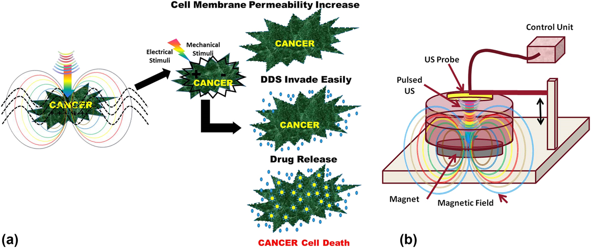
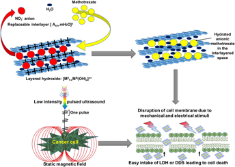
The rationale behind our proposed approach of combing LIPUS and SMF is that the charged ions of physiological/cell media or tissue experience Lorentz force Reference Montalibet, Jossinet, Matias and Cathignol34 when they are exposed to ultrasound in the presence of magnetic field. This ultrasound-induced Lorentz force rapidly oscillates the ions/tissue leading to the generation of local electrical currents/fields proportional to the electrical conductivity of the medium. Reference Montalibet, Jossinet, Matias and Cathignol34 We hypothesize that these noninvasively generated electrical currents/fields can potentially alter several cellular processes Reference Batista Napotnik, Reberšek, Vernier, Mali and Miklavčič30,Reference Nuccitelli35 and the ultrasound generated mechanical stresses can play a synergic role in cancer cell inhibition. A schematic diagram showing our hypothesis is presented in Fig. 1(a). The use of LIPUS can oscillate the intra-/inter-cellular charged ions and generate mechanical stresses on the cells/tissues. However, when the cells are exposed to LIPUS + SMF, they rapidly oscillate, due to Lorentz force, and generate local electrical currents/fields. Reference Montalibet, Jossinet, Matias and Cathignol34 The synergy of mechanical and electrical stimulations thus generated can potentially increase the cell membrane permeability, ROS, and oxidative stress, and break DNA of cancer cells Reference Furusawa, Fujiwara, Campbell, Zhao, Ogawa, Hassan, Tabuchi, Takasaki, Takahashi and Kondo36 leading to cell death. In the presence of DDS, the above processes can increase the drug intake by cells and thus enhances the cell inhibition. We believe that the proposed approach can potentially increase the treatment efficiency and reduce the drug dosage and associated health hazards. However, within our knowledge, the use of LIPUS and SMF to improve the efficiency of anticancer DDS is not yet reported. Therefore, in this work, an attempt has been be made to study the influence of LIPUS and SMF on the in vitro efficiency of anticancer drug (methotrexate, MTX) loaded layered double hydroxide (LDH) NPs. Detailed in vitro influence of this treatment on colon cancer cell (HCT116) inhibition in the presence of DDS is presented.
(a) Schematic representation of proposed hypothesis on the influence of SMF + LIPUS treatment on cancer cells. (b) Experimental scheme for in vitro cancer cell treatment using LIPUS + SMF. The cells were exposed to LIPUS from top using a probe that is in contact with cell media in a Petri dish. A permanent magnet placed below the Petri dish exposed the cell to SMF simultaneously.

II. EXPERIMENTAL
A. Synthesis and characterization of Mg–Al LDH intercalated with MTX
LDH was chemically synthesized following an ion-exchange method. Reference Chakraborty, Roychowdhury, Sengupta and Ghosh37 Methotrexate (Sigma Aldrich, Bangalore, India) solution (0.3987 gm/10 mL) was added to LDH suspension at pH 10 with continuous stirring for 12 h to get LDH–MTX composite (DD + MT), which was washed 3–4 times by dispersing in water followed by centrifugation. The washed DD + MT precipitate was freeze-dried (EYELA, FDU2200, Tokyo, Japan) at −82 °C and 20 Pa pressure to get dry LDH–MTX nanopowder. X-ray diffraction (XRD) analysis of pristine and DD + MT powder was performed using an X’Pert Pro MPD diffractometer (Panalytical, Almelo, The Netherlands) with Cu Kα radiation at 40 mA, 40 kV. Particle morphology of the powder was studied using scanning electron microscopy (SEM; Supra 35 VP, Carl Zeiss, Oberkochen, Germany). Thermogravimetric analysis (TGA/DTA, NETZSCH STA 409 CD, Gebrüder-Netzsch-Straße Germany) of the powder was carried out with a heating rate of 10 °C/min, in air, up to 1000 °C using simultaneous thermal analyzer. Particle size distribution was assessed using photon correlation spectroscopy (Zetatrac, Microtrac, Montgomeryville, Pennsylvania) with dilute suspensions of LDH and DD + MT in water, phosphate buffer saline (PBS), and a mixed solvent X [0.3 M HCL and PBS in the 1:9 volume ratio]. Reference Chakraborty, Roychowdhury, Sengupta and Ghosh37
B. In vitro tissue culture experiments
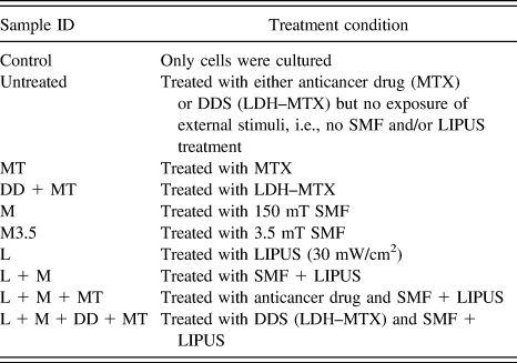
Human colon carcinoma cancer cells (HCT116) (ATCC, Rockville, MD) were routinely cultured in DMEM (Invitrogen, Carlsbad, CA) supplemented with 10% heat-inactivated fetal bovine serum, 1 U/mL penicillin G, and 1 mg/mL streptomycin, in 10 cm (diameter) plastic Petri dishes in a CO2 incubator (ESCO, Singapore) at 37 °C with 5% CO2. The cells were subcultured using trypsin-EDTA (Invitrogen, Carlsbad, California) when they were 85–90% confluent. The cultured cells were trypsinized and plated in 35-mm cell culture plates (2 × 10⁴ cells) (Nunc™, Thermo Fisher Scientific, Waltham, Massachusetts). The cells were exposed (15 min/day) to a treatment of static magnetic field (SMF, 150 mT), LIPUS (LIPUS, Frequency: 1.5 MHz, Pulse width: 200 μs, repeated at 1 kHz, Intensity: 30 mW/cm2) (Bone Heal 100, Technomed Electronics, Chennai, India), and their combination. The size of the permanent magnet was ∅ 30 mm with 5–6 mm thick and the LIPUS probe diameter was 22.22 mm. Initially two different SMF strengths were used (3.5 and 150 mT) to determine suitable SMF strength that can inhibit HCT116 in the presence of LIPUS. As the inhibition was more under 150 mT SMF with 30 mW/cm2 LIPUS, all other cell culture studies, with bare MTX as well as LDH–MTX, were performed under this condition to evaluate the combined effect of LIPUS + SMF (hereafter denoted as L + M) treatment. The details of sample notations used in the present investigation are presented in Table I. All tissue culture experiments were carried out till 72 h of incubation and the typical experimental setup is shown in Fig. 1(b).
Sample notations and treatment conditions used for HCT116 cell proliferation analysis, using MTT assay, in the present investigation.

C. In vitro cell proliferation assay
Initially, to determine the desired drug concentration, the dose response curve was generated, where the IC50 (half maximal inhibitory concentration) of MTX was evaluated using six concentrations in the range of 5 and 100 ng/mL with a stock solution of MTX (10 mg/ml) in dimethyl sulphoxide (DMSO; Sigma Aldrich, Bangalore, India). The cells plated in a 96-well plate (5 × 10³ cells/mL per well) were then incubated at 37 °C in a CO2 incubator for 24 h to allow sufficient cell adhesion. After addition of MTX, the incubation was continued for a total duration of 72 h (n = 3). MTT assay was performed by adding 10 µL of 3-(4,5-dimethylthiazole-2-yl)-2,5-phenyltetrazolium bromide (MTT) reagent, with a concentration of 1 mg/mL (Sigma Aldrich, St. Louis, Missouri) in 1:9 ratio (MTT:DMEM), to all wells and incubated in the dark for 4 h at 37 °C. Then the solution was removed and 100 µL DMSO was added to each well. The absorbance of the solution was measured at 550 nm on an ELISA reader (Bio-Rad, Hercules, California) to generate the dose response curve to determine IC50 of MTX.
The cultured HCT116 cells were plated in 35-mm plates (2 × 10⁴ cells) (Nunc™, Thermo Fisher Scientific, Waltham, Massachusetts) and incubated 24 h for adherence. Then MTX (IC50) or LDH–MTX (with IC50 concentration of MTX) was added to the cells (n = 12), followed by different treatments. To assess if the proposed treatment conditions have any detrimental effect on healthy cells, we have performed similar proliferation assay using mouse fibroblast cells (NIH3T3) (ATCC, Rockville, Maryland) as model cells. These cells were also cultured routinely following the standard protocol described above. The NIH3T3 cells were plated in 35-mm plates (2 × 10⁴ cells) and incubated 24 h for adherence, followed by exposure to identical L + M treatment for 15 min/day after treating the cells with MTX (IC50) or LDH–MTX (n = 3). After treatment, the cells were incubated for 72 h and the cell viability was measured using MTT assay.
D. Cell apoptosis and cell cycle analysis
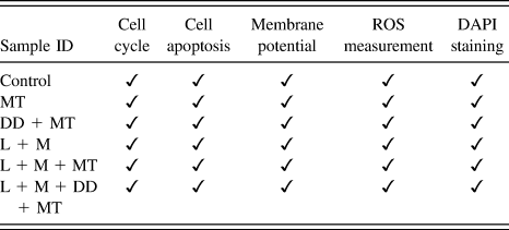
The cell apoptosis was determined using Annexin V/PI double staining. After desired culture period, the cells were trypsinized, washed with 1× PBS, and the suspended pellet was analyzed using a flow cytometer (CyFlow Cube 6, SysmexPartec GmbH, Münster, Germany). The standard protocol specified by the manufacturer of the cell apoptosis kit (BD bioscience, Franklin Lakes, New Jersey, Cat No: 556570) was followed. Similarly, the cycle analysis was performed using Cycle TEST™ PLUS DNA Reagent kit (BD Cat No: 340242). As the cell inhibition was prominent at 72 h of incubation, the cell cycle analysis was carried out at this culture duration. The treatment conditions used for cell apoptosis and cell cycle analysis are presented in Table II.
Treatment conditions used for cell cycle, apoptosis, cell membrane potential, ROS, and DAPI staining. Culture duration = 72 h.

E. Cell membrane potential and ROS measurement
The changes in the plasma membrane potential of HCT116 cells exposed to different treatments (Table II) were measured by flow cytometry using bis(1,3-dibutylbarbituric acid) trimethineoxonol [DiBAC4(3), Invitrogen, Carlsbad, California]. 100 nM DiBAC4(3) was prepared using DMSO according to the manufacturer’s instructions. This solution was added to the treated cells after 72 h of incubation. Then the cells were incubated for 10 min in the dark and the mean fluorescence intensity of the cell population was analyzed using a flow cytometer at an excitation maxima of 490 nm. For ROS determination, the treated cells (1 × 10⁵) were trypsinized after 72 h incubation, washed in 1× PBS and resuspended in 1 mL of 1× PBS to measure ROS using a detection assay kit (ab186029, Abcam, Cambridge, Massachusetts) and flow cytometer at an excitation wave length of 650 nm.
F. Fluorescence microscopy
To evaluate the effect of different treatments on HCT116 cells, in terms of their nucleus and nuclear membrane structure, qualitative analysis of the cells was performed using fluorescence microscopy. After desired culture duration, under different treatment conditions (Table II), the cells were stained using diluted 4′,6-diamidino-2-phenylindole (DAPI) (Catalog No: D1306, Thermo Fisher, Waltham, Massachusetts) stock solution (with PBS at 1:5000). Then the cells were fixed for 10 min in 3.7% formaldehyde and after removing the fixative, the permeabilized cells were immersed in 0.2% Triton X-100 for 5 min. The cells were then incubated for 1–5 min at room temperature in diluted DAPI solution followed by PBS washing. Finally, the cells were imaged using a fluorescence microscope (BX51, Olympus, Tokyo, Japan).
G. Estimation of mechanical and electrical stimuli
The mechanical stimulus which is the effective pressure induced by LIPUS at the cell/soft tissue was estimated as follows:
 $$p = \sqrt {I \cdot Z} \quad .$$
Similarly, the electrical filed generated (E), when the cell/soft tissue oscillates (due to ultrasound) under magnetic field, was estimated as follows:
 $$E = \left( {{1 \over C}} \right) \cdot \left( {{p \over Z}} \right) \cdot B\quad ,$$
where p is the effective pressure (Pa), I is the intensity of ultrasound (W/cm2), Z is the acoustic impedance of the cell or soft tissue (kg/m2/s), C is the velocity of light (m/s), and B is the magnetic field strength (G). E is measured in electrostatic units esu (1 esu = 300 V/cm). The acoustic impedance data have been taken from the “Database of Tissue Properties” available at https://www.itis.ethz.ch/virtual-population/tissue-properties/database/.
H. Statistical analysis
The data obtained in cell proliferation, cell apoptosis, and cell cycle tests were statistically analyzed using Students t-test and p < 0.05 was considered statistically significant.
III. RESULTS
A. Characterization of Mg–Al LDH intercalated with MTX
The XRD patterns of LDH and LDH–MTX, Fig. 2(a), showed characteristic well-defined (001) reflection without any impurity peaks, demonstrating the formation of single-phase LDH (JCPDS No. 35-0964). The (001) reflection in the pristine LDH was shifted to lower angle in LDH–MTX signifying drug intercalation. Reference Chakraborty, Dasgupta, Sengupta, Chakraborty and Basu38 As shown in Fig. 2(b), the weight loss in the pristine LDH increased to 57% on heating, up to 518 °C, due to interlayer nitrate anion decomposition. At the same temperature, the LDH–MTX sample showed a weight loss of 50% because of decarboxylation of MTX, decomposition of nitrate and other anions. Thermogravimetric analysis of LDH–MTX indicated that the loading of MTX into the LDH matrix was 44%. The particle size of LDH after intercalation of MTX into interlayers of the LDH matrix was found to be between 30 and 70 nm when assessed using SEM, Fig. 2(c). The dose response curve of bare MTX for HCT116 cell line, shown in Fig. 2(d), indicates that the IC50 under present experimental conditions was 10 ng/mL.
(a) Powder XRD patterns of LDH and LDH–MTX (inset). (b) Thermal analysis curves of (A) MTX, (B) LDH–MTX, (C) LDH. (c) SEM image of Mg–Al LDH–MTX nanopowder. (d) Dose response curve showing the influence of MTX concentration on HCT116 cell viability (n = 3).

B. In vitro cell proliferation assay under the influence of LIPUS + SMF
Initial treatments were carried out using different SMF strengths along with 30 mW/cm2 LIPUS to check HCT116 cell inhibition, and the range of SMF strengths (3.5 and 150 mT) used in the present investigation has been shown to be safe for healthy cells. Reference Shirasaka39 The results shown in Fig. 3(a) indicate that LIPUS with 3.5 mT SMF (L + M 3.5) exposure to cancer cells can inhibit the cells up to 40% during 72 h of incubation. The inhibition was further increased to ∼80% when the SMF intensity was increased to 150 mT with LIPUS intensity of 30 mW/cm2 (L + M 150). Therefore, further experiments were carried out using 150 mT SMF + 30 mW/cm2 LIPUS (L + M).
(a) Proliferation of HCT116 cells exposed to daily 15-min treatment of 30 mW/cm2 LIPUS (L), 30 mW/cm2 LIPUS along with 3.5 mT SMF (L + M 3.5), and 30 mW/cm2 LIPUS along with 150 mT SMF (L + M), (P < 0.05 between Control and Treated samples at all culture durations) (b and c) Proliferation of HCT116 cells exposed to daily 15-min treatment of 150 mT SMF (M), 30 mW/cm2 LIPUS (L) and their combination (L + M). (b) Cells treated in the presence of MTX (10 ng/mL) alone (n = 12). (c) Cells treated in the presence of LDH–MTX (DD + MT) (n = 12) (P < 0.05 between Untreated and Treated samples at all culture durations).

Figure 3(b) shows the effect of M, L, and L + M treatments on HCT116 cell inhibition in the presence of anticancer drug (MTX). The “Untreated” group (without L or M or L + M treatment) achieved approximately 50% reduction in cell proliferation at 72 h while the individual treatments (L or M) also exhibited similar reduction in the proliferation with LIPUS being marginally better than other two groups at all culture durations, as shown in Fig. 3(b). However, the L + M treatment significantly decreased the proliferation and approximately 50% of the cells were inhibited at 24 h culture duration. By 48 h of culture, the cell proliferation reduced to 28% which was further decreased to 16% at the end of 72 h culture. A clear increase in the inhibition of cancer cells, up to 30%, was observed with L + M treatment compared to “Untreated” and “M” conditions after 72 h.
To investigate whether the L + M treatment can enhance the efficacy of LDH–MTX, the proliferation of HCT116 cells under L + M treatment in the presence of DD + MT (LDH–MTX) was carried out. The proliferation of “Untreated” group reached approximately 50% in 48 h when the MTX was delivered as DD + MT, Fig. 3(c), which is 24 h earlier than the same “Untreated” group when bare MTX was used [Fig. 3(b)]. This observation suggests enhanced efficiency (bioavailability) of anticancer drug when delivered using LDH carrier. Similar reduction in HCT116 cell proliferation was observed with “M” treatment. However, the LIPUS treatment (L) appears to accelerate inhibitory effect and up to 60% inhibition was recorded in 24 h. Figure 3(c) also shows that the cells failed to proliferate beyond 17% with an increase in the culture duration up to 72 h under L + M treatment in the presence of MTX. The use of L + M treatment restricted the cancer cell proliferation to 20% in 24 h and maximum proliferation of 5% was achieved at 72 h. These results demonstrate that the L + M treatment can improve the efficacy of both drug and DDS.
C. Changes in the cell cycle and cell apoptosis
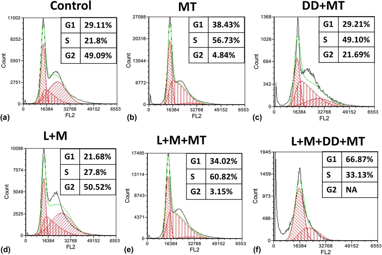
To understand the influence of the L + M treatment on HCT116 cellular activities, cell cycle analysis with PI stain and apoptosis assessment with Annexin V–PI stains were performed at 72 h culture duration where maximum cell inhibition was observed. The gated cell population in different phases of cell cycle is shown in Fig. 4. It is well established that the MTX drug is S-phase blocker Reference Yao, Feng and Chen4,Reference Savage and Prasad40 and therefore we focused on gated cell population in the G1 phase. As expected, the results, Fig. 4(b), revealed that the cells treated with MTX stagnate in G1 as well as in S phase, compare to Control group [Fig. 4(a)], where significant amount of cells were observed in G2 phase. However, with DD + MT treatment, the cell cycle arrest was only marginal, as shown in Fig. 4(c). Interestingly, the L + M treatment alone did not show any restriction on cell cycle [Fig. 4(d)]. The restriction of cell population in G1 and S phases was more (60% of cells in S phase and 34% in G1, P < 0.05) when the cells were treated with drug along L + M [Fig. 4(e)]. The restriction was more prominent when DDS (DD + MT) was used in conjunction with L + M treatment, Fig. 4(f), where 67% of cells were in G1 phase and 33% in S, P < 0.05. Surprisingly, no G2 phase was observed in the L + M + DD + MT treatment.
(a–f) HCT116 cell cycle assessment showing gated cell population in different phases at 72 h (n = 6). (a) Control (only cells), (b) MT treated cells, (c) DD + MT treated cells, (d) L + M treated cells, (e) L + M treated in the presence of MT, and (f) L + M treated in the presence of DD + MT.

The assessment of cell apoptosis, shown in Fig. 5, corroborated the cell cycle analysis. The apoptosis cells constituted 31% when treated with MTX [Fig. 5(b)] and it increased to 59% when the MTX was delivered in the form of DD + MT (LDH–MTX), as shown in Fig. 5(c). The apoptotic cell concentration increased considerably with L + M treatment. However, the severity of apoptosis increased to 64% with L + M treatment without any drug or drug carrier, Fig. 5(d). While, in the presence of MTX and DD + MT (LDH–MTX), the L + M treatment resulted in 71% [Fig. 5(e)] and 99% [Fig. 5(f)] apoptotic cells, respectively. Interestingly, the amount of cell transition from early to late apoptosis was found to be different under these treatment conditions. For example, when the cells were treated with MTX alone, the amount of cells in the later stage of apoptosis was only 9%, which increased to 67% with L + M treatment as shown in Figs. 5(b) and 5(e). Similarly, the early apoptotic cells increased to 80% from 31% when the L + M treatment was applied to the cells in the presence of DD + MT (LDH–MTX), Figs. 5(c) and 5(f).
(a–d) Apoptosis assessment of HCT116 cells with Annexin V–PI stains after 72 h of incubation (n = 6). (a) Control (only cells). (b) MT (MTX only) treated cells. (c) DD + MT (LDH–MTX) treated cells. (d) L + M (30 mW/cm2 LIPUS + 150 mT SMF). (e) L + M + MT (L + M in the presence of MTX) (* P < 0.05 between Control and L + M + MT). (f) L + M + DD + MT (L + M in the presence of LDH–MTX) (* P < 0.05 between Control and L + M + DD + MT).

D. Cancer cell membrane potential and ROS production
The HCT116 cell membrane potential, under MTX (MT) and LDH–MT (DD + MT) treatment, determined using flow cytometry is shown in Fig. 6(a), which demonstrates marginal hyperpolarization of cancer cells when treated with MTX or LDH–MTX (small peak shift toward left) compared to “Control” group (cells only). A similar trend was observed when the cells were treated with 30 mW/cm2 LIPUS + 150 mT SMF (L + M), Fig. 6(b). However, the peak shift was relatively high when the L + M treatment was used in the presence of MTX (L + M + MT) or LDH–MTX (L + M + DD + MT). This indicates more cell membrane hyperpolarization leading to increased transmembrane transport of several ions followed by cell death Reference Yang and Brackenbury19,Reference Li and Huang41,Reference Soto-Cerrato, Manuel-Manresa, Hernando, Calabuig-Fariñas, Martínez-Romero, Fernández-Dueñas, Sahlholm, Knöpfel, García-Valverde, Rodilla, Jantus-Lewintre, Farrás, Ciruela, Pérez-Tomás and Quesada42 under these treatment conditions. It is believed that the LIPUS + SMF (L + M) treatment induced cell membrane changes possibly increased the intake of anionic drug by the cells, when treated with MTX or LDH–MTX thus resulting in overall enhancement in HCT116 cellular inhibition. Furthermore, the LIPUS alone has been shown to induce micromotions (∼50 µm/s) in the soft tissue, Reference Bolander, Greenleaf, Bronk and Kinnick43 which can potentially destabilize the cell membrane followed by cell death.
(a and b) Flow cytometric estimation of the HCT116 cell membrane potential using DiBAC4(3), a membrane potential sensitive fluorescent probe (n = 6). (a) Cells treated with MTX (MT) and LDH–MTX (DD + MT) were compared with Control (only cells). (b) 30 mW/cm2 LIPUS + 150 mT SMF (L + M) treatment applied in the presence MT and DD + MT.

It is known that the oxidative stress, which is related to elevated intracellular levels of ROS, can damage cell structure, DNA, proteins, and lipids leading to cell death. Therefore, we have measured the oxidative stress generated under different treatment conditions in terms of ROS and the results are presented in Fig. 7. The ROS intensity increased from 122 to 259 when the HCT116 cells were treated with anticancer drug (MT) and DDS (DD-MT) as shown in Fig. 7(a). The use of drug carrier increased the ROS by 47%. The L + M treatment also found to increase the ROS level, Fig. 7(b), to 820 compared to bare drug with an intensity of 122. Almost seven times increase in the ROS production was noted with L + M treatment in the presence of anticancer drug (L + M + MT) and DDS (L + M + DD + MT).
Measurement of intracellular ROS: (a) Cells treated with MTX (MT) and LDH–MTX (DD + MT) were compared with Control (only cells). (b) Cells treated with 30 mW/cm2 LIPUS + 150 mT SMF (L + M) treatment in the presence MT (L + M + MT) and DD + MT (L + M + DD + MT).

E. Nuclear membrane characteristics
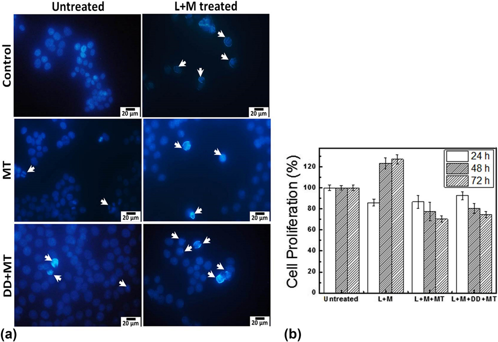
To examine the initiation of changes in the HCT116 cell nucleus, due to 30 mW/cm2 LIPUS + 150 mT SMF (L + M) treatment, we have used DAPI staining after 24 h of culture. The morphology of DAPI-stained HCT116 cell nuclei exposed to different treatments is shown in Fig. 8(a). In general, the DAPI can stain live cells as well as fixed cells. However, the morphology of HCT116 cells treated with either MTX (M) or LDH–MTX (DD + MT) showed damaged cell nucleus which is an indication of apoptosis. Reference Okkenhaug, Bilancio, Emery and Vanhaesebroeck44 Furthermore, the damage appears to be relatively more in the cells exposed to the L + M treatment indicating the effectiveness of this treatment in inhibiting HCT116 cell proliferation. The damaged cell nucleus directly led to cell inhibition thus providing overall improvement in the treatment efficacy.
(a) Morphology of DAPI-stained HCT116 cell nuclei with and without 30 mW/cm2 LIPUS + 150 mT SMF (L + M) treatment after 24 h incubation. Arrows indicate the cells with damaged cell nucleus, which are more prominent in treated cells. (b) Proliferation of NIH3T3 cells (n = 3) exposed to daily 15-min treatment of 30 mW/cm2 LIPUS + 150 mT SMF (L + M) in the presence of MTX (L + M + MT) and LDH–MTX (L + M + DD + MT) (P < 0.05 between Untreated and Treated samples).

F. Influence of the L + M treatment on healthy cells
Although the proposed L + M treatment found to have positive influence on cancer cell inhibition, it is important to know how this treatment would affect normal/healthy cells. Therefore, we have performed similar MTT proliferation assay on healthy mouse fibroblast (NIH3T3) cells. The results, Fig. 8(b), show that the treatment (L + M) marginally inhibits the proliferation of NIH3T3 cells at 24 h. However, with increase in the culture duration, the cells proliferated and their number increased rapidly at 48 h. In the presence of drug (L + M + MT), the treatment reduced NIH3T3 cell proliferation but the inhibition was significantly lower than observed with HCT116 cells [Fig. 3(b)]. Similarly, the inhibition of healthy cells under L + M treatment in the presence of drug carrier (L + M + DD + MT) was also marginal and lower than HCT116 cells shown in Fig. 3(c). One interesting observation is that the L + M treatment alone (without anticancer drug) increased the NIH3T3 cell proliferation, with culture duration, suggesting its beneficial effects on these healthy cells.
IV. DISCUSSION
There is an unmet need for newer, alternative, and adjuvant cancer treatments with increased therapeutic efficacy, reduced side effects, and capability to provide personalized cancer treatment to reduce hospital visits/cost. In this work, we have demonstrated that the in vitro efficiency of the cancer treatment can be improved by 40% minimum (i.e., for the same amount of drug, the amount of cancer cell inhibition can be improved by 40%) with the combined use of LIPUS (30 mW/cm2) and SMF (150 mT) in the presence of drug and/or DDS. Alternatively, with this treatment, one can decrease the drug dosage and associated health hazards, while achieving similar cell inhibition that can be achieved with direct use of anticancer drug or DDS. The capacity of this new treatment is the noninvasive generation of synergy between LIPUS induced mechanical stresses Reference Mo, Coussios, Seymour and Carlisle15,Reference Hoffman, Grashoff and Schwartz45 and LIPUS + SMF generated local electrical fields Reference Montalibet, Jossinet, Matias and Cathignol34 that would negatively regulate cancer cell activities via altering cell membrane characteristics Reference Batista Napotnik, Reberšek, Vernier, Mali and Miklavčič30 (permeability, potential). Furthermore, this new treatment accelerated cancer cellular activities leading to early inhibition, i.e., more inhibition of cells at least 24 h earlier than observed without this treatment.
The role of membrane potential in controlling cellular activities, more specifically their proliferation and differentiation, is well known. Reference Yang and Brackenbury19 The hallmark of rapidly proliferating tumor cells is that they are characterized by depolarized membrane potential (less negative membrane potential). Reference Hanahan and Weinberg20 Therefore, these cells contain higher intracellular Na+ levels compared to noncancerous cells and are believed to trigger initiation of mitosis and DNA synthesis leading to rapid advancement in their cell cycle thereby sustained tumor growth. Reference Binggeli and Weinstein46 A recent in vivo study also demonstrated that the membrane depolarization alone can increase cancerous cell proliferation and tumor growth via enhanced angiogenesis. Reference Lobikin, Chernet, Lobo and Levin47 Our results show that HCT116 cells are hyperpolarized (more negative cell membrane) when exposed to different treatments. Such hyperpolarization of cancer cells can be viewed as efflux of K+ ions or influx of Cl− ions leading to accumulation of net negative charge across the cell membrane. This flow of ions across the cell membrane depends on the cell membrane permeability, Reference Yang and Brackenbury19 which can be altered using drugs, and mechanical and electrical stimuli such as present LIPUS + SMF treatment. The hyperpolarization of HCT116 cells due to LIPUS + SMF treatment, with or without MTX and LDH–MTX, is believed to arrest the cell cycle. Reference Cone and Tongier48 The restriction of cell transition (G1 → S phase) in the cycle increased significantly (>66% in G1 phase, P < 0.05) due to the application LIPUS + SMF in the presence of DDS. Moreover, the interaction between 30 mW/cm2 LIPUS + 150 mT SMF treatment and the polar, ionic molecules of cellular compartment produced significantly high amount of ROS because of pro-inflammatory changes inside the cancer cell Reference Montalibet, Jossinet, Matias and Cathignol34 thus inhibited their growth and proliferation. It has been observed that the SMF interacts with the charges (ions, proteins, etc.) of biological system through several physical mechanisms 49 and alters the activity, concentration, and life time of paramagnetic free radicals, i.e., ROS, reactive nitrogen species, which cause oxidative stress, genetic mutation, and apoptosis in the cancer cells. Reference Ghodbane, Lahbib, Sakly and Abdelmelek50 Therefore, elevated production of ROS in HCT116 cells due to SMF + LIPUS treatment could play an important role in the natural immunological defense Reference Nathan51 of the body against cancer, through intracellular signaling pathways, Reference Lander52 and found to damage ion channel of cancer cells Reference Berk, Dodd and Henry53 leading to changes in their morphology and apoptosis. It is believed that the LIPUS + SMF treatments increased the cell membrane permeability and allowed intracellular action of the drug or DDS. The results of cell cycle, apoptosis and membrane potential analysis suggest that the LIPUS + SMF treatment increases HCT116 cell inhibition via similar mechanisms as observed when drug or DDS was used. For example, both treatments showed (i) stagnation of HCT116 cells in G1 and S phase (G1 → S restriction) and (ii) similar change in cell membrane potential (hyperpolarization). However, the use of LIPUS + SMF treatment accelerated the cellular activities of HCT116 as evidenced by the increase in late apoptotic cells (43%). The increase in the late apoptotic cell population also indicates DNA fragmentation and loss of plasma membrane integrity of these cells, which occurs in the later stages of apoptosis. In addition, the SMF would also have assisted in destabilizing the cell membrane leading to apoptosis. Reference Ghibelli, Cerella, Cordisco, Clavarino, Marazzi, De Nicola, Nuccitelli, D’Alessio, Magrini, Bergamaschi, Guerrisi and Porfiri23,Reference Teodori, Grabarek, Smolewski, Ghibelli, Bergamaschi, De Nicola and Darzynkiewicz25
The observed influence of 30 mW/cm2 LIPUS +150 mT SMF on HCT116 cell inhibition is believed to be due to noninvasively generated electrical and mechanical stimuli. The mechanical stimulus, which is the effective pressure induced by LIPUS at the cell/soft tissue and the electrical filed generated (E), when the cell or soft tissue oscillates (due to ultrasound) under magnetic field, was estimated using Eqs. (1) and (2), respectively. Under present experimental conditions of 30 mW/cm2 LIPUS and 150 mT (1500 G) SMF, the average effective pressure generated in the soft tissues would be 20–22 kPa and the electrical field generated would be 19–23 µV/cm. Although the effective pressure may change under in vivo conditions, due to attenuation of ultrasound by different tissues and their location within the body, these LIPUS-induced mechanical stresses can either elastically or plastically deform the cells/tissues leading to physical changes in the cell membrane, which have been confirmed by cell membrane potential measurements. The elastic deformation of cell/tissue has been found to form stomas/lacunars in the cell membranes Reference Mo, Coussios, Seymour and Carlisle15,Reference Tachibana, Uchida, Ogawa, Yamashita and Tamura54,Reference Baker, Robertson and Duck55 which can increase the drug intake or transmembrane ion flow leading to cell death. On the other hand, the plastic deformation of the cell membrane can break the membrane and kill the cells. Furthermore, the LIPUS alone has been shown to induce micromotions (∼50 µm/s) in the soft tissues, Reference Bolander, Greenleaf, Bronk and Kinnick43 which can potentially destabilize the diseased cell membrane leading to cell death. Similarly, the electrical field generated due to LIPUS + SMF treatment would have synergistically disturbed the stability of cancer cell activities leading to early and increased cancer cell death. Similar observations have been reported when the cells were exposed to external electrical pulses, Reference Ghibelli, Cerella, Cordisco, Clavarino, Marazzi, De Nicola, Nuccitelli, D’Alessio, Magrini, Bergamaschi, Guerrisi and Porfiri23,Reference Batista Napotnik, Reberšek, Vernier, Mali and Miklavčič30,Reference Nuccitelli, Huynh, Lui, Wood, Kreis, Athos and Nuccitelli56 where the electrical fields were generated by inserting electrodes, contrary to the noninvasive, in situ generation in the present work.
Our experimental results demonstrate that the proposed noninvasive treatment of using LIPUS + SMF can stimulate cancer cells (HCT116) death via hyperpolarization of cell membranes to the extent equal to that of widely used anticancer drug methotrexate. Furthermore, this treatment has been found to be safe for healthy cells (NIH3T3). However, further studies are advocated to confirm the effectiveness of LIPUS + SMF treatment for cancer therapy. Alternatively, this new approach can be used either to decrease the drug dosage, thereby decreasing the side effects associated with anticancer drugs. The therapeutic efficiency of the current LIPUS + SMF treatment can be further enhanced through tailoring the treatment exposure time, which was 15 min/day in the present investigation. However, the synergetic effect of noninvasively generated in vitro mechanical and electrical stimuli can change under in vivo conditions, due to variations in the attenuation of LIPUS power by different tissues depending on the distance between the LIPUS or SMF source and the location of the cancer cells/tumor within the body. Therefore, further studies are required to establish the observed in vitro benefits in other cancer cell lines and through in vivo studies.
V. CONCLUSIONS
Present preliminary in vitro results demonstrate that the efficiency of cancer treatment, with direct drug or DDS, can be improved by 40% minimum when LIPUS of 30 mW/cm2 is used in the presence of 150 mT SMF. It is believed that the synergy between LIPUS-induced mechanical stimuli (20–22 kPa) and LIPUS + SMF generated local electrical stimuli (19–23 µV/cm) would have downregulated the natural activities of cancer cells by altering cell membrane characteristics such as permeability, potential, and generation of ROS. These results demonstrate that this new approach can be used either to decrease the drug dosage, thereby decreasing the side effects associated with anticancer drugs, or overall treatment time.
ACKNOWLEDGMENTS
The authors acknowledge financial support from Council of Scientific and Industrial Research (CSIR) India under grants ESC 0103 and SRF fellowship to S.S. The help rendered by Dr. Subhadip Bodhak during technical discussions is greatly acknowledged.