Abnormal flow into the pulmonary artery because of patent ductus arteriosus is commonly encountered in paediatric echocardiography. The coronary arteriovenous fistulae and anomalous left coronary artery from the pulmonary artery can also cause similar flow into the pulmonary artery. Rarely, isolation of the left subclavian artery can present with abnormal flow into the pulmonary artery in children with cyanotic CHDs. Here we describe a child who was incidentally detected to have isolation of the left subclavian artery during evaluation of atrial septal defect for device closure.
Case report
An 8-year-old girl with recurrent respiratory tract infections was referred for cardiac evaluation. Clinical examination showed equal pulses in all four limbs, mild cardiomegaly, ejection systolic murmur at the pulmonary area, and a wide, fixed, second heart sound, suggesting the diagnosis of an atrial septal defect. Saturation was 99% in all four limbs by finger pulse oximetry. Chest roentgenogram showed cardiomegaly, pulmonary plethora, and a right-sided aortic arch. Transthoracic echocardiography confirmed the diagnosis of atrial septal defect, which was suitable for percutaneous device closure. Although the right-sided aortic arch was confirmed, the arch branching pattern was not clear. Unusually a flow into the pulmonary artery was seen from a neck vessel. With the possibilities of an abnormally oriented patent ductus arteriosus in a right arch and a rare systemic artery to the pulmonary artery fistula, it was decided to delineate the communication by transoesophageal echocardiography and cardiac catheterisation before proceeding with device closure.
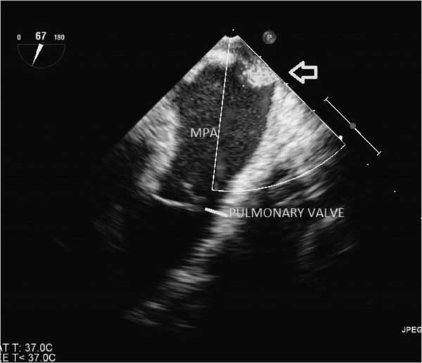
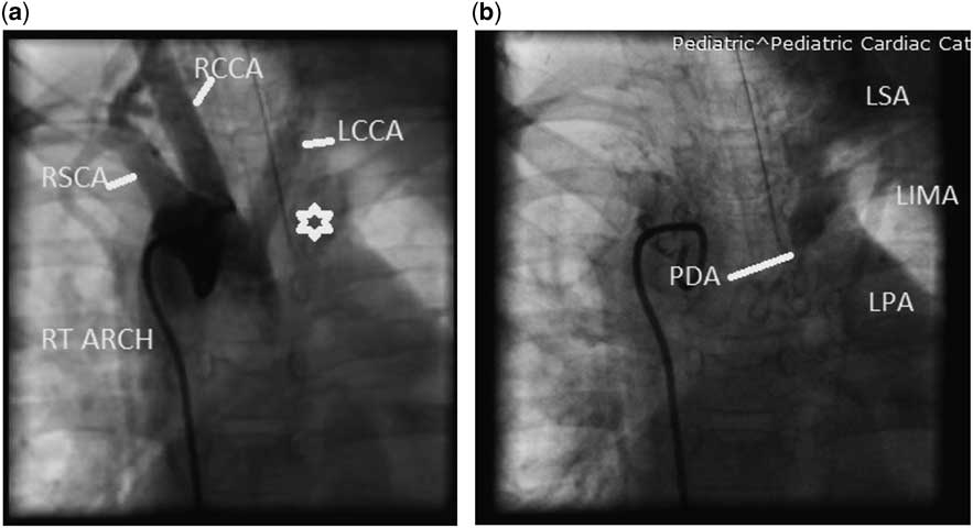
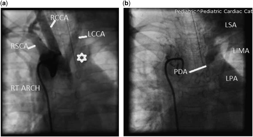
Transoesophageal echocardiography showed a 13-mm ostium secundum atrial septal defect, confirmed the presence of an abnormal flow into the pulmonary artery (Fig 1 and Supplementary video S1), and ruled out other intracardiac abnormalities. Cardiac catheterisation revealed normal right heart pressures and saturation step up at both the right atrial and pulmonary arterial level – mixed venous, right ventricle, and left pulmonary artery saturations of 71, 82, and 83.5%, respectively. Ascending aortic angiogram in an anteroposterior view showed a right aortic arch with the first branch not giving rise to the left subclavian artery (Fig 2a). On delayed image, retrograde flow from the left vertebral artery and collaterals from the other side of the neck filled the left subclavian artery, which in turn filled the pulmonary artery through the stenosed patent ductus arteriosus (Fig 2b and Supplementary video S2), confirming the diagnosis of isolation of the left subclavian artery. Digital subtraction angiography also showed the same findings. As isolation of the left subclavian artery does not result in a vascular ring, and the child was asymptomatic for vertebral steal or limb ischaemia, it was decided to percutaneously close the atrial septal defect with a 14-mm HEART R atrial septal occluder (Lifetech Scientific, Shenzhen, China) in the same sitting and follow-up the child.
Transoesophageal echocardiography image showing abnormal flow into the PA at the level of PA bifurcation. MPA=main pulmonary artery; PA=pulmonary artery.

( a and b ) Aortic arch angiogram in anteroposterior view showing the right aortic arch with the left subclavian artery (site shown by star) not arising from the first branch as normally expected. Delayed image of the same angiogram in ( a ) showing filling of the left subclavian artery from the vertebral artery and collaterals from the other side of the neck, which in turn drains through the PDA into the pulmonary artery. LCCA=left common carotid artery; LIMA=left internal mammary artery; LPA=left pulmonary artery; LSA=left subclavian artery; PDA=patent ductus arteriosus; RCCA=right common carotid artery; RSA=right subclavian artery.
Discussion
Abnormal flow into the pulmonary artery with step up in saturation can be due to patent ductus arteriosus, aorto pulmonary window, supracristal ventricular septal defect, anomalous left coronary artery from pulmonary artery, coronary artery to pulmonary artery fistula, and systemic artery to pulmonary artery fistula. Isolation of the left subclavian artery with patent ductus arteriosus, illustrated in this case report is a very rare cause of abnormal flow into the pulmonary artery in acyanotic CHDs with the right aortic arch. Isolation refers to the origin of a neck vessel, commonly the subclavian artery, from the pulmonary artery via patent ductus arteriosus with no communication with the aortic arch.
Origin of isolation of the left subclavian artery can be embryologically explained by two ipsilateral interruptions in the aortic arch system, namely, the left dorsal aorta distal to the seventh intersegmental artery and left fourth arch resulting in the left subclavian artery connected to the pulmonary artery via the sixth aortic arch. This condition can either be asymptomatic or can present with left upper limb ischaemia and subclavian steal-related neurological symptoms.Reference Killen, Battersby and Klatte 1 , Reference Mar Anho, Gooch, Ablaza, Nakhjaha and Goldberg 2 It does not constitute a vascular ring as there is no vessel posterior to the oesophagus.Reference Stewart, Kincaid and Edwards 3
Review of the literature by Leutmer et alReference Leutmer and Miller 4 showed that more than half of the reported cases of isolation of the left subclavian artery were associated with cyanotic CHDs commonly tetrology of Fallot; only 13% of these patients had symptoms related to this condition. Diagnosis should be suspected in a patient with right aortic arch, with the abnormal arch branching pattern or symptoms of pulmonary or subclavian run off. Management of this condition is directed at correcting the underlying heart disease and implantation of the left subclavian artery into the left common carotid artery or the aortic arch on cardiopulmonary bypass.
In the present case, if the child develops symptoms related to isolation of the left subclavian artery on follow-up, device closure of the ductus is an option that can mitigate symptoms of subclavian steal from the cerebral circulation. In addition, surgical implantation of the subclavian artery to the aortic arch or its branches off cardiopulmonary bypass, as the intracardiac abnormality has already been corrected, can be performed.Reference Schena and Gandhi 5 , Reference Konstantinov, Saxena and d’Udekem 6
Acknowledgements
None.
Financial Support
This research received no specific grant from any funding agency, commercial, or not-for-profit sectors.
Conflicts of Interest
None.