Introduction
Mars is one of the main targets for the ongoing and in development life-detection missions beyond Earth, due to its potential past habitability. It is known that Mars actually hosts carbon, energy sources and different water reservoirs (Malin and Edgett, Reference Malin and Edgett2000; Rasmussen et al., Reference Rasmussen, Blake, Fletcher and Kilburn2009). Nevertheless, the current atmospheric and surface conditions are characterized by a CO2-rich atmosphere (Owen et al., Reference Owen, Biemann, Rushneck, Biller, Howarth and Lafleur1977), oxidizing substances, such as perchlorates (Lasne et al., Reference Lasne, Noblet, Szopa, Navarro-González, Cabane, Poch and Coll2016), oxygen peroxide (H2O2) and a hostile surface environment due to the presence of radiation (ionizing and non-ionizing radiation; Hassler et al., Reference Hassler, Zeitlin, Wimmer-Schweingruber, Ehresmann, Rafkin, Eigenbrode and Berger2014). This highly radiative environment is one of the main challenges both for life survival and biomolecule integrity, reducing the chance for robotic exploration to detect traces of life. Ionizing radiation can penetrate several metres into the Martian soil causing structural and chemical damages to biological molecules (Dartnell et al., Reference Dartnell, Desorgher, Ward and Coates2007). Indeed, no organic matter has been found in Martian regolith by the previously Viking missions on Mars (Biemann et al., Reference Biemann, Oro, Toulmin, Orgel, Nier, Anderson and Lafleur1977), even though the high infall meteorites should have accumulated detectable amounts of organics (Drake et al., Reference Drake, Greeley, McKay, Blanchard, Carr, Gooding, McKay, Spudis and Squyres1988). The exploration missions focus their investigation on the Martian subsurface being the Martian surface hostile for life as we know it.
Traces of life beyond Earth, the so-called biosignatures are defined as ‘an object, molecules or pattern of exclusively biological origin’ (des Marais et al., Reference Des Marais, Nuth, Allamandola, Boss, Farmer, Hoehler and Spormann2008). Among biosignatures, different biomolecules could be considered as direct and unambiguous signs of extant or extinct life, due to chemical and structural complexity and to the specificity that allows to assume that they can be only synthesized by living organisms (Pace, Reference Pace2001; Summons et al., Reference Summons, Albrecht, McDonald, Moldowan, Botta, Bada, Gomez-Elvira, Javaux, Selsis and Summons2008; Davila and McKay, Reference Davila and McKay2014). Terrestrial biomolecules that store and transfer genetic information (nucleic acids and proteins) are considered the most unambiguous signs of life, given the implausibility that these can be produced abiotically in a natural environment (Neveu et al., Reference Neveu, Hays, Voytek, New and Schulte2018). Besides, intra and extra-cellular molecules (e.g. pigments) that acts as protective molecules against external stresses are considered good biosignatures.
Considering the previously mentioned radiation environment beyond Earth, which biosignatures are effectively stable and, therefore, recognizable during life-detection missions? The answer to this question is investigated in the frame of the STARLIFE project that aimed at understanding the effect of radiation on selected microorganisms and their associated molecules, by exposing them to different space-relevant radiations (Moeller et al., Reference Moeller, Raguse, Leuko, Berger, Hellweg and Fujimori2017). The evaluation of the stability/degradation of organics in conditions similar to those experienced beyond Earth's magnetic field protection, is of outmost importance to support the in situ life-detection missions. Among the microorganisms and biomolecules tested in the STARLIFE project, we have chosen the cryptoendolithic black fungus Cryomyces antarcticus, isolated from McMurdo Dry Valleys (Antarctica), one of the best terrestrial analogues of the Martian environment (Cassaro et al., Reference Cassaro, Pacelli, Aureli, Catanzaro, Leo and Onofri2021a).
The fungus has been chosen for its proven ability to resist radiation stresses (Selbmann et al., Reference Selbmann, Pacelli, Zucconi, Dadachova, Moeller, de Vera and Onofri2018). It survived gamma rays (up to 55.61 kGy, 60Co, Pacelli et al., Reference Pacelli, Selbmann, Zucconi, Raguse, Moeller, Shuryak and Onofri2017a), X-rays (up to 300 Gy, Pacelli et al., Reference Pacelli, Bryan, Onofri, Selbmann, Shuryak and Dadachova2017b), heavy ions in de-hydrated and hydrated conditions (iron ions up to 1000 and 2000 Gy, respectively; Aureli et al., Reference Aureli, Pacelli, Cassaro, Fujimori, Moeller and Onofri2020; Pacelli et al., Reference Pacelli, Alessia, Siong, Lorenzo, Moeller, Fujimori and Silvano2021a) and helium ions (up to 1000 Gy, Pacelli et al., Reference Pacelli, Cassaro, Aureli, Moeller, Fujimori and Onofri2020a). Melanin, as constituent of the fungal cell-wall, was previously identified as the main responsible for the fungal ability to resist radiation stresses. In particular, it was recently demonstrated that the fungus C. antarcticus has two types of melanins produced from two different metabolic pathways: L-DOPA and DHN melanin. This peculiarity could contribute to its enhanced resistance to radiation (Pacelli et al., Reference Pacelli, Cassaro, Maturilli, Timperio, Gevi, Cavalazzi and Onofri2020b).
The melanin was previously reported as highly resistant to the damaging conditions encountered in space and therefore proposed as a promising biosignature for life detection (Pacelli et al., Reference Pacelli, Cassaro, Baqué, Selbmann, Zucconi, Maturilli and Onofri2021b). Furthermore, its spectroscopic stability after ionizing and non-ionizing radiation (UV, X and gamma rays, iron and helium ions) exposure was largely demonstrated (Pacelli et al., Reference Pacelli, Selbmann, Zucconi, Raguse, Moeller, Shuryak and Onofri2017a, Reference Pacelli, Bryan, Onofri, Selbmann, Zucconi, Shuryak and Dadachova2018, Reference Pacelli, Selbmann, Zucconi, Coleine, de Vera, Rabbow and Onofri2019, Reference Pacelli, Alessia, Siong, Lorenzo, Moeller, Fujimori and Silvano2021a, Reference Pacelli, Cassaro, Baqué, Selbmann, Zucconi, Maturilli and Onofri2021b, Reference Pacelli, Cassaro, Catanzaro, Baqué, Maturilli, Böttger, Botta, Saladino, Rabbow and Onofri2021c; Aureli et al., Reference Aureli, Pacelli, Cassaro, Fujimori, Moeller and Onofri2020). Melanic pigments are ancient pigments widespread in all living kingdoms. Many extremophilic organisms, living in high altitude habitats and Arctic and Antarctic regions, synthetize melanic pigments gaining protection against UV and solar radiation (Nosanchuk and Casadevall, Reference Nosanchuk and Casadevall2003). In some cases, fungi, even in living condition, have been isolated from highly radiative environments (e.g. cooling pools of nuclear reactors, the stratosphere, the International Space Station (ISS), and inside the damaged nuclear reactor at Chernobyl; Dadachova and Casadevall, Reference Dadachova and Casadevall2008), suggesting the role of pigments in radioprotection and microbial survivability mechanisms. In this context, de-hydrated colonies of the fungus C. antarcticus have been exposed to increasing doses of gamma rays (60Co irradiation doses, up to ~113 kGy) as one of the constituents of the Galactic Cosmic Rays (GCRs; Simpson, Reference Simpson1983) with the aim to investigate the stability of its associated biomolecules through quantitative amplification methods and spectroscopic analyses.
Materials and methods
Fungal colonies preparation and irradiation conditions
In this experiment, the meristematic black fungus C. antarcticus MNA-CCFEE (Italian National Antarctic Museum's Culture Collection of Fungi from Extreme Environment) 515 isolated from sandstone rocks collected by L. Vishniac at Linnaeus Terrace (McMurdo Dry Valleys, Southern Victoria Land, Antarctica) during the expedition in 1980–1981, was used as test organism. The fungus was stored in the Fungal Culture Collection of the Antarctic National Museum of Mycological Section of the University of Tuscia (Viterbo, Italy). Colonies for the experiment were obtained by spreading 2000 Colony-Forming Units (CFU) on MEA medium (malt extract, powdered 30 g L−; agar 15 g L−; Applichem, GmbH) on Petri dishes. Fungal samples were incubated at 15 °C for 3 months. After growth, the colonies were de-hydrated under a laminar flow in sterile conditions and exposed to gamma rays, generated from a 60Co source (gamma rays at 1.17 MeV, low linear energy transfer of 0.2 keV μm−e; Table 1), provided by Beta-Gamma-Service GmbH in Cologne (Germany). Irradiation received doses and the total irradiation time are reported in Table 1.
Samples description and radiation values experienced during STARLIFE experiments. Samples were exposed at the dose rate of 100 Gymin−1 (Moeller et al., Reference Moeller, Raguse, Leuko, Berger, Hellweg and Fujimori2017)

Nucleic acid extractions
DNA was extracted from de-hydrated fungal colonies, by using the Nucleospin Plant kit (Macherey-Nagel, Düren, Germany) following the protocol optimized for black fungi as reported in Selbmann et al. (Reference Selbmann, De Hoog, Mazzaglia, Friedmann and Onofri2005). Before amplification, DNA was quantified using QUBIT system and diluted at the concentration of 0.1 ngμL−1 for the following analyses.
Quantitative PCR (qPCR) analysis of C. antarcticus nucleic acids
After extraction and quantification process two gene clusters, a long-repeated fragment (LSU gene of 939 bp) and a short and non-repeated fragment in the genome (β-actin gene of 330 bp); respectively, were amplified. qPCR analysis was performed with a BioRad CFX96 real-time PCR detection system (BioRad, Hercules, CA) using primers targeting the fungal LSU rRNA gene and the β-actin genes: LR0R (ACCCGCTGAACTTAAGC) (Cubeta et al., Reference Cubeta, Echandi, Abernethy and Vilgalys1991) and LR5 (TCCTGAGGGAAACTTC) (Vilgalys and Hester, Reference Vilgalys and Hester1990), ACT512-F (ATGTGCAAGGCCGGTTTCGC) and ACT783-R (TACGAGTCCTTCTGGCCCAT) (Carbone and Kohn, Reference Carbone and Kohn1999), each at 5 pmol final concentration, following the optimized protocol reported by Pacelli et al. (Reference Pacelli, Cassaro, Baqué, Selbmann, Zucconi, Maturilli and Onofri2021b).
Statistical analyses
For multiple data points, mean and standard deviation were calculated. Statistical analyses were performed by one-way analysis of variance (Anova) and pair wise multiple comparison procedure (t test), carried out using the statistical software SigmaStat 2.0 (Jandel, USA).
Spectrophotometric analysis
Melanin pigments were purified from irradiated and non-irradiated (Lab Ctr) fungal colonies as previously described in Pacelli et al. (Reference Pacelli, Cassaro, Aureli, Moeller, Fujimori and Onofri2020a, Reference Pacelli, Cassaro, Maturilli, Timperio, Gevi, Cavalazzi and Onofri2020b). After three day of lyophilization, purified pigments were solubilized in 500 μL of NaOH 1 M and its UV-Visible spectrum was measured by the use of a VWR-UV 1600 PC Spectrophotometer. NaOH 1 M was used as a zero and the instrument was set in a range of 200–800 nm for the analysis. The acquired data were analysed using Spectragryph software version 1.2.14. In order to quantify the extracted fungal melanin, a calibration line using the absorbance at 650 nm of the synthetic DHN (1,8-DiHydroxyNaphthalene, Thermo-Fisher scientific) melanin was performed, as stated in Raman and Ramasamy (Reference Raman and Ramasamy2017).
Confocal Raman spectroscopy analyses
Confocal Raman spectroscopy was performed at the German Aerospace Center in Berlin, using a 532 nm excitation laser, with a WITec alpha300 Confocal Raman microscope, at room temperature, under ambient atmospheric conditions. Raman spectrometer equipped with 532 nm green laser excitation line in the 4000–100 cm−1 region, with 4–5 cm−1 resolution with a diode laser of 1.5 μm laser spot size. Magnification was 10 × via microscope. Image scans were performed at 0.7 mW using 1 accumulation and 1s for each measurement. In order to reduce saturation or damaging effects, measurement is repeated on three distinct areas of each sample up to 100 μm × 100 μm and up to 500 image points, thus collecting a minimum of 1000 measurements per sample. All obtained data were analysed with the WITec Project FIVE software, using the optimized protocol described by Pacelli et al. (Reference Pacelli, Cassaro, Baqué, Selbmann, Zucconi, Maturilli and Onofri2021b).
Fourier transform infrared resonance spectroscopy analysis
Fourier Transform Infrared Resonance (FTIR) spectroscopy was used to identify the major functional groups, as a part of fungal biomolecules, through their characteristic absorption bands in defined regions of the spectrum.
A pool of colonies of non-irradiated and irradiated samples were placed into aluminium supports without any preparation and the analyses were carried out at room temperature under evacuated conditions. A FTIR spectrometer (Bruker Vertex80 V, Germany) at the Planetary Spectroscopy Laboratory (PSL) of the German Aerospace Center (DLR) in Berlin, was used to record the spectra of fungal samples region at resolution of 4–5 cm−1 over 250 scans in 10 000–400 cm−1 wavenumbers range. The same protocol is followed for each measurement. To compare data, spectra were analysed using Spectragryph software version 1.2.14. Bands assignment were performed with a 3-threshold value; 3% of maximum intensity and a noise factor of 2.
Results
Dna analyses by qPCR assay after gamma rays exposure
The integrity of fungal DNA after exposure to increasing doses of gamma rays was investigated amplifying two distinct regions (the ribosomal LSU region and the housekeeping β-actin genes) through qPCR assay. DNA amplification revealed a good nucleic acid integrity, with a difference in copy numbers amplification due to the gene repetition in the fungal genome. Overall, the amplification shows a similar trend for both LSU (Fig. 1A) and β-actin (Fig. 1B) genes, with a decrease in the number of amplified copy numbers as the radiation dose increases (up to ~113 kGy). Summarizing, we obtained an average of 930 and 370 DNA copies amplified; in particular, approximately 85 DNA copy numbers were obtained from samples at ~113 kGy whose LSU gene has been amplified (Fig. 1A) and 200 DNA copies from the same samples whose β-actin gene has been amplified (Fig. 1B).
(a) Quantitative qPCR of a 939 bp target gene (LSU); (b) a 330 bp target gene (β-actin) of C. antarcticus DNA, after exposure to increasing doses of γ-rays. On the axis of the ordinates the number of amplified copies on a logarithmic scale is shown; on the abscissa axis the treatments, expressed in kGy. Lab Ctr: DNA of C. antarcticus de-hydrated colonies not exposed to the treatments. Pos Ctr: DNA of C. antarcticus hydrated colonies. The same letters above the bars indicate that the values are not significantly different according to the t test (P ≤ 0.05).

Spectrophotometric analysis of C. antarcticus melanin pigments
Spectrophotometric analyses were performed on extracted melanin pigments from C. antarcticus irradiated colonies in comparison with relative laboratory control (Lab Ctr). The obtained UV-Vis spectra (Fig. 2) showed a similarity among irradiated and control samples, with the highest absorption peak near 230 nm in the UV region and a decrease in absorption with increasing wavelength in the visible region (Pacelli et al., Reference Pacelli, Cassaro, Aureli, Moeller, Fujimori and Onofri2020a, Reference Pacelli, Cassaro, Maturilli, Timperio, Gevi, Cavalazzi and Onofri2020b). However, a slight bulge was highlighted near 290–330 nm in all tested samples. In addition, a clear peak at 430 nm has been found in samples exposed to 19.04 kGy of gamma radiation. Overall, no evident changes in absorbed spectra have been reported (Fig. 2).
(a) UV-VIS absorbance spectra (200–800 nm) of extracted melanin of C. antarcticus after exposure to increasing doses of γ-rays; on x-axis, UV-VIS wavelengths (in nm); on y-axis, absorbance values (in arbitrary unit). (b) Amount of the extracted melanin from each sample.

Confocal Raman spectroscopy analyses
Raman spectroscopy is an optical technique that is one of the powerful techniques in biopolymer characterization (Krafft, Reference Krafft2004), largely used in space science experiments. Based on inelastic light scattering resulting from the vibration modes in a molecule, it provides specific biochemical information, which allows to discriminate different substances alone or in a mixture (Rebrošová et al., Reference Rebrošová, Šiler, Samek, Růžička, Bernatová, Holá and Petráš2017) and to investigate possible changes at the molecular level (Ferrara et al., Reference Ferrara, De Angelis, De Luca, Coppola, Dale and Coppola2016).
In this work, we performed confocal Raman spectroscopy analyses in order to investigate similarities and differences among melanic pigments of fungal colonies exposed to increasing doses of γ-rays (results of 6.66 and 55.81 kGy are not show), in comparison with the laboratory controls (Lab Ctr).
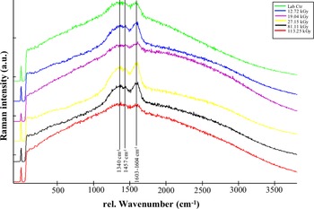
The spectra acquired directly from fungal colonies showed two broad bands at 1590–1605 cm−1 and a second lower intensity band around 1340 cm−1, which correspond to the melanin spectrum (Culka et al., Reference Culka, Jehlička, Ascaso, Artieda, Casero and Wierzchos2017; Pacelli et al., Reference Pacelli, Alessia, Siong, Lorenzo, Moeller, Fujimori and Silvano2021a) (Fig. 3). A third band near 1457 cm−1, which is also a characteristic Raman band of melanic pigments of the black fungus C. antarcticus (Pacelli et al., Reference Pacelli, Cassaro, Baqué, Selbmann, Zucconi, Maturilli and Onofri2021b), was found. Overall, no evident differences were reported among the different exposure conditions (Fig. S1A). As reported in Fig. S1B, a similar band position (near 1601 cm−1) was revealed for control samples (Lab Ctr) and the lower irradiation dose (12.72 kGy). On the contrary, a slight shift (around 1603–1604 cm−1) appeared for the higher irradiation doses (up to ~113 kGy). Moreover, a slight depletion was revealed only in samples exposed to ~113 kGy, but, nevertheless, the three bands that characterize the fungal melanin are still evident, even at the highest dose. In conclusion, the characteristic melanin Raman bands are detectable in all the exposure conditions.
Raman spectra comparison of C. antarcticus colonies exposed to increasing doses of γ-rays (generated from a 60Co source). Melanin main bands are located at 1340 and 1603–1604 cm−1.

Fourier transformed infrared resonance spectroscopy analysis
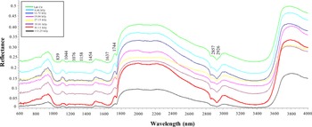
FTIR analysis is one of the most powerful techniques to detect the integrity of biomolecules since it provides an assignment of the spectral characteristic of different functional groups and the absence of a band in the spectrum of a treated sample may indicate a change in the structure of the molecule. In the analysed material, chemical bonds are able to vibrate at a characteristic frequency representative of the material structure and in a FTIR plot, for each absorption band individual chemical bonds can be identified and assigned. In this work, the useful spectral information was obtained between 600 and 4000 cm−1. Overall, each spectrum is characterized by a similar trend if compared with relative control (Lab Ctr), no evident changes were reported in the acquired spectra (Fig. 4). In particular, all the spectra show strong characteristic stretching bands for C = C bond [1750 cm−1 associated to lipids and fatty acids; Fig. 4 (Movasaghi et al., Reference Movasaghi, Rehman and Rehman2008)]. The bands observed at 1454 cm−1 and near 1158 cm−1 revealed the detection of chitin characteristic vibrations (Salman et al., Reference Salman, Shufan, Tsror, Moreh, Mordechai and Huleihel2014; Forfang et al., Reference Forfang, Zimmermann, Kosa, Kohler and Shapaval2017). The bands near 1637 and 1044 cm−1 [due to the bending vibration modes of aromatic ring C = C or C = N (Mbonyiryivuze et al., Reference Mbonyiryivuze, Mwakikunga, Dhlamini and Maaza2015) and to carbon ring breathing vibrations in cyclic compounds (Shurvell, Reference Shurvell, Chalmers and Griffiths2006; Pal et al., Reference Pal, Gajjar and Vasavada2013)], could be associated with the presence of melanin pigments (Fig. 4). The band at 2857 cm−1 is clearly attributed to fungal melanins (Paim et al., Reference Paim, Linhares, Mangrich and Martin1990; Pal et al., Reference Pal, Gajjar and Vasavada2013; Sun et al., Reference Sun, Zhang, Sun, Zhang, Shan and Zhu2016; Raman and Ramasamy, Reference Raman and Ramasamy2017); while band at 2926 cm−1 (Fig. 4) corresponds to C. antarcticus melanin (Pacelli et al., Reference Pacelli, Cassaro, Aureli, Moeller, Fujimori and Onofri2020a, Reference Pacelli, Cassaro, Maturilli, Timperio, Gevi, Cavalazzi and Onofri2020b).
FT-IR spectra in the 600–4000 nm spectral range of de-hydrated C. antarcticus colonies exposed to increasing doses of γ-rays (generated from a 60Co source).

Discussion
Over the years, the search for life on other planets has undergone changes. During the Viking missions to Mars, research studies aimed at searching for microorganisms in Martian soils; a method that requires the viability of microorganisms and a good growth rate (McKay, Reference McKay2020). Nowadays, life-detection missions encompass different measurements, including the search of biosignatures, which could represent unambiguously the sign of extant or recently extinct life (McKay, Reference McKay2004; Neveu et al., Reference Neveu, Hays, Voytek, New and Schulte2018). One of the main hurdles in the search for life is the highly radiative environment that could destroy organic molecules or their remains, not allowing their detection during in situ life-detection mission. Among the GCRs, gamma-rays are the highest-energy form of light and have deleterious effects on biological molecules. Some are generated by transient events, such as solar flares and the huge star explosions known as supernovas. Others are produced by steady sources like the supermassive black holes at the hearts of galaxies.
Previous studies on the same samples showed a high resistance of the black fungus to this type of radiation in terms of cell survival and ultrastructure integrity (Pacelli et al., Reference Pacelli, Selbmann, Zucconi, Raguse, Moeller, Shuryak and Onofri2017a). Here, we aimed at investigating the effects of gamma rays on two classes of fungal biomolecules, nucleic acids and pigments, in order to provide support for life-detection missions on Mars and for the future analyses of the Mars Samples Return. Based on the idea that life shares a similar biochemistry in the Universe and on the biogenicity requirement for a biosignature, we firstly focused our attention on the stability of nucleic acids after gamma rays' exposure. The choice of this macromolecule is determined by a multitude of factors. First of all, DNA is widespread in all life forms on Earth; the formation of double-stranded DNA does not occur under abiotic conditions, differently from other biological molecules, such as amino acids, polycyclic aromatic hydrocarbons, etc. (Trevors, Reference Trevors2003; Berger et al., Reference Berger, Bilski, Hajek, Puchalska and Reitz2013). And secondly, PCR-based detection methods are highly specific and sensitive, allowing the detection of a single DNA molecule (Briggs et al., Reference Briggs, Good, Green, Krause, Maricic, Stenzel and Pääbo2009). By using a qPCR assay, we reported that the fungal DNA is always amplifiable, and therefore detectable, even after the ~113 kGy dose of exposure inside the fungal cells (Fig. 1A and B). In particular, we amplified 930 and 370 DNA copy numbers by amplifying LSU and β-actin genes; respectively.
Similar results have been obtained for DNA of the cyanobacterium Chroococcidiopsis exposed to gamma radiation, in the frame of the same irradiation campaign: a reduction of the amplifiable target gene was detected in hydrated and de-hydrated samples exposed to 113.25 kGy (Verseux et al., Reference Verseux, Baqué, Cifariello, Fagliarone, Raguse, Moeller and Billi2017). Additional studies on hydrated colonies of C. antarcticus exposed to gamma rays will be carried out, in order to evaluate if protection against radiation may be enhanced from the desiccated condition. Preliminary studies on hydrated and metabolically active colonies of this fungus exposed to accelerated iron ions showed a good DNA amplification up to 2000 Gy (Pacelli et al., Reference Pacelli, Alessia, Siong, Lorenzo, Moeller, Fujimori and Silvano2021a).
The resistance of C. antarcticus' DNA under different stressors has been extensively reported (Onofri et al., Reference Onofri, de la Torre, de Vera, Ott, Zucconi, Selbmann and Horneck2012, Reference Onofri, de Vera, Zucconi, Selbmann, Scalzi, Venkateswaran and Horneck2015, Reference Onofri, Selbmann, Pacelli, Zucconi, Rabbow and de Vera2019; Pacelli et al., Reference Pacelli, Selbmann, Zucconi, Raguse, Moeller, Shuryak and Onofri2017a, Reference Pacelli, Bryan, Onofri, Selbmann, Shuryak and Dadachova2017b, Reference Pacelli, Selbmann, Zucconi, Coleine, de Vera, Rabbow and Onofri2019, Reference Pacelli, Cassaro, Aureli, Moeller, Fujimori and Onofri2020a, Reference Pacelli, Cassaro, Maturilli, Timperio, Gevi, Cavalazzi and Onofri2020b, Reference Pacelli, Alessia, Siong, Lorenzo, Moeller, Fujimori and Silvano2021a, Reference Pacelli, Cassaro, Catanzaro, Baqué, Maturilli, Böttger, Botta, Saladino, Rabbow and Onofri2021c; Aureli et al., Reference Aureli, Pacelli, Cassaro, Fujimori, Moeller and Onofri2020; Cassaro et al., Reference Cassaro, Pacelli, Baqué, de Vera, Böttger, Botta and Onofri2021b). Also, DNA was successfully extracted and amplified in C. antarcticus samples exposed to space conditions in Low Earth orbit (LEO) in the presence of lunar regolith analogue (Cassaro et al., Reference Cassaro, Pacelli, Baqué, Cavalazzi, Gasparotto, Saladino, Botta, Böttger, Rabbow, de Vera and Onofri2022) and of Martian regolith analogues (Pacelli et al., Reference Pacelli, Cassaro, Baqué, Selbmann, Zucconi, Maturilli and Onofri2021b).
These results suggest the need to accelerate the development of miniaturized amplification instruments, as part of the instrumentation onboard the next generation of rovers. For example, Oxford Nanopore Technologies MinION instrument, which can detect and sequence nucleic acids, has been already tested (Raymond-Bouchard et al., Reference Raymond-Bouchard, Maggiori, Brennan, Altshuler, Manchado, Parro and Whyte2022). One of the main challenges in using these devices on the surface of planetary bodies, could be represented by the disturbing compounds in the soil, that may prevent the amplification. However, recent studies reported that the sequencing of two different types of extract (purified and unpurified) DNA revealed a comparable community composition in Permafrost-associated soil samples (Raymond-Bouchard et al., Reference Raymond-Bouchard, Maggiori, Brennan, Altshuler, Manchado, Parro and Whyte2022).
Then, we focused on the stability of melanin pigments. This class of pigments is widespread in all living kingdoms and often dominating extremophilic species, suggesting that these pigments are used as a protective strategy in their lifecycle (Dadachova et al., Reference Dadachova, Bryan, Huang, Moadel, Schweitzer, Aisen and Casadevall2007). Here, we investigated potential melanin modifications through a multidisciplinary analysis (UV-Vis spectrophotometry, and FT-IR and Raman spectroscopies). The results of UV-Vis analysis revealed a good stability of melanic pigments even at the ~113 kGy dose of exposure: an absorption peak at 230 nm and a decrease in absorbance at higher wavelength was detected (Fig. 2, Pacelli et al., Reference Pacelli, Cassaro, Aureli, Moeller, Fujimori and Onofri2020a, Reference Pacelli, Cassaro, Maturilli, Timperio, Gevi, Cavalazzi and Onofri2020b). No additional peaks or bulges were detected in the acquired spectra, in contrast with fungal samples exposed to Mars-like conditions (Cassaro et al., Reference Cassaro, Pacelli, Baqué, de Vera, Böttger, Botta and Onofri2021b). These results are in accordance with Meeßen et al. (Reference Meeßen, Sánchez, Sadowsky, de la Torre, Ott and de Vera2013), where UV/VIS-spectrometry data supported the identification of melanin in lichens of astrobiological interest, confirming its role in photoprotection.
Raman spectra showed the three characteristic bands of melanin (Fig. 3): a major band at higher wavenumber (1590–1605 cm−1), associated to C-N bonds (Samokhvalov et al., Reference Samokhvalov, Liu and Simon2004) and a second band at 1340 cm−1, due to the stretching of the C-C bonds within the rings of the aromatic melanin monomers (Galván et al., Reference Galván, Jorge, Ito, Tabuchi, Solano and Wakamatsu2013). The third band (1457 cm−1, Fig. 3), distinctive of the fungal melanin pigments (Pacelli et al., Reference Pacelli, Cassaro, Aureli, Moeller, Fujimori and Onofri2020a, Reference Pacelli, Cassaro, Maturilli, Timperio, Gevi, Cavalazzi and Onofri2020b) allows to discriminate melanin from (i) any thermal degradation or (ii) amorphous carbon. Our results, confirmed that fungal melanin maintains its stability also at the irradiation dose of 113 kGy, although with a slight thinning of the bands (Fig. 3, red line), and a slight shift of the main peak position at the higher doses (Fig. S1B). These results are in accordance with previous analyses on fungal melanin after exposure to space conditions in Low Earth Orbit (LEO) (Pacelli et al., Reference Pacelli, Cassaro, Baqué, Selbmann, Zucconi, Maturilli and Onofri2021b) and to simulated space conditions (Pacelli et al., Reference Pacelli, Alessia, Siong, Lorenzo, Moeller, Fujimori and Silvano2021a, Reference Pacelli, Cassaro, Catanzaro, Baqué, Maturilli, Böttger, Botta, Saladino, Rabbow and Onofri2021c; Cassaro et al., Reference Cassaro, Pacelli, Baqué, de Vera, Böttger, Botta and Onofri2021b).
On the contrary, the carotenoid Raman signals of the photobiont part of the lichen Circinaria gyrosa exposed to gamma radiation, showed alterations at the dose of 113 kGy (Meeßen et al., Reference Meeßen, Backhaus, Brandt, Raguse, Böttger, de Vera and de la Torre2017); while the carotenoid signal of de-hydrated cells of the cyanobacterium Nostoc sp., have been detected up to 27 kGy of gamma rays when exposed alone and to ~113 kGy, when protected by Martian regolith simulants (Baqué et al., Reference Baqué, Hanke, Böttger, Leya, Moeller and de Vera2018). Photosynthetic pigments and nucleic acids of Chroococcidiopsis cells also resulted to be detectable even at the dose of 113.25 kGy of gamma rays (Verseux et al., Reference Verseux, Baqué, Cifariello, Fagliarone, Raguse, Moeller and Billi2017).
Melanin also exhibits unaltered absorption bands in the 600–4000 cm−1 region of FT-IR spectra (Fig. 4). In particular, bands referred to melanin pigments have been detected at different wavelength: the bands at 2857, 2926, 1637 and 1044 cm−1 are indicative of phenols or carboxylic groups, which characterize the absorption of fungal melanin (Sun et al., Reference Sun, Zhang, Sun, Zhang, Shan and Zhu2016; Pacelli et al., Reference Pacelli, Cassaro, Aureli, Moeller, Fujimori and Onofri2020a, Reference Pacelli, Cassaro, Maturilli, Timperio, Gevi, Cavalazzi and Onofri2020b). The FT-IR spectra showed identical spectroscopic spectra among irradiated and control samples (Fig. 4).
The evaluation of the stability of melanins through three different and complementary techniques, supports its use as a promising biosignature.
The hypothetical finding of a spectral signature of pigment would be a possible sign of life, since no abiotic and uncatalyzed pigment synthesis is known so far in environmental conditions (Neveu et al., Reference Neveu, Hays, Voytek, New and Schulte2018). The reported stability of melanin pigments against various types of ionizing and non-ionizing radiation (Robinson, Reference Robinson2001; Vember and Zhdanova, Reference Vember and Zhdanova2001; Nosanchuk and Casadevall, Reference Nosanchuk and Casadevall2003; Cassaro et al., Reference Cassaro, Pacelli, Baqué, de Vera, Böttger, Botta and Onofri2021b; Pacelli et al., Reference Pacelli, Cassaro, Baqué, Selbmann, Zucconi, Maturilli and Onofri2021b, Reference Pacelli, Cassaro, Catanzaro, Baqué, Maturilli, Böttger, Botta, Saladino, Rabbow and Onofri2021c) could be exploited also in different scenarios of space exploration. As demonstrated in Turick et al. (Reference Turick, Ekechukwu, Milliken, Casadevall and Dadachova2011), ionizing radiations interact with melanin altering its oxidation/reduction potential. This alteration results in electric current production and further study on the interaction between melanic pigments and ionizing radiation could offer new insights on the application of melanin pigments in the synthetic biology field. Besides, the role of melanin as energy generators could be useful also to generate melanin-based products, especially important to reduce global warming changes (Malo and Dadachova, Reference Malo, Dadachova, Tiquia-Arashiro and Grube2019). When exposed to ionizing radiation, melanized fungal cells showed an increment of the growth rate in comparison to non-melanized cells, probably due to the capability of these pigments to capture electromagnetic radiation, which could be used by microorganisms as metabolic energy, suggesting a potential role in energy capture (Dadachova et al., Reference Dadachova, Bryan, Huang, Moadel, Schweitzer, Aisen and Casadevall2007).
The FT-IR analyses revealed additional bands related to lipids and fatty acids (near 1750 cm−1, Movasaghi et al., Reference Movasaghi, Rehman and Rehman2008). The identification of chemical bonds belonging to chitin was detected near 1454 and 1158 cm−1 (Fig. 4, Movasaghi et al., Reference Movasaghi, Rehman and Rehman2008). Chitin is one of the main constituents of the fungal cell wall and one of the most abundant compounds on Earth (Hunt, Reference Hunt1970). It is a compound formed of β 1–4–bonded N-acetylglucosamine. The hypothesis to consider chitin as a biosignature for the search of life elsewhere has been already reported in Pacelli et al. (Reference Pacelli, Cassaro, Catanzaro, Baqué, Maturilli, Böttger, Botta, Saladino, Rabbow and Onofri2021c). In this work, we confirmed the stability of chitin after the exposure to ~113 kGy of gamma rays. The characterization of suitable candidates for a good biosignature could be difficult considering the preservation of certain molecules over geological time scale and the environmental conditions (Summons et al., Reference Summons, Albrecht, McDonald, Moldowan, Botta, Bada, Gomez-Elvira, Javaux, Selsis and Summons2008). However, chitin may be preserved over geological time period: it has been detected in fungi preserved in Cretaceous amber (Speranza et al., Reference Speranza, Ascaso, Delclòs Martínez and Peñalver2015) and in a 50-Ma-old marine sponge (Ehrlich et al., Reference Ehrlich, Kaluzhnaya, Brunner, Tsurkan, Ereskovsky, Ilan and Wörheide2013). Recently, chitin has been also discovered in fungal fossil filaments in a Neoproterozoic shale rock (Bonneville et al., Reference Bonneville, Delpomdor, Préat, Chevalier, Araki, Kazemian and Benning2020), supporting its utilization as a biosignature in space exploration.
In conclusion, this work aimed to investigate the persistence of fungal biomolecules to space-relevant radiation over time, considering that the maximum tested dose of ~113 kGy may be compared to an exposure of 1.5 million years on the Martian surface and of 13 million at two metres-depth in the subsurface (extrapolated from Hassler et al., Reference Hassler, Zeitlin, Wimmer-Schweingruber, Ehresmann, Rafkin, Eigenbrode and Berger2014). The study about the stability of biosignatures is of outmost importance for the in-situ life detection missions on other planets, such as Mars. Through this work, we confirmed the persistence of melanin pigments and nucleic acids and we highlighted chitin as a promising biosignature. Further investigations through the application of -omics analyses could represent a challenging task to deepen the molecular mechanisms at the basis of the nucleic acid stability of this fungus.
Acknowledgements
We thank the PNRA (Italian National Program for Antarctic Research) for supporting sample collection in Antarctica, and the Italian National Antarctic Museum ‘Felice Ippolito’, for funding MNA-CCFEE. We acknowledge the HIMAC team at the NIRS in Chiba, Japan for the irradiation. We thank the Italian Space Agency for the grant n. 2018-6- U.0 (BioSigN MicroFossils).
Author contributions
CP, SO, JPPdV, RM, designed the research; AC, CP, MB, AF performed the experiments. AC, CP, MB, AM analysed data. AC drafted the paper with inputs from all other authors. All authors approved the submitted version.
Financial support
This research was financially supported by the Italian Space Agency (ASI), BioSigN MicroFossils- ASI grant n. 2018-6-U.0). STARLIFE project was supported by the MEXT Grant-in-Aid for Scientific Research on Innovative Areas ‘Living in Space’ (Grant Numbers: 15H05935 and 15K21745). RM was supported by the DLR grant FuE-Projekt ‘ISS LIFE’ (Programm RF-FuW, TP 475). MB acknowledges the support of the Deutsche Forschungsgemeinschaft (DFG – German Research Foundation) for the project ‘Raman Biosignatures for Astrobiology Research’ (RaBioFAM; project number: 426601242).
Conflict of interest
The authors declare that they have no competing financial interests.
Conflict of interest
The authors declare they have not conflicts to disclose.