

Fatigue has been identified as the most important untreated symptom in cancer today (Curt, Reference Curt2001). Despite its prevalence and impacts, in many cases, the etiology of cancer-related fatigue is not clearly understood.
Recent research shows that static and functional makers of inflammatory activity are positively related to fatigue reports in cancer survivors (Bower et al., Reference Bower, Ganz and Aziz2002; Collado-Hidalgo et al., Reference Collado-Hidalgo, Bower and Ganz2006). However, to date, the association between fatigue and inflammatory activation in patients with advanced cancer has not been examined. In investigating this issue, the clinical utility of potential findings will be strengthened if the biomarkers identified are among those parameters assessed during routine patient care.
In patients with tumor burdens, hematological abnormalities may develop as a result of cytokine secretion (Ruka et al., Reference Ruka, Rutkowski and Kaminska2001). For example, granulocytosis is often observed in cancer patients, being particularly prevalent in patients with lung or colorectal cancers (Jameson & Johnson, Reference Jameson, Johnson, Fauci, Hauser and Longo2005). In addition to impacts on immune status, the cytokine dysregulation that induces granulocytosis may have consequences for patients’ subjective experiences (Maier & Watkins, Reference Maier and Watkins1998). Sickness behaviors, including experiences of fatigue, occur as part of central nervous system responses to peripheral inflammation (Watkins & Maier, Reference Watkins and Maier1999; Krueger & Majde, Reference Krueger and Majde2003; Irwin, Reference Irwin2008). Thus, in patients with active cancer, fatigue reports may result from tumor-induced cytokine alterations.
On the premise that circulating concentrations of myeloid-origin cells may provide information about the intensity of the inflammatory state, this exploratory study investigated whether routinely collected cellular immune data were associated with the severity of fatigue reported by advanced lung cancer patients. In addition, this study examined whether the severity of fatigue was related to survival. Specifically, it was hypothesized that in advanced-stage non-small-cell lung cancer (NSCLN) patients, reports of more severe fatigue would be associated with (1) evidence of a more aggressive inflammatory state and (2) shorter survival.
METHOD
The fatigue data for this analysis was extracted from a larger data set that examined the effects of early referral to the Palliative Care Service at Massachusetts General Hospital on patient and caregiver satisfaction (described by Temel et al., Reference Temel, Jackson and Billings2007). Each of the 44 participants who contributed data to the current study held a diagnosis of either Stage IIIb with effusion or Stage IV NSCLC.
Fatigue Assessment
Fatigue was assessed using a composite of three items. These items were “I have a lack of energy” and “I am forced to spend time in bed” from the Functional Assessment of Chronic Illness Therapy–Fatigue Scale (FACIT) and “I feel as if I am slowed down” from the depression subscale of the Hospital Anxiety and Depression Scale. The interitem reliabilities of these three items ranged from r = .56 (p = .001) to r = .65 (p = .001).
Hematologic Variables
Results from routinely drawn blood tests were retrospectively extracted from electronic medical records. White blood cell count, absolute neutrophil, monocyte, and lymphocyte counts and hemoglobin concentration were recorded as continuous variables.
Statistical Analysis
Data were analyzed using a series of linear and binomial regressions. Covariates were entered in the first stage of each model followed by the variable of primary interest.
Due to the nonnormality of the survival distribution, this variable was categorized using the cutoff of survival of greater or less than 90 days from the fatigue observation (N = 20, N = 24, respectively). This categorized variable was used as the subject of binomial regression. The covariates entered at Stage 1 were age, gender, and time until treatment termination; fatigue data were entered at Stage 2.
The “time until treatment termination” variable was entered to serve two purposes. The primary aim of this covariate was to factor out of the analysis differences in fatigue, survival, and immune status that were systemically related to whether participants were still undergoing treatment or had terminated their chemotherapy. The variable was constructed by subtracting the date of the last chemotherapy dose from the date of the fatigue assessment. This formed a continuous variable that was either negative or positive depending on whether treatment was terminated before or after the date of the fatigue and immune assessments. In addition to accounting for effects that withdrawal of treatment per se may have had on the fatigue and immune variables of interest, in advanced lung cancer the decision to discontinue chemotherapy likely signals that a patient's condition appears sufficiently severe that further treatment will not offer survival or quality of life benefits. With this relationship in mind, the covariate constructed provides the analysis with a measure that takes into account the medically apparent illness of participants.
Linear regression was used to assess the association between the continuous variables, hemoglobin concentration, and fatigue (covariates age, gender, and time until treatment termination). Finally, a series of four linear regressions was used to investigate the relationship between fatigue reports and cellular immune status. Stage 1 covariates for these analyses included age, gender, hemoglobin concentration, and days until treatment termination.
RESULTS
Participant Characteristics
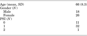
All participants enrolled held the diagnosis of Stage IIIb with effusion or Stage IV NSCLC. The mean age of participants was 66, and 60% of the sample were women (Table 1). At recruitment, most patients held at Performance Status Index score of one.
Participant Characteristics
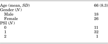
PSI: Eastern Cooperative Oncology Group Performance Status Index.
Description of Outcome, Covariate, and Predictor Variables
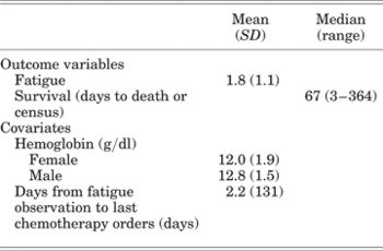
The mean fatigue summary score was 1.8 (Table 2). Average scores for the individual items equated to a mean response of “somewhat” in answer to the item “I have a lack of energy” (2.1, range 0–4), average responses between “a little bit” and “somewhat” in answer to the item “I am forced to spend time in bed” (1.4, range 0–4), and an average answer of “very often” (1.9, range 0–3) in response to the item “I feel as if I am slowed down.” Examination of the score distribution for the summary variable showed a normal score distribution, and so parametric statistical tests were used in subsequent analyses.
Description of Outcome and Covariate Variables

N = 44.
The median participant survival was 67 days. Fifty-four percent of participants died within 3 months of the fatigue observation and 80% within 9 months. At the date of the audit, the 7 patients who remained alive had survived for an average of 322 days (range 267–364 days) since their fatigue and blood data were recorded.
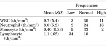
For the hematological variables, average participant values are reported in Table 3. Within these averages, 40% of participants showed a neutrophil count above the adult norm (Table 3), whereas 21% showed monocytes counts and 77% lymphocyte counts that were below adult norms. Fifty-two percent (n = 23) of participants showed hemoglobin concentrations that were below gender-adjusted norms.
Description of Predictor Variables

WBC: white blood cell. N = 44. WBC, <4.0 th/mm3 = low; >11.0 th/mm3 = high. Neutrophil, <2.0 th/mm3 = low; >7.5 th/mm3 = high. Monocyte, <0.2 th/mm3 = low; >0.8 th/mm3 = high. Lymphocyte, <1.5 th/mm3 = low; >4.0 th/mm3 = high.
Regression Analyses
Fatigue Intensity Associated with Survival Time
Binomial regression controlling for effects related to age, gender, and time until treatment termination showed that those who died within 90 days of answering the fatigue questions reported significantly more severe fatigue than that reported by participants who continued to live for longer than 3 months (β = −.34, p = .03).
Association between Fatigue and Hemeoglobin Concentration
Linear regression showed that, when we controlled for effects due to gender, age, and the time of the fatigue assessment relative to the date at which chemotherapy was discontinued, lower hemoglobin concentrations were associated with reports of more intense fatigue. Variations in hemoglobin concentration accounted for 10% of the variance in the fatigue data (standardized β = −.36, p = .04).
Associations between Fatigue and Components of White Blood Cell Counts
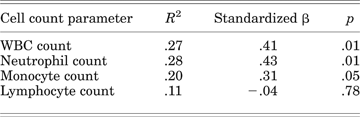
Linear regression showed that after we controlled for effects due to gender, age, hemoglobin levels, and discontinuation of chemotherapy, fatigue was significantly related to white blood cell count (WBC; Table 4). This model accounted for nearly 30% of the variance in fatigue reports.
Regressions of Fatigue Severity and Immune Parameters
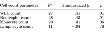
Degrees of freedom = 43. Covariates present in each model were hemoglobin concentration, age, gender, and timing of chemotherapy termination relative to the fatigue observation.
To investigate whether specific components of the WBC count were responsible for the relationship between immune cell count and fatigue reports, separate regression analyses were run for each of the main components of the WBC count. These three analyses showed that fatigue severity was significantly associated with variations in the concentrations of cells of myeloid but not lymphoid origin.
Addition of the neutrophil data into the basic regression model increased the proportion of variance explained nearly threefold (from 11% to 27%). Further, the standardized beta coefficients were −.39 for hemoglobin concentration and .43 for neutrophil concentration. The similar magnitudes of these effects show that neutrophil count plays as strong a role as the established variable, hemoglobin, in accounting for the fatigue reported by this sample of advanced-stage lung cancer patients.
In a separate model, addition of the monocyte data to the basic regression model doubled the proportion of variance explained (from 11% to 20%). Again, effect sizes of similar magnitude were observed between hemoglobin (standardized β = −.26) and monocytes concentrations (standardized β = .31). In contrast to the effects just described, addition of lymphocyte data did not change the proportion of variance explained. Thus, our analyses suggest that the relationship between fatigue and immune cell counts is carried by cells of myeloid origin. Further, these analyses show that increases in circulating concentrations of myeloid-origin cells are associated with fatigue-related effects that are of a similar magnitude to the effects exerted by hemoglobin deficits.
DISCUSSION
Using simple questions of the type that are routinely asked during clinical interviews, this exploratory study showed that the severity of fatigue reported by advanced-stage lung cancer patients was associated with the length of their survival. As expected, those patients who reported more severe fatigue died more rapidly. The aim of this study was to investigate how routinely assessed hematological parameters relate to patient fatigue reports.
In keeping with existing research (Kimel et al., Reference Kimel, Leidy and Mannix2008), fatigue in our sample was significantly related to hemoglobin concentration, with lower hemoglobin levels associated with more severe fatigue reports. Although identification of this relationship does not contribute new information to the understanding of how hematological state relates to fatigue reports, it does establish that the assessment tool used is sufficiently sensitive to capture relationships that are known to operate and are of a magnitude that is clinically meaningful. Over and above effects related to hemoglobin level, our analyses show that fatigue in advanced cancer patients is significantly associated with other hematological parameters measured during routine assessment.
A growing body of sickness–behavior research suggests that cancer-related fatigue may be related to the inflammatory state (Bower et al., Reference Bower, Ganz and Aziz2002; Collado-Hidalgo et al., Reference Collado-Hidalgo, Bower and Ganz2006). However, relationships between fatigue and the intensity of inflammatory state have not yet been investigated in advanced-stage cancer patients. In this exploratory investigation we used information from routine white blood cell counts to provide an indirect measure of inflammatory state. These analyses showed that, over and above effects due to age, gender, hemoglobin differences, or changes that may have been induced by decisions to discontinue chemotherapy, reports of more severe fatigue were associated with evidence of a more intense inflammatory milieu.
As a whole, the WBC count showed a statistically significant association with fatigue severity. When the cell subtypes were considered individually, it appeared that it was cells of myeloid origin that were carrying most of the fatigue-related effect. Variation in lymphocyte count was not associated with fatigue reports. Given that blood concentrations of cells of myeloid origin (neutrophils and monocytes) are associated more strongly with inflammatory processes than cells of lymphoid origin, the pattern of association identified is in keeping with the exploratory hypothesis that fatigue intensity may be associated with the indirect measure of inflammatory state that is provided by immune cell counts.
The addition of WBC count, neutrophil, or monocyte data into the regression models contributed to explanation of a substantial proportion of variance in fatigue reports that was not captured when hemoglobin data were entered as the only hematological variable. Further, the effect sizes associated with entry of the whole white blood cell count, but more specifically with entry of myeloid-origin cell counts, were of a similar magnitude to the effect sizes associated with hemoglobin concentrations. The fatigue-inducing effects of hemoglobin deficiencies are well accepted; our analyses here suggest that consideration of the fatigue-related effects that are associated with myeloid-cell line variations may also be of clinical importance.
Although this exploratory analysis used routinely collected but indirect measures to assess inflammatory state, future research should investigate whether measures of inflammatory activation such as C-reactive protein or direct cytokine/soluble cytokine receptor assays are associated with the fatigue experienced by advanced-stage lung cancer patients. If the association is found to persist, grounds will be built for clinical management to consider whether modification of the inflammatory state in advanced-stage cancer patients may be an avenue, in addition to current practices of hemoglobin deficit management, through which to alleviate the profound fatigue that reduces the quality of life of advanced stage cancer patients.
ACKNOWLEDGMENT
Johanna Paddison was supported by a Fulbright Fellowship which enabled her to spend time with the Division of Psychiatry and Medicine and the Benson Henry Institute, both at Massachusetts General Hospital.