The congenitally corrected transposition of the great arteries is a rare congenital heart disease characterised by atrio-ventricular and ventriculo-arterial discordance. The pulmonary veins regularly connect to the left atrium. From here, the blood enters the morphologic right ventricle, which supplies the aorta. Thus, the right ventricle is in systemic circulation.Reference Connelly, Liu, Williams, Webb, Robertson and McLaughlin 1 Therefore, although the blood flow is normal and oxygenated blood is pumped into the body, the right ventricle encounters an unphysiological high pressure load. Associated cardiac anomalies are common findingsReference Presbitero, Somerville, Rabajoli, Stone and Conte 2 , Reference Rutledge, Nihill, Fraser, Smith, McMahon and Bezold 3 and determine the clinical presentation and long-term survival.Reference Graham, Bernard and Mellen 4 An increasing number of complications, such as congestive heart failure and complete heart block, occur in adulthood, whereas there is only a low prevalence of complications, mainly arrhythmias, within the first two decades.Reference Presbitero, Somerville, Rabajoli, Stone and Conte 2 Most patients present with symptoms of congestive heart failure in adulthood, and, even in isolated congenitally corrected transposition, life expectancy is reduced.Reference Voskuil, Hazekamp and Kroft 5 Several studies describe progressive right ventricular failure dependent on age and the near-universal presence of tricuspid insufficiency.Reference Graham, Bernard and Mellen 4 , Reference Beauchesne, Warnes, Connolly, Ammash, Tajik and Danielson 6 – Reference Van Praagh, Papagiannis, Grunenfelder, Bartram and Martanovic 9 In addition, a mismatch between increased oxygen demand of the hypertrophic right ventricular myocardium and decreased oxygen supply by the single right coronary artery has been suggested to cause systemic right ventricular failure.Reference Hauser, Bengel and Hager 10
Dextro-transposition of the great arteries shows an atrio-ventricular concordance and only a ventriculo-arterial discordance, meaning that the left atrium is connected to the left ventricle, from which the pulmonary artery arises. Before the anatomical correction became the procedure of choice for surgical treatment of dextro-transposition, the atrial switch operation was performed to achieve a physiological correction of blood flow on the atrial level. Also in this situation the right ventricle works as the systemic ventricle and shows late insufficiency as a common sequel related to excessive systemic hypertrophy.Reference Hornung, Kilner, Davlouros, Grothues, Li and Gatzoulis 11
Cardiac magnetic resonance imaging has been established as a powerful and precise tool for the assessment of biventricular morphology and function in patients with congenital heart disease and systemic right ventricles.Reference Hornung, Kilner, Davlouros, Grothues, Li and Gatzoulis 11 – Reference Gutberlet, Hoffmann and Kunzel 14 In the present study, we sought to compare ventricular volumes and function of systemic right ventricles in congenitally corrected and dextro-transposition of the great arteries after atrial switch, to evaluate the determinants of systemic right ventricular function in congenitally corrected transposition and compare patient parameters with those of a group of healthy controls.
Methods
Patient population, control group, and study design
We retrospectively included 19 (9 male) consecutive patients with congenitally corrected transposition and 31 (21 male) consecutive patients after an atrial switch procedure for correction of dextro-transposition of the great arteries. All patients came for regular follow-up outpatient visits to our tertiary care institution and were referred to cardiac magnetic resonance imaging. Exclusion criteria were usual magnetic resonance contraindications, such as implanted defibrillators or pacemakers or ferromagnetic intra-cranial metallic implants. On echocardiography, performed within 2 days of the cardiac magnetic resonance examination, tricuspid insufficiency was categorised as absent, mild, moderate, or severe. The New York Heart Association functional class was obtained from clinical examination the same day that the cardiac magnetic resonance examination was performed. Patient parameters were compared with those of a control group of 25 (12 male) volunteers without any symptoms or history of cardiac disease.
All patients or parents provided written informed consent. The study was approved by the local ethics committee and complied with the Declaration of Helsinki.
Cardiac magnetic resonance imaging
All examinations were performed on a 1.5-tesla scanner (Intera CV, Philips Medical Systems, Best, The Netherlands) in the supine position using a dedicated five-channel phased-array surface cardiac coil. Black-blood images in axial orientation were acquired to visualise morphology. Standard breath-hold cine steady-state free-precession sequences in the short-axis, four-chamber view and right ventricular vertical long-axis orientation were acquired for volumetric and functional imaging. In congenitally corrected transposition, an additional coronal cine sequence was acquired to visualise the parallel position of the aorta and the pulmonary trunk. Short-axis imaging was planned on the four-chamber view parallel to the atrioventricular valve covering the whole heart, gapless from the apex to the base, with a slice thickness of 8 millimetres. The echo time was 1.8 milliseconds, the repetition time was 3.6 milliseconds, the flip angle was 50°, and the matrix size was 256 × 128. Typical in-plane resolution was 1.2 × 1 millimetre. We acquired 30 phases per heart cycle.
Cardiac magnetic resonance image analysis was independently performed by two fully blinded observers (A.F. and M.G.) with 7 and 10 years of experience in cardiac magnetic resonance imaging, respectively, using a Philips View Forum workstation (Best, The Netherlands; version 4.2-Cardiac Evaluation Package) in our cardiac magnetic resonance core laboratory, which has expertise in congenital heart disease imaging and has proven low intra- and inter-observer variability for assessments of cardiac biventricular volumes and function.Reference Beerbaum, Barth and Kropf 15 , Reference Grothoff, Hoffmann and Lehmkuhl 16 Biventricular endocardial and epicardial borders were manually traced in end-systole and end-diastole in each slice. In planimetry of the systemic RV myocardial mass, the results show a better reproducibility when RV trabeculation is excluded from the myocardial mass and counted as a part of the RV cavity. When RV is included in RV myocardial mass, that means extensively outlined and counted as a part of the myocardium, reproducibility is worse.Reference Winter, Bernink and Groenink 17
The following parameters were calculated for both the right and left ventricles:
-
• end-diastolic volume;
-
• end-systolic volume;
-
• stroke volume;
-
• myocardial mass; and
-
• ejection fraction.
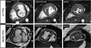
The volumes and mass were normalised to the body surface areaReference Mosteller 18 (Fig 3). The specific density of 1.05 gram per millilitre was used to calculate the myocardial mass. The inter-ventricular septum was assessed separately.
Statistics
Categorical variables are expressed as number and percentage. Owing to the inhomogeneity of the relatively small patient cohort, continuous data are given as medians [25%; 75% percentile]. To compare the two patient groups and the control group, we used Tamhane's T2 multiple comparison test. Differences between categorical variables were assessed by the Chi square test. Relationships between different parameters were examined using linear and quadratic regression analyses. Differences between variables are demonstrated using Box-Whisker-plot diagrams; regression analysis is demonstrated using a scatterplot diagram.
All tests were two-sided, with a significance level of α = 0.05. For statistical analysis, Statistical Package for the Social Sciences software version 16.0 (SPSS Incorporation, Chicago, Illinois, United States of America) was used.
Results
Cardiac magnetic resonance image quality was diagnostic in all 19 patients with congenitally corrected transposition, in 31 patients with dextro-transposition, and in 25 controls. Maximum scan time was 70 minutes. There were no indeterminate results or missing data.
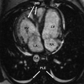
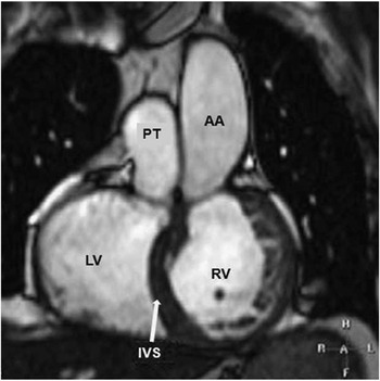
Patients presented with large and hypertrophic systemic right ventricles (Figs 1–3) and a wide range of left ventricular myocardial masses (Table 2).
Four-chamber view steady-state free-precession image in congenitally corrected transposition of the great arteries with situs inversus. In this situation the systemic right ventricle, usually in the left position, is in the right position again. Note the massive hypertrophy of the right ventricular free wall, the moderator band, and the thin left ventricular myocardium. A situs inversus can be found in approximately 5% of congenitally corrected transposition patients. DA = descending aorta; LA = left atrium; LV = left ventricular; MB = moderator band; PLE = pleural effusions; RA = right atrium; RV =right ventricle.

Steady-state free-precession image in congenitally corrected transposition of the great arteries in a coronal orientation demonstrating the “side by side” position of the ascending aorta and the pulmonary trunk. The inter-ventricular septum is hypertrophic with a convex shape towards the left ventricular cavity. AA = ascending aorta; IVS = inter-ventricular septum; PT = pulmonary trunk; RV = right ventricle.

End-diastolic steady-state free-precession basal, midventricular, and apical short-axis images with manually traced endocardial and epicardial borders. In congenitally corrected transposition of the great arteries the systemic right ventricle is in the left position, and in dextro-transposition the systemic right ventricle is in the normal right position. The right ventricular trabeculation and the papillary muscles are counted as left ventricular cavity. This leads to an underestimation of the right ventricular myocardial mass, but provides a better reproducibility. In this example, the inter-ventricular septum is counted as a part of the right ventricle. TGA = transposition of the great arteries.

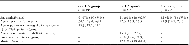
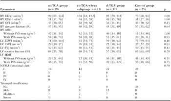
Patient and control group characteristics are presented in Table 1. The distribution of patient and control group volumetric and functional parameters has been summarised in Table 2.
Patient and control group characteristics.

cc = congenitally corrected; d = dextro; PV = pulmonary valve; TGA = transposition of the great arteries
Categorical data are presented as frequency and percentage. Continuous data are presented as median [25%; 75% percentile] or list of numbers
Patient and control group parameters.

cc = congenitally corrected; d = dextro; EDVI = end-diastolic volume index; ESVI = end-systolic volume index; IVS = inter-ventricular septum; LV = left ventricle; MMI = myocardial mass index; NYHA = New York Heart Association; RV = right ventricle; SVI = stroke volume index; whraa = without haemodynamically relevant associated anomalies
Data are presented as median [25%; 75% percentile]
p-values indicate level of significance between the cc-TGA and the d-TGA group
Congenitally corrected transposition of the great arteries and associated anomalies
An isolated congenitally corrected transposition without associated anomalies was found in five patients, and the other 14 patients presented with cardiac anomalies: a ventricular septal defect was found in seven patients (37%), a subpulmonary or pulmonary stenosis was found in six patients (32%), an atrial septal defect in three patients (16%), a patent foramen ovale was found in two patients (11%), and Ebstein's anomaly and a persistent left superior caval vein was found in one patient (5%). In all, three patients had undergone surgery as a result of associated anomalies: in two patients a pulmonary homograft was implanted, and one patient underwent a pulmonary valve replacement. Pulmonary artery hypertension was present in two cases, of which one was severe. On echocardiography, 17 patients showed tricuspid insufficiency: nine mild, seven moderate, one severe.
In six patients, anomalies were regarded as haemodynamically relevant: two patients with homograft stenoses, one patient with pulmonary insufficiency, one with severe pulmonary artery hypertension, one with ventricular septal defects, and one patient with severe tricuspid insufficiency. We created a subgroup of patients without haemodynamically relevant associated anomalies (n = 13; see below).
Dextro-transposition of the great arteries, associated anomalies, and post-operative complications
Of 31 patients who had undergone atrial switch for correction of dextro-transposition, 12 had undergone Mustard, and 19 had undergone Senning procedures. An initial ventricular septal defect had been closed in five patients without a residual shunt, and two patients had a low-grade pulmonary arterial hypertension.
Tricuspid insufficiency was absent in nine patients, was mild in 15 patients, and was moderate in seven patients with dextro-transposition (Table 2).
Congenitally corrected transposition compared with dextro-transposition and controls
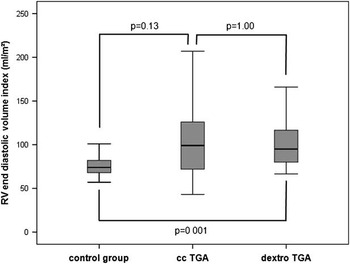
In patients with congenitally corrected transposition there was a clear trend towards higher right ventricular end-diastolic volumes when compared with controls (99 [65; 134] millilitres per square metre versus 74 [68; 84] millilitres per square metre, p = 0.13 (Fig 4); missing statistical significance owing to the small sample size). There was no difference compared with dextro-transposition (99 [65; 134] millilitres per square metre versus 95 [79; 118] millilitres per square metre, p = 1.00). Right ventricular end-diastolic volume index in both patient groups had increased compared with left ventricular end-diastolic volume index in controls (99 [65; 134] millilitres per square metre versus 77 [69; 84] millilitres per square metre, p = 0.02, for congenitally corrected transposition patients; 95 [79; 118] millilitres per square metre versus 77 [69; 84] millilitres per square metre, p < 0.001, for dextro-transposition patients).
Boxplot diagram illustrating right ventricular end-diastolic volume indices in controls, congenitally corrected transposition of the great arteries, and dextro-transposition patients after atrial switch. Both patient groups show larger ventricles when compared with controls; however, there is no difference in RV size between both patient groups. RV = right ventricular; TGA = transposition of the great arteries.

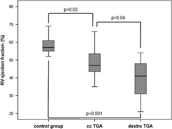
Both patient groups showed impaired right ventricular ejection fractions compared with the right ventricular ejection fractions of controls (47 [43; 55] % versus 57 [55; 62] %, p = 0.02, for congenitally corrected transposition patients; 41 [31; 49] % versus 57 [55; 62] %, p < 0.001, for dextro-transposition patients; Fig 5) and the left ventricular ejection fractions of controls (47 [43; 55] % versus 65 [63; 69] %, p < 0.001, for congenitally corrected transposition patients; 41 [31; 49] % versus 65 [63; 69] %, p < 0.001, for dextro-transposition patients). Right ventricular ejection fraction in the congenitally corrected transposition group was significantly higher compared with right ventricular ejection fraction in the dextro-transposition group (47 [43; 55] % versus 41 [31; 49] %, p = 0.04; Fig 5). The right ventricular ejection fraction of the subgroup of congenitally corrected patients without associated haemodynamically relevant findings was not significantly different from the ejection fraction of all patients with congenitally corrected transposition of the great arteries.
Boxplot diagram for comparison of right ventricular ejection fractions in controls, congenitally corrected transposition of the great arteries, and dextro-transposition patients after atrial switch. Systolic function of the systemic ventricle is impaired in both patient groups; in congenitally corrected transposition patients, ejection fraction is higher compared with that in patients who had undergone dextro-transposition. RV = right ventricular; TGA = transposition of the great arteries.

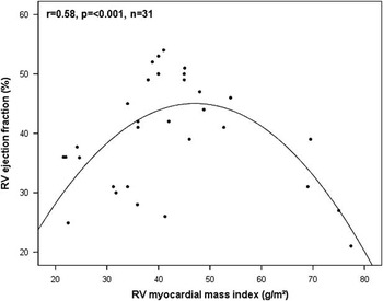
Patients’ right ventricular myocardial mass index including the inter-ventricular septum was equal to the left ventricular myocardial mass index of controls (58 [48; 74] grams per square metre versus 53 [48; 66] grams per square metre, p < 0.39, for congenitally corrected transposition patients; 54 [44; 61] grams per square metre versus 53 [48; 66] grams per square metre, p < 0.59, for dextro-transposition patients). There was no difference in right ventricular myocardial mass index between the congenitally corrected transposition group and the dextro-transposition group (58 [48; 74] grams per square metre versus 54 [44; 61] grams per square metre, p = 0.84). In dextro-transposition, there was a quadratic correlation between right ventricular ejection fraction and right ventricular myocardial mass index (p < 0.001, r = 0.58), meaning that patients with lower myocardial masses showed a positive correlation and patients with higher myocardial masses showed a negative correlation with systemic ventricular function (Fig 6).
Scatterplot diagram demonstrating a quadratic correlation between right ventricular myocardial mass index including the inter-ventricular septum and right ventricular ejection fraction in dextro-transposition of the great arteries. There is impaired right ventricular ejection fraction with very low and very high right ventricular myocardial mass. RV = right ventricular.

Congenitally corrected transposition and determinants of systemic function
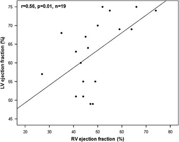
Patients with congenitally corrected transposition of the great arteries showed a weak negative correlation between age at examination and right ventricular ejection fraction (r = −0.18, p = 0.04). The right ventricular ejection fraction of congenitally corrected transposition patients with a severe or moderate tricuspid insufficiency was only marginally lower than that in congenitally corrected transposition patients with a mild tricuspid insufficiency (p = 0.79). There was a correlation between right ventricular ejection fraction and left ventricular ejection fraction (r = 0.56, p = 0.01; Fig 7). Right ventricular myocardial mass index did not correlate with right ventricular ejection fraction (r = −0.12, p = 0.63). There was also no significant correlation between right ventricular end-diastolic volume index and right ventricular ejection fraction (r = 0.05, p = 0.83).
Scatterplot diagram illustrating the inter-ventricular interaction in patients with congenitally corrected transposition of the great arteries. Impaired right ventricular ejection fraction associated with impaired left ventricular ejection fraction suggesting a functional unit of both ventricles. RV = right ventricular; LV = left ventricular.

Discussion
Associated cardiac anomalies are common in congenitally corrected transposition of the great arteries and could also be found in most patients in the present study. The mean age of these patients was within the fourth decade, which agrees with previous studies and reflects the natural history of the disease.Reference Dimas, Moodie, Sterba and Gill 7 , Reference Bos, Hagler and Silvilairat 19 In our study group, there were six patients in whom associated anomalies were regarded as haemodynamically relevant. These patients showed alterations of the pulmonary ventricle in terms of left ventricular “hypertrophy” and dilatation rather than changes in the right systemic ventricle, as the underlying pathology was left ventricular pressure or volume overload. Pulmonary stenosis is a well-known finding that occurs in approximately 40% of patients with congenitally corrected transposition and can be helpful in supporting right ventricular function.Reference Warnes 20 Nevertheless, these patients need close cardiac magnetic resonance follow-up examinations to repair high-degree stenosis in a timely manner and optimise interventional or surgical therapy.
We demonstrated that right ventricular myocardial mass index excluding the septum was almost three times higher in patients with congenitally corrected transposition of the great arteries compared with right ventricles of controls and was within the same range when compared with the left ventricular myocardial mass index of controls. Nevertheless, systemic ejection fraction was lower, which can be explained by the inability of the right ventricle to fully compensate for the morphological and structural disadvantages in systemic circulation. Macroscopic and microscopic architecture of the right ventricle is different from that of the left ventricle: the proportion of non-compacted myocardium is higher, and the irregular shape hinders high-pressure build-up.Reference Greenbaum, Ho, Gibson, Becker and Anderson 21 , Reference Ohuchi, Beighley, Dong, Zamir and Ritman 22 These findings are also observed in patients with dextro-transposition after atrial switch, as confirmed in the present and a previous study.Reference Hornung, Kilner, Davlouros, Grothues, Li and Gatzoulis 11 Compared with dextro-transposition, however, right ventricular myocardial mass was even higher in the congenitally corrected transposition group, probably owing to the longer duration of pressure load, as the age at examination for this group was significantly higher than the post-operative interval in the dextro-transposition group.
When comparing the two patient groups, one has to be aware of the differences with regard to mean patient age and number of previous operations. Patients with congenitally corrected transposition of the great arteries were older and had fewer operations, which might bias the volumetric and functional results.
Systemic ejection fraction was reduced in both patient groups. It was, however, higher in patients with congenitally corrected transposition than in those with dextro-transposition. Although in patients with congenitally corrected transposition the mean duration of pressure load was approximately 15 years longer, these patients could preserve a better systemic function. The reason for this result might be the post-operative situation after atrial switch in dextro-transposition of the great arteries, which results in rigidness of the atria with an impaired contribution to ventricular filling.Reference Derrick and Cullen 23 , Reference Tulevski, Lee and Groenink 24 In patients with congenitally corrected transposition, however, atrial function is undamaged, and the intact physiology of atrioventricular interaction results in higher ejection fractions. Furthermore, after atrial switch, patients are reported to show fibrosis of the systemic right ventricular myocardium in cardiac magnetic resonance delayed-enhancement imaging, which is probably a result of peri-operative ischaemia and which additionally impairs ejection fraction.Reference Babu-Narayan, Goktekin and Moon 25
To date, few data are available on cardiac magnetic resonance volumetry and function in patients with congenitally corrected transposition. In a previous study on 13 patients, the researchers found right ventricular ejection fraction within a normal range.Reference van der Zedde, Oosterhof, Tulevski, Vliegen and Mulder 26 However, most studies using cardiac magnetic resonance imaging, echocardiography, or a cardiac catheter support our results of impaired systemic right ventricular function in adulthood, and clinical observations in our tertiary care centre underscore this finding.Reference Dimas, Moodie, Sterba and Gill 7 , Reference Benson, Burns and Schwaiger 27 , Reference Giardini, Lovato and Donti 28 Normal systemic function in late adulthood should be regarded as an exception rather than a characteristic of congenitally corrected transposition of the great arteries.
Compared with the right ventricle, our data show a good function of the subpulmonary left ventricle in both patient groups. In two patients with pulmonary hypertension, the left ventricle could easily compensate for the increased pressure load and keep ejection fraction within a normal range.
There is an on-going discussion on the causes of systemic right ventricular failure in congenitally corrected transposition. Abnormalities of the tricuspid valve can be found in almost all these patients.Reference Warnes 20 Tricuspid insufficiency is considered one of the main reasons for the deterioration of systemic ventricular function and is the only independently significant factor of death.Reference Prieto, Hordof, Secic, Rosenbaum and Gersony 8 In our patient cohort, the majority of patients showed tricuspid insufficiency as well, mostly mild to moderate, which also led to right ventricular enlargement. A comparison of mild insufficiency with moderate and severe tricuspid insufficiency, however, demonstrated no significant differences in systemic right ventricular ejection fraction.
In dextro-transposition after atrial switch, excessive right ventricular hypertrophy is inversely correlated with right ventricular ejection fraction and has been identified as a risk factor for right ventricular failure, probably via demand ischaemia.Reference Hornung, Kilner, Davlouros, Grothues, Li and Gatzoulis 11 We can confirm this finding, as right ventricular myocardial mass index was quadratically correlated with right ventricular ejection fraction, and patients with low-degree or excessive hypertrophy demonstrated impaired systemic right ventricular function (Fig 6). In patients with congenitally corrected transposition, however, right ventricular myocardial mass index and right ventricular ejection fraction did not correlate, although the right ventricle in both patient groups functioned in the systemic circulation, and right ventricular hypertrophy in congenitally corrected patients was even slightly greater compared with that in patients with dextro-transposition. Therefore, it is unclear why the pathophysiological mechanisms should be substantially different. One possible explanation might be the greater inhomogeneity of the cohort with congenitally corrected transposition with respect to associated cardiac anomalies. The relatively small size of our patient cohort might be a further reason. Future studies with larger patient groups will show whether the combination of hypertrophy and failure is a general mechanism in the adaptational processes of systemic right ventricles or a specific problem after atrial switch procedure.
The only single parameter correlating with right ventricular ejection fraction was age at examination. This is in line with the clinical findings of previous studies.Reference Graham, Bernard and Mellen 4 , Reference Hornung, Bernard, Jaeggi, Howman-Giles, Celermajer and Hawker 29 Nevertheless, we hypothesise that systemic right ventricular systolic impairment is a multifactorial process, with the time factor impairing systolic function by long-lasting pressure and volume overload. Our patient cohort might be too small to identify further independent predictors of right ventricular function.
Inter-ventricular interaction as found in the present study is a well-known observation in patients with altered right ventricular anatomy and function.Reference Grothoff, Spors and Abdul-Khaliq 12 Anatomical findings demonstrate an inter-woven myocardial structure, making both ventricles a functional unit.Reference Sedmera 30 , Reference Sedmera and McQuinn 31 Excessive hypertrophy of the right ventricular myocardium might impair left ventricular function either by deformation of the left ventricle and a resulting non-physiological contraction pattern or by coronary steal, or by both. Modern techniques such as diffusion tensor imagingReference Tseng, Wedeen, Reese, Smith and Halpern 32 should provide deeper insights into the pathophysiological mechanisms.
Limitations
To date, this is one of the largest patient cohorts with congenitally corrected transposition of the great arteries examined by cardiac magnetic resonance imaging. Absolute numbers in this rare congenital heart disease are small, however, resulting in limited statistical power.
A further major limitation of this study is the fact that both age and number of operations were different between the two patient groups. This is again a result of the rarity of these malformations and should be considered when interpreting the data.
We did not perform delayed-enhancement imaging. Myocardial scarring, however, might play a role in right ventricular performance and could be an independent predictor of ejection fraction and/or prognosis.
As our patient numbers were small, it is difficult to measure the clinical impact of cardiac parameters. Hard clinical endpoints such as death or congestive heart failure are rare events; most patients are in New York Heart Association functional class I or II, and surrogate endpoints such as quality of life are fault prone and difficult to measure.
Conclusions
Although the right ventricle is in a systemic position in both congenitally corrected and dextro-transposition of the great arteries, the pathophysiology of right ventricular failure is at least partially different, and patients with congenitally corrected transposition can preserve a better function over a longer period of time. As cardiac magnetic resonance imaging is an ideal tool for assessing ventricular volumes and function in patients with congenital heart defects, close cardiac magnetic resonance follow-up examinations should be performed to start medicamentous, interventional, or surgical therapies in a timely manner.