Hypoplastic left heart syndrome is a complex CHD that comprises a small left ventricle, aortic valve atresia or severe stenosis, mitral valve atresia or severe stenosis, and hypoplastic aortic arch with coarctation of the aorta.Reference Noonan and Nadas 1 Among children with hypoplastic left heart syndrome, dysfunction of the morphologically right ventricle identified soon after initial presentation is associated with higher intermediate – before stage II operative repair – and overall mortality.Reference Altmann, Printz, Solowiejczky, Gersony, Quaegebeur and Apfel 2 Therefore, accurate assessment of intrinsic right ventricle function is critical in this population. The echocardiographic assessment of single right ventricle function is fraught with difficulty, however, because of its unique complex geometry. Traditional assessment has involved subjective qualitative methods or quantitative systolic ejection phase indices based on geometric assumptions.
The ratio of ventricular systolic and diastolic durations and the myocardial performance index have recently emerged as indices of ventricular function independent of ventricular geometry.Reference Williams, Ritter, Tani, Pagoto and Minich 3 , Reference Friedberg and Silverman 4 Myocardial performance index utilises Doppler-derived systolic and diastolic time intervals to generate a combined index of global ventricular function.Reference Tei 5 , Reference Tei, Ling and Hodge 6 The myocardial performance index has been validated in children with Ebstein’s anomaly and hypoplastic left heart syndrome as an objective index of ventricular function.Reference Williams, Ritter, Tani, Pagoto and Minich 3 , Reference Eidem, Tei, O’Leary, Cetta and Seward 7 The ratio of systolic and diastolic durations has been shown to be an indicator of global function in children with idiopathic dilated cardiomyopathy and right ventricle function in hypoplastic left heart syndrome.Reference Friedberg and Silverman 4 , Reference Friedberg and Silverman 8 Increased values of the myocardial performance index and the ratio of systolic and diastolic durations indicate decreased ventricular function.
Therapeutic options for hypoplastic left heart syndrome include staged Norwood procedure, Sano modification procedure, Hybrid procedure, and heart transplantation. Low cardiac output following Norwood procedure has been described, and may be secondary to low pump function or high pulmonary-to-systemic blood flow ratio or both.Reference Friedberg and Silverman 8 Hybrid stage 1 procedure (Hybrid), which consists of bilateral pulmonary artery banding, patent ductus arteriosus stenting, and atrial septostomy, is increasingly being performed as a first-stage palliation for hypoplastic left heart syndrome.Reference Akintuerk, Michel-Behnke and Valeske 9 , Reference Galantowicz, Cheatham and Phillips 10 The goals of Hybrid palliation are to provide reliable systemic cardiac output through the patent ductus arteriosus, control pulmonary blood flow, and create unobstructed flow from the left atrium, yielding a stable balanced circulation without the use of cardiopulmonary bypass.Reference Fenstermaker, Berger and Rowland 11 , Reference Galantowicz and Cheatham 12 In theory, Hybrid palliation can shift the risk of major open-heart surgery to later in life, with a potential beneficial impact on neurological development.Reference Galantowicz, Cheatham and Phillips 10 The intermediate outcome of the Hybrid approach to hypoplastic left heart syndrome has been reported as comparable with a traditional Norwood strategy.Reference Galantowicz, Cheatham and Phillips 10 A recent study using mathematical modelling compared the haemodynamic effects of Hybrid palliation and Norwood strategy for hypoplastic left heart syndrome.Reference Hsia, Cosentino, Corsini, Pennati, Dubini and Migliavacca 13 The computational results suggested inferior systemic oxygen delivery, coronary blood flow, and perfusion pressure following Hybrid palliation. This was the rationale for the current study’s hypothesis of a detrimental effect of Hybrid palliation on single ventricular function. Data on the effect of Hybrid stage I palliation on right ventricular function are currently scarce.
The purposes of this study were as follows: to compare the right ventricular myocardial performance index and the ratio of systolic and diastolic durations in groups of normal neonates and those with hypoplastic left heart syndrome before any intervention; to compare these right ventricular functional indices before and at specified time points following Hybrid palliation for hypoplastic left heart syndrome; and to compare right ventricular functional indices in a sub-group of infants with hypoplastic left heart syndrome who survived and those who died before the next stage of the surgery. We hypothesised that infants with hypoplastic left heart syndrome will have increased right ventricular myocardial performance index and ratio of systolic and diastolic durations compared with normal individuals in the early postnatal period; infants with hypoplastic left heart syndrome will develop right ventricular dysfunction after Hybrid; and infants with hypoplastic left heart syndrome who died before the next stage of surgery would have had significantly increased right ventricular dysfunction after Hybrid compared with survivors.
Methods
Patients
This was a retrospective study conducted in a tertiary Children’s hospital. The inclusion criterion was infants with hypoplastic left heart syndrome who underwent Hybrid palliation. The cardiac catheterisation database was searched to identify patients who underwent Hybrid procedure between October, 2006 and September, 2011. Controls were weight-matched infants who underwent echocardiography for evaluation of heart murmur and had no structural heart defects or other concurrent medical issues. Exclusion criteria were infants with other CHDs who underwent Hybrid procedure; two-dimensional and Doppler echocardiographic data were collected before Hybrid and after Hybrid – post 1, 0–4 days; post 2, 5–9 days; post 3, 2–3 weeks; post 4, 1–1.5 months. If an infant had multiple echocardiograms before Hybrid, the study closest to the Hybrid procedure was taken. A chart review was performed for infants with hypoplastic left heart syndrome to obtain demographic and clinical data. Cardiac catheterisations before the next stage surgery were reviewed in all patients. The study was approved by the Institutional Research Board of Wayne State University.
Two-dimensional and Doppler echocardiography
All echocardiographic examinations were performed using an IE-33 imaging system (Philips Medical, Andover, Massachusetts, United States of America). Doppler time intervals were obtained from the apical images and parasternal short-axis views. Right ventricle ejection time was obtained from the parasternal short-axis view using pulse-wave Doppler interrogation of the pulmonary valve with the sample volume positioned just above the pulmonary valve. The time interval between the two tricuspid inflows was obtained from the apical view using pulse-wave Doppler interrogation of the right ventricle inflow with the sample volume placed at the tips of the tricuspid valve. The degree of right ventricle dysfunction and tricuspid regurgitation were also qualitatively assessed. As simultaneous inflow and outflow Doppler recordings were not available in right ventricle morphology, the inflow and outflow recordings were obtained separately, but the data were only included when the RR interval between the Doppler tracings showed <5% difference. A maximal systolic pulmonary valvular annulus diameter was measured from the long-axis or apical views and pulmonary valve area was derived from π×diameter×diameter/4. The velocity time integral of the systolic flow jet was measured at the pulmonary annulus using pulsed Doppler from the parasternal short-axis view or apical view. Cardiac index was calculated as (pulmonary velocity time integral)×(pulmonary valvular annulus)×(heart rate) indexed to body surface area. Images were analysed offline by a single reader blinded to the clinical information (D.K.).
Measurement of myocardial performance index and ratio of systolic and diastolic durations
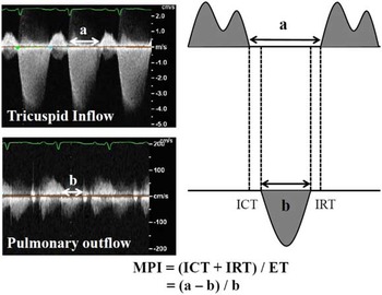
The myocardial performance index and the ratio of systolic and diastolic durations were calculated as previously described.Reference Friedberg and Silverman 4 – Reference Tei, Ling and Hodge 6 In brief, ejection time was measured from the onset to the termination of right ventricular outflow. The time spent in the isovolumetric phase and isovolumetric contraction and relaxation times was calculated by subtracting ejection time from the termination to onset of tricuspid inflow. Myocardial performance index was calculated by dividing isovolumetric times by ejection time (Fig 1). Systolic duration was derived from the duration of tricuspid regurgitation, whereas diastolic duration was derived from the time from the termination to onset of tricuspid regurgitation jet. The ratio of systolic and diastolic durations was calculated from systolic duration divided by diastolic duration. Patients with insufficient tricuspid regurgitation jet were excluded from the study. In controls, myocardial performance index and systolic and diastolic durations ratio was measured for the right ventricle in the same manner. Inter- and intra-observer variability of the myocardial performance index and the ratio of systolic and diastolic durations were tested in 23% of measurements in randomly selected patients from our cohort.
MPI. Doppler interrogations for tricuspid inflow and pulmonary outflow were obtained from the apical images and parasternal short-axis views, respectively. ET=ejection time; ICT=isovolumic contraction time; IRT=isovolumic relaxation time; MPI=myocardial performance index.

Statistical analysis
Statistical analysis was performed using SPSS software for PC (version 17.0) (SPSS Inc., Chicago, Illinois, United States of America). Continuous variables were reported as mean±SD, whereas nominal variables were reported as numbers (percentage). Independent Student’s t-test and the χ2 test were used to compare demographic and haemodynamic parameters between groups of infants with normal anatomy and hypoplastic left heart syndrome before the Hybrid palliation. Analysis of variance and the χ2 test were used to analyse various echo parameters in infants with hypoplastic left heart syndrome before and at various time points after Hybrid palliation. Statistical significance was defined as p<0.05.
Results
A total of 23 infants with hypoplastic left heart syndrome and 35 controls comprised the study cohort; six infants with hypoplastic left heart syndrome and eight controls were excluded due to insufficient tricuspid regurgitation. Infants with hypoplastic left heart syndrome had a diagnosis of aortic atresia – mitral atresia, 10 and mitral stenosis, three – or aortic stenosis – mitral atresia, two and mitral stenosis, eight. Antenatal diagnosis of the lesion was available in the majority (74%) of cases with hypoplastic left heart syndrome. The mode of delivery was via caesarean section in 43%, and the mean±SD maternal age was 26.9±5.4 years. The mean±SD gestational age was 38.3±1.7 weeks with preterm delivery before 37 completed weeks in 13%. The mean±SD birth weight was 3.08±0.60 kg, and 5-minute Apgar score in all patients was ⩾8. Our cohort included 52% black, 26% white, and 22% infants of other races. The medical issues before Hybrid included gastrointestinal in two infants – necrotising enterocolitis in one and duodenal atresia requiring Ladd procedure in one – renal in three infants – hydronephrosis in two and renal insufficiency associated with left small solitary kidney in one – infection – pneumonia in one and urinary tract infection in one – severe haemodynamic compromise in two infants – shock with closing ductus and intact atrial septum in one and severe ventricular dysfunction requiring extracorporeal membrane oxygenation in one. The baseline echocardiography (pre-Hybrid) was performed 7.7±6.2 days before Hybrid. Infants with hypoplastic left heart syndrome underwent Hybrid at a mean±SD age of 12.0±7.7 days and weight of 3.1±0.7 kg. Post-Hybrid 1, 2, 3, and 4 echocardiographies were performed 2.0±1.1, 6.7±2.0, 17.4±4.3, and 34.0±3.1 days after Hybrid, respectively. Inotropic medications were used in 9% pre-Hybrid, 88% at post 1, 37% at post 2, 38% at post 3, and 22% at post 4. Inter-stage transcatheter interventions included balloon atrial septostomy with and without atrial stent placement (n=4), balloon angioplasty of the right pulmonary artery banding followed by repeat stent placement of patent ductus arteriosus (n=1) before post 4, and stent placement for retrograde coarctation (n=2) after post 4. Inter-observer correlation coefficients (R) of the myocardial performance index and ratio of systolic and diastolic durations ratio tested by two readers (S.A. and D.K.) were 0.763 and 0.820, respectively. Intra-observer variability of myocardial performance index and ratio of systolic and diastolic durations of the same reader was also high – R of 0.813 and 0.927, respectively.
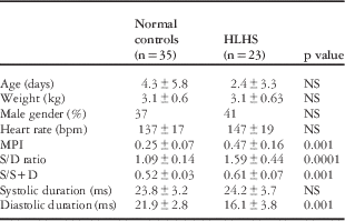
Normal controls versus hypoplastic left heart syndrome patients (Table 1)
The mean age, weight, and gender distribution were comparable between normal controls (n=35) and infants with hypoplastic left heart syndrome (n=23). Myocardial performance index was significantly higher in infants with hypoplastic left heart syndrome – 0.47±0.16 versus 0.25±0.07, p<0.001. The ratio of systolic and diastolic durations was also significantly higher in infants with hypoplastic left heart syndrome – 1.59±0.44 versus 1.09±0.14, p<0.0001. There was no difference in the systolic duration between groups, whereas the diastolic duration was significantly lower in infants with hypoplastic left heart syndrome compared with controls – 16.1±3.8 versus 21.9±2.8, p<0.0001.
Demographics and echocardiographic indices in normal controls and infants with hypoplastic left heart syndrome (pre-intervention).

HLHS=hypoplastic left heart syndrome; MPI=myocardial performance index; S/D ratio=systolic/diastolic ratio
Values are expressed as mean±SD
Infants with hypoplastic left heart syndrome before and after Hybrid stage 1 procedure
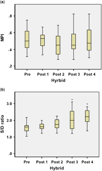
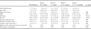
Table 2 compares the echocardiographic indices at the specified time points before and until 1.5 months after Hybrid. Cardiac index, myocardial performance index, ratio of systolic and diastolic durations, and ratio of systolic duration/systolic duration+diastolic duration were comparable between pre-Hybrid and post 1 Hybrid (NS). Myocardial performance index did not change until 1 month after Hybrid (Fig 2), whereas the ratio of systolic and diastolic durations increased significantly (p=0.043) from pre-Hybrid to 2−3 and 4−6 weeks following the procedure, signifying worsening right ventricular function.
Box plot of myocardial performance index ( a ) and ratio of systolic and diastolic durations ( b ) in five groups (pre, post 1–4 Hybrid). The lower and upper bounds of the boxes indicate the 25th and 75th percentile values, respectively. The horizontal line indicates the median, and the whisker bars represent the 10th and 90th percentiles. *Significant p value is compared with pre-Hybrid value. MPI=myocardial performance index; S/D ratio=systolic/diastolic ratio.

Echocardiographic indices in infants with hypoplastic left heart syndrome before and after Hybrid stage 1 procedure.

MPI=myocardial performance index; S/D ratio=systolic/diastolic ratio
*Significant p value is compared to pre-Hybrid value
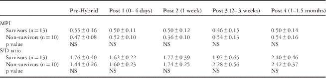
Infants with hypoplastic left heart syndrome who survived and died before the next stage of surgery (Table 3)
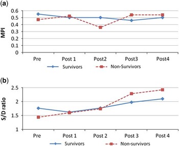
Myocardial performance index and ratio of systolic and diastolic durations ratio were not significantly different between infants with hypoplastic left heart syndrome who survived (n=13) and those who died (n=10) before the next stage of surgery consisting of arch reconstruction and cavopulmonary anastomosis (Fig 3).
Comparison of MPI and S/D ratio in infants with hypoplasitc left heart syndrome who survived and died before the next stage surgery (pre, post 1−4 Hybrid).

MPI=myocardial performance index; S/D ratio=systolic/diastolic ratio
The change of myocardial performance index ( a ) and ratio of systolic and diastolic durations ( b ) in infants with hypoplastic left heart syndrome who survived and died before the next stage surgery (pre, post 1−4 Hybrid). There is no significant difference in the myocardial performance index and the ratio of systolic and diastolic durations between groups (p=NS). MPI=myocardial performance index; S/D ratio=systolic/diastolic ratio.

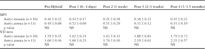
Infants with hypoplastic left heart syndrome who had a diagnosis of aortic stenosis and aortic atresia (Table 4)
Myocardial performance index and ratio of systolic and diastolic durations were not significantly different between infants with hypoplastic left heart syndrome who had a diagnosis of aortic stenosis (n=10) and aortic atresia (n=13); however, both the myocardial performance index and the ratio of systolic and diastolic durations tended to be higher in infants with aortic atresia at 2–3 and 4–6 weeks following the Hybrid procedure. The degree of tricuspid regurgitation and qualitative right ventricular dysfunction did not correlate with either the myocardial performance index or the ratio of systolic and diastolic durations.
Comparison of MPI and S/D ratio in infants with hypoplasitc left heart syndrome who had a diagnosis of aortic stenosis and aortic atresia (pre, post 1−4 Hybrid).

MPI=myocardial performance index; S/D ratio=systolic/diastolic ratio
Discussion
To our knowledge, this is the first study to evaluate the effect of Hybrid on right ventricular function in infants with hypoplastic left heart syndrome using validated quantitative echocardiographic parameters. Our results demonstrate that the right ventricular myocardial performance index and the ratio of systolic and diastolic durations are significantly higher, and the diastolic duration is significantly shorter in infants with hypoplastic left heart syndrome even before any intervention, compared with controls, suggesting intrinsic right ventricular dysfunction in this population. We speculate that the shorter diastolic time may be associated with a decrease in coronary flow and may be pathogenic in the ventricular dysfunction associated with hypoplastic left heart syndrome. In addition, the right ventricular myocardial performance index remained unchanged immediately and up to 1 month after Hybrid palliation, but the ratio of systolic and diastolic durations increased significantly after 2 weeks of Hybrid palliation. This may suggest that right ventricular dysfunction develops later after Hybrid or that inotropes used in the early period following the Hybrid procedure may have masked a significant difference in the echocardiographic parameters.
Children with hypoplastic left heart syndrome are at risk for right ventricular dysfunction even before the first-stage palliation, because the right ventricle is the systemic pumping chamber, needs to eject blood against systemic-level resistance, and has a higher pre-load due to elevated pulmonary-to-systemic blood flow ratio.Reference Friedberg and Silverman 8 Iannettoni et alReference Iannettoni, Bove and Mosca 14 reported right ventricular function to be fair or poor in 10/73 patients before Norwood stage I palliation. Forbess et alReference Forbess, Cook, Roth, Serraf, Mayer and Jonas 15 reported that 19, 15, and 5% of 127 patients with hypoplastic left heart syndrome before Norwood procedure had mild, moderate, and severe decrease in right ventricular function, respectively. Altman et alReference Altmann, Printz, Solowiejczky, Gersony, Quaegebeur and Apfel 2 found that, among 60 patients, 13, 16, and 5% had mild, moderate, and severe right ventricular dysfunction, respectively, as assessed by apical area shortening fraction and biplane pyramidal ejection fraction on two-dimensional echocardiography. In 33 children with hypoplastic left heart syndrome – mean age of 3.12 years, at various palliation stages – the ratio of systolic and diastolic durations was found to be significantly higher compared with controls – 1.65±0.85 versus 0.85±0.2, p<0.001.Reference Friedberg and Silverman 8 The ratio of systolic and diastolic durations was significantly increased in patients with Norwood stage 1 versus Stages 2 and 3–2.16 versus 1.4 and 1.32, respectively. In our cohort, the systolic/diastolic ratio 4–6 weeks after the Hybrid procedure was 2.21 and was comparable with the reported value of post-Norwood stage 1.
Myocardial performance index was first introduced by Tei et alReference Tei 5 , Reference Tei, Ling and Hodge 6 in 1995. Williams et alReference Williams, Ritter, Tani, Pagoto and Minich 3 first reported the utility of the myocardial performance index in children with single ventricle. In a study of 41 children with single ventricle, three of whom had hypoplastic left heart syndrome – mean age of 11.0 months, before bilateral cavopulmonary anastomosis – the myocardial performance index was higher compared with 30 age-matched controls – 0.67±0.14 versus 0.32±0.10, p<0.001. There was a weak but significant correlation between ventricular end-diastolic pressure and the myocardial performance index in children with single ventricle. Zhang et al reported myocardial performance index values in 161 children with single ventricle before and after total cavopulmonary connection compared with 80 controls without CHDs. Myocardial performance index was significantly higher in children with single ventricle – 0.54±0.11 versus normal left ventricle 0.30±0.08, normal right ventricle 0.26±0.08, p<0.001.Reference Forbess, Cook, Roth, Serraf, Mayer and Jonas 15 The myocardial performance index was well correlated with catheterisation-derived maximal dp/dt.Reference Zhang, Sun and Zhu 16 Our findings of elevated myocardial performance index and ratio of systolic and diastolic durations in infants with hypoplastic left heart syndrome, compared with controls, are consistent with previous data. In our cohort, the myocardial performance index did not change over time, whereas the ratio of systolic and diastolic durations increased significantly after Hybrid palliation. Friedberg et alReference Friedberg and Silverman 8 suggested that the ratio of systolic and diastolic durations may be a better echocardiographic marker to discriminate between patients with abnormal and normal ventricular function in children with hypoplastic left heart syndrome. It was speculated that the right ventricular dysfunction may not be completely accounted for by the myocardial performance index, because only isovolumic relaxation is incorporated into the diastolic phase for the calculation of myocardial performance index.
Interestingly, initial right ventricular dysfunction did not impact early survival after Norwood stage 1 operation but was associated with decreased intermediate and overall survival.Reference Altmann, Printz, Solowiejczky, Gersony, Quaegebeur and Apfel 2 Although the physiology after Norwood is different from that after Hybrid stage 1, such an association between pre-existing ventricular dysfunction and outcome following Hybrid has not been examined. In our study, compared with infants with hypoplastic left heart syndrome who survived, those who died before the next stage of surgery appeared to show a further increase in the ratio of systolic and diastolic durations 2 weeks after Hybrid; however, this was not statistically different (p=0.14), likely due to a small sample size.
Following the Hybrid, significant physiological changes occur with modified afterload and pulmonary-to-systemic blood flow ratio due to pulmonary artery banding and ductus arteriosus stenting; however, owing to the lack of cardiopulmonary bypass, cardiac function was expected to be unchanged following Hybrid.Reference Galantowicz, Cheatham and Phillips 10 The haemodynamic effects of Hybrid and surgical Norwood palliations for hypoplastic left heart syndrome were assessed recently by computational models.Reference Hsia, Cosentino, Corsini, Pennati, Dubini and Migliavacca 13 Compared with the Norwood models, the Hybrid approach had inferior systemic and cerebral oxygen deliveries with higher pulmonary-to-systemic blood flow ratio and lower cardiac output. Furthermore, lower systemic blood flow and the obligatory retrograde perfusion through an unreconstructed aortic arch led to substantially less coronary blood flows in the Hybrid model. In addition, a diastolic runoff through the ductal stent was exhibited in the Hybrid model, which resulted in flow reversal in the brachiocephalic artery. Right ventricular performance was evaluated from pressure–volume loops, and Norwood models showed higher ejection fraction and mechanical efficiency than the Hybrid models. The authors speculated that the poorer ventricular performance in the Hybrid model would be explained by the combination of the obligatory volume load on the single ventricle and the additional afterload by pulmonary artery banding. In a single previous study on 30 patients, Fenstermaker et alReference Fenstermaker, Berger and Rowland 11 reported inter-stage echocardiographic changes in children with hypoplastic left heart syndrome who underwent Hybrid palliation. Echocardiographic data were compared between post-Hybrid stage 1 procedure, before and after inter-stage interventions, and before comprehensive stage II procedure. In this study, tricuspid regurgitation and right ventricular dysfunction qualitatively increased compared with post-Hybrid stage 1 procedure in patients requiring an intervention and improved after an intervention – for example, in cases such as additional stent placement in the ductus arteriosus, dilations of the ductus arteriosus stent, repeat balloon atrial septostomy, stent placement in the distal arch, stent in atrial septum, dilation of the descending aorta, and left pulmonary artery angioplasty. The authors speculated that increasing afterload by stenosis across the ductal stent or retro-aortic arch would result in decreased coronary perfusion, especially in patients with aortic atresia, leading to ventricular dysfunction and tricuspid regurgitation. Our data suggested that right ventricular function assessed by the ratio of systolic and diastolic durations may get worse after Hybrid palliation. Furthermore, both the myocardial performance index and the ratio of systolic and diastolic durations tended to be higher in infants with aortic atresia compared with aortic stenosis, although these differences were not statistically significant, probably due to the small sample size. We speculate that the combination of volume ventricular overload, pulmonary over-circulation, and sub-optimal coronary perfusion may cause right ventricular dysfunction after Hybrid palliation.
Study limitations
There were certain limitations to our study. Our sample size was relatively small. There was 17% mortality (4/23) before post 4 Hybrid, which may underestimate the right ventricular dysfunction, because patients who died might have had more right ventricular dysfunction. Right ventricular function can be modified by inotropic support, stenosis across the ductal stent, and retrograde coarctation. The degree of inotropic support use and inter-stage transcatheter intervention was not accounted for in this study. This was a retrospective study at a single institution with its inherent limitations. In addition, Hybrid approach is a newly introduced procedure and has its own learning curve in each institution. Our time points of assessment, while reflecting usual clinical practice, were arbitrary. We could not compare infants who underwent Hybrid and Norwood, because, at least in the initial period, infants who underwent Hybrid were deemed to be poor surgical candidates. The possibility that baseline medical condition and pre-existing ventricular dysfunction contributed to the progression of ventricular function cannot be ignored. There is a need to compare these indices between Hybrid and Norwood palliation. The myocardial performance index and the ratio of systolic and diastolic durations are load-dependent to some extent so that change of loading condition could contribute to abnormal values of those markers. Whether an increase in myocardial performance index and ratio of systolic and diastolic durations following Hybrid palliation is a reflection of the abnormal intrinsic contractility or worsening of the loading condition is unclear. Although only trivial tricuspid regurgitation on Doppler evaluation is needed to measure systolic and diastolic durations, absence of tricuspid regurgitation may limit the use of the ratio of systolic and diastolic durations. We included two patients with hypoplastic left heart syndrome variants, who had mitral stenosis, aortic atresia, and ventricular septal defect, in our cohort.
Nonetheless, this systematic echo exploration of the haemodynamic effects of Hybrid palliation in a homogeneous cohort of neonates with hypoplastic left heart syndrome, using objective echocardiographic indices, provides insight into the physiology following Hybrid palliation. These data are potentially important in the identification of infants with right ventricle dysfunction and management of post-Hybrid right ventricular dysfunction.
Conclusions
The right ventricular myocardial performance index and the ratio of systolic and diastolic durations was significantly impaired in infants with hypoplastic left heart syndrome even before any intervention compared with controls. The right ventricular function did not change immediately after Hybrid in infants with hypoplastic left heart syndrome, but the ratio of systolic and diastolic durations increased significantly 2 weeks after Hybrid. Further investigation to assess the risk factors and implications of ventricular dysfunction is warranted.
Acknowledgements
Authors’ contribution: D.K. designed the study, analysed the data, and drafted the paper. S.A. interpreted the data and revised and finally approved the manuscript. G.N., D.R.T., T.J.F., R.E.D., and H.L.W. contributed to the study design and revised the paper critically.
Financial Support
This research received no specific grant from any funding agency, commercial, or not-for-profit sectors.
Conflicts of Interest
None.
Ethical Standards
The authors assert that all procedures contributing to this work comply with the ethical standards of the relevant national guidelines on human experimentation (Human Investigation Committee at Wayne State University) and with the Helsinki Declaration of 1975, as revised in 2008, and has been approved by the institutional committees of Wayne State University.