Introduction
Phosphatic or phosphatized small skeletal fossils are generally dominant components among early Cambrian faunal assemblages. These fossils, including spicules, tubes, plates, cap-shaped shells, and disarticulated sclerites, are usually millimetric in size and are collectively described as ‘small shelly fossils (SSFs)’ (Matthews and Missarzhevsky, Reference Matthews and Missarzhevsky1975). Although the taxonomic affinities of many disarticulated sclerites remain unresolved, the discovery of extraordinarily well-preserved, articulated, multiplated, soft-bodied specimens in Burgess Shale–type Lagerstätten can provide significant insights into the functional morphology and phylogenetic position of SSF taxa. Perhaps the most influential examples are the descriptions of Microdictyon sinicum Chen, Hou, and Lu, Reference Chen, Hou and Lu1989 from the Chengjiang Biota (Chen et al., Reference Chen, Zhou and Ramsköld1995), Halkieria evangelista Conway Morris and Peel, Reference Conway Morris and Peel1995 from North Greenland (Conway Morris and Peel, Reference Conway Morris and Peel1990, Reference Conway Morris and Peel1995; Vinther and Nielsen, Reference Vinther and Nielsen2005), and Wiwaxia corrugata (Matthew, Reference Matthew1899) from the Burgess Shale (Conway Morris, Reference Conway Morris1985; Smith, Reference Smith2014).
Minute net-like phosphatic plates were first reported by Matthews and Missarzhevsky (Reference Matthews and Missarzhevsky1975) from the Strenuella Limestone of Comley, England, and were described as problematic SSFs. The generic name, Microdictyon Bengtson, Matthews, and Missarzhevsky, 1981 in Missarzhevsky and Mambetov, Reference Missarzhevsky and Mambetov1981, was formally introduced on the basis of isolated reticulated sclerites from the lower Cambrian of Malyi Karatau, South Kazakhstan. To date, isolated Microdictyon plates have been reported from South Australia (Bengtson et al., Reference Bengtson, Matthews and Missarzhevsky1986, Reference Bengtson, Conway Morris, Cooper, Jell and Runnegar1990; Gravestock et al., Reference Gravestock, Alexander, Jago, Rozanov and Zhuravlev2001; Topper et al., Reference Topper, Brock, Skovsted and Paterson2009, Reference Topper, Brock, Skovsted and Paterson2011), South China (Hao and Shu, Reference Hao and Shu1987; Shu and Chen, Reference Shu and Chen1989; Tong, Reference Tong1989; Li and Zhu, Reference Li and Zhu2001; Zhang and Aldridge, Reference Zhang and Aldridge2007; Yang et al., Reference Yang, Steiner and Keupp2015), Kazakhstan (Missarzhevsky and Mambetov, Reference Missarzhevsky and Mambetov1981; Bengtson et al., Reference Bengtson, Matthews and Missarzhevsky1986; Dzik, Reference Dzik2003), Uzbekistan (Bengtson et al., Reference Bengtson, Matthews and Missarzhevsky1986), Mongolia (Esakova and Zhegallo, Reference Esakova and Zhegallo1996), Siberia (Bengtson et al., Reference Bengtson, Matthews and Missarzhevsky1986; Missarzhevsky, Reference Missarzhevsky1989; Demidenko, Reference Demidenko2006; Varlamov et al., Reference Varlamov, Rozanov, Khomentovsky, Shabanov and Abaimova2008; Kouchinsky et al., Reference Kouchinsky, Bengtson, Clausen, Gubanov, Malinky and Peel2011, Reference Kouchinsky, Bengtson, Clausen and Vendrasco2015), Turkey (Sarmiento et al., Reference Sarmiento, Fernández-Remolar and Göncüoglu2001), England (Shropshire, Avalonia; Matthews and Missarzhevsky, Reference Matthews and Missarzhevsky1975; Bengtson et al., Reference Bengtson, Matthews and Missarzhevsky1986; Hinz, Reference Hinz1987), Baltica (Bengtson et al., Reference Bengtson, Matthews and Missarzhevsky1986), Northeast Greenland (Laurentia; Skovsted, Reference Skovsted2006), Canada (British Columbia, Laurentia; Bengtson et al., Reference Bengtson, Matthews and Missarzhevsky1986), United States (New York, California, Nevada, Utah, Laurentia; Bengtson et al., Reference Bengtson, Matthews and Missarzhevsky1986; eastern Massachusetts, Avalonia; Landing, Reference Landing1988), and Mexico (Laurentia; McMenamin, Reference McMenamin1984; McMenamin and McMenamin, Reference McMenamin and McMenamin1990). Although Microdictyon had a near cosmopolitan distribution in the lower and middle Cambrian, the large majority of species have been established exclusively on the basis of disarticulated sclerites; Microdictyon sinicum from the Chengjiang Biota (Chen et al., Reference Chen, Zhou and Ramsköld1995; Hou and Bergström, Reference Hou and Bergström1995) and Microdictyon sp. from the Kaili Biota (Zhao et al., Reference Zhao, Yuan, Zhu, Yang, Guo, Qian, Huang and Pan1999, Reference Zhao, Yuan, Zhu, Yang, Guo, Peng and Yang2002, Reference Zhao, Zhu, Babcock, Yuan, Parsley, Peng and Wang2005) remain the only well-known articulated species.
These delicate specimens from the Chengjiang Lagerstätte show that Microdictyon is an elongated caterpillar-like animal possessing paired uniramous lobopod limbs with nine pairs of reticulated plates positioned along both sides of the subcylindrical trunk (Chen et al., Reference Chen, Hou and Lu1989, Reference Chen, Zhou and Ramsköld1995; Ramsköld and Hou, Reference Ramsköld and Hou1991; Bergström and Hou, Reference Bergström and Hou2001; Hou et al., Reference Hou, Aldridge, Bergström, Siveter and Feng2004). These soft-bodied specimens indicate that Microdictyon is quite similar to other fossils with uniramous lobopods, including Aysheaia Walcott, Reference Walcott1911 (Whittington, Reference Whittington1978), Hallucigenia Walcott, Reference Walcott1911 (Conway Morris, Reference Conway Morris1977), Xenusion Pompeckj, Reference Pompeckj1927 (Dzik and Krumbiegel, Reference Dzik and Krumbiegel1989), Luolishania Hou and Chen, Reference Hou and Chen1989, Onychodictyon Hou, Ramsköld, and Bergström, Reference Hou, Ramsköld and Bergström1991, and Cardiodictyon Hou, Ramsköld, and Bergström, Reference Hou, Ramsköld and Bergström1991. In general, these fossils are assigned to Lobopodia (Hou and Bergström, Reference Hou and Bergström1995; Ramsköld and Chen, Reference Ramsköld and Chen1998), and comparisons of soft-part anatomy show great similarity to extant onychophorans (Ramsköld and Hou, Reference Ramsköld and Hou1991; Ramsköld and Chen, Reference Ramsköld and Chen1998; Smith and OrtegaHernández, Reference Smith and Ortega-Hernández2014). Some lobopodians with mosaic features of both lobopods and arthropods, such as Miraluolishania haikouensis Liu et al., Reference Liu, Shu, Han and Zhang2004, Jianshanopodia decora Liu et al., Reference Liu, Shu, Han, Zhang and Zhang2006, and Diania Liu et al., Reference Liu, Steiner, Dunlop, Keupp, Shu, Ou, Han, Zhang and Zhang2011, indicate that well-preserved lobopodian fossils can potentially shed light on the origin of arthropods (Liu et al., Reference Liu, Shu, Han and Zhang2004, Reference Liu, Shu, Han, Zhang and Zhang2006, Reference Liu, Steiner, Dunlop, Keupp, Shu, Ou, Han, Zhang and Zhang2011; Ma et al., Reference Ma, Hou and Bergström2009).
The high diversity and morphological disparity of disarticulated phosphatic lobopodian plates represent an excellent complement to soft-bodied Lagerstätte specimens. Despite their near cosmopolitan distribution in the Cambrian, Microdictyon plates had not been previously documented from the North China Platform. Here we report the first occurrence of Microdictyon plates from the Xinji Formation on the North China Platform, extending the paleogeographic distribution of this genus. The aim of this paper is to describe and illustrate the isolated plates from the North China Platform and to provide an overview of the paleogeographic and stratigraphic distribution of Microdictyon and taxonomic problems associated with these net-like sclerites.
Geologic setting
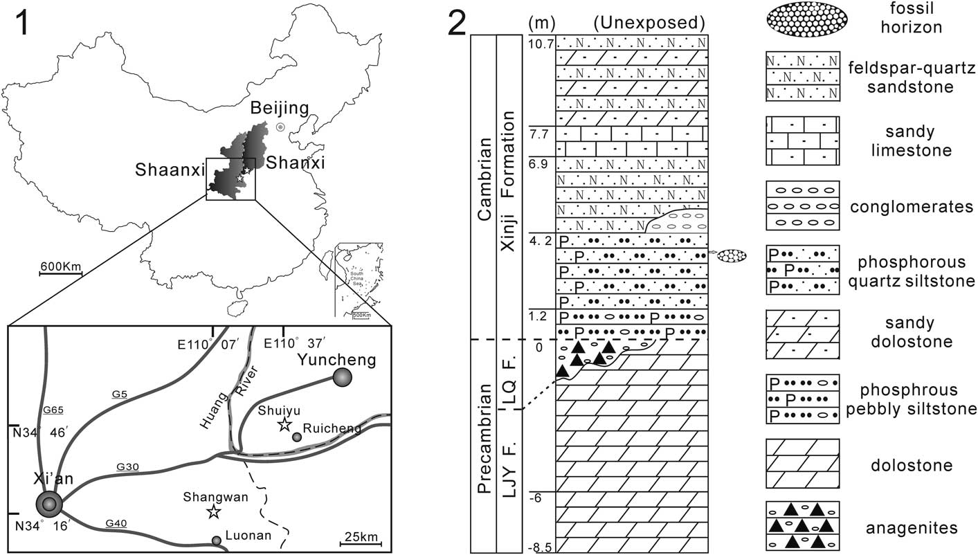
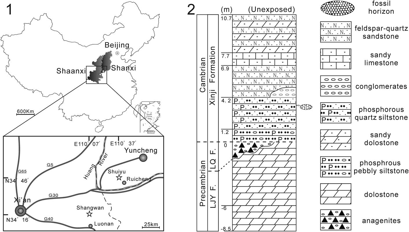
Specimens of Microdictyon described herein were collected from the Xinji Formation at the Shuiyu section, Ruicheng County, Shanxi Province, and at the Shangwan section, Luonan County, Shaanxi Province (Fig. 1.1), along the south margin of the North China Platform (Li et al., Reference Li, Zhang, Hua and Yang2014).
Locality map of sampled sections and the lithostratigraphic column of Shuiyu section. (1) Locality map showing the positions of Shuiyu section and Shangwan section; (2) lithostratigraphic column of the Xinji Formation at Shuiyu section, Ruicheng County, Shanxi Province. LQ F.=Luoquan Formation; LJY F.=Longjiayuan Formation.
The Xinji Formation is regionally the lowermost Cambrian strata, disconformably overlying Precambrian strata. It consists of siliciclastic rocks intercalated with carbonates and is mainly distributed on the southwestern to southern margin of the North China Platform (Liu et al., Reference Liu, Wang, Zhang and Du1991; Liu et al., Reference Liu, Ma, Zhu, Jin, Dai, Chen, Fan, Zhang and Zhang1994). The thickness of the Xinji Formation decreases from the south to the north along the south margin of the North China Platform, and the lithology varies (Liu, Reference Liu1986; Liu et al., Reference Liu, Wang, Zhang and Du1991; Yan et al., Reference Yan, Zhang and Wang1993). Due to its diverse and well-preserved small shelly fossils, the Xinji Formation has attracted considerable attention (He et al., Reference He, Pei and Fu1984; He and Pei, Reference He and Pei1985; Feng et al., Reference Feng, Qian and Rong1994; Pei and Feng, Reference Pei and Feng2005; Li et al., Reference Li, Zhang, Hua and Yang2014; Pan et al., Reference Pan, Miao, Yang and Li2015; Li et al., Reference Li, Zhang, Yun and Li2016; Skovsted et al., Reference Skovsted, Pan, Topper, Betts, Li and Brock2016), though isolated plates of Microdictyon were not recovered previously. Trilobites were also reported from the Xinji Formation (Zhang and Zhu, Reference Zhang and Zhu1979; Zhang et al., Reference Zhang, Zhu, Yuan, Lin, Qian, Wu and Yuan1979).
In the Shangwan section, the Xinji Formation disconformably overlies the Luoquan Formation (Ediacaran), which is composed predominantly of conglomerates, gravel-bearing siltstones, and shales (for stratigraphic column, see fig. 1 in Li et al., Reference Li, Zhang, Hua and Yang2014). The Xinji Formation is approximately 20 m thick and can be subdivided into two parts. The 11 m thick lower part of the formation consists mainly of sandy phosphoric limestones, shales, and siltstones. The ~0.8 m thick basal bed consists mainly of phosphoric sandy limestones and contains abundant small shelly fossils. One isolated plate of Microdictyon described herein was collected from this bed. The upper part of the Xinji Formation consists mostly of calcareous sandstones and dolomitic limestones with trilobites, but scarce small shelly fossils. The Xinji Formation is conformably overlain by the Zhushadong Formation (Li et al., Reference Li, Zhang, Hua and Yang2014).
In the Shuiyu section, the Xinji Formation disconformably overlies the Precambrian strata, either the Luoquan Formation (Ediacaran, predominantly conglomerates) or the Longjiayuan Formation (Paleoproterozoic or Mesoproterozoic, gray dolostones) (Fig. 1.2). There is a karst surface between the Luoquan Formation and the Longjiayuan Formation. According to a local geological survey report, the Xinji Formation at Shuiyu section is about 39.8 m thick and can be subdivided into two parts: the lower part is dominated by gray-black phosphatic conglomerates, purple-red shale, and phosphoric sandstone, and the upper part consists of red quartz sandstone intercalated with argillaceous dolostone (Bureau of Geology and Mineral Resources of Shanxi Province, 1989). Here, only the basal part (10.7 m) has been observed, with the upper part of the section covered by vegetation. There are abundant trace fossils in the basal rocks, and the trace fossil assemblage belongs to a typical Cruziana ichnofacies (Miao and Zhu, Reference Miao and Zhu2014), indicating a subtidal environment for the basal Xinji Formation at the Shuiyu section. Several isolated plates of Microdictyon described herein were collected from a strongly weathered bed of calcic phosphoric quartz siltstone, 3.4 m above the base of the Xinji Formation (Fig. 1.2).
The Xinji Formation yields a small trilobite fauna, consisting of Estaingia (Bergeroniellus) lonanensis Hsiang, 1965 in Lu et al., Reference Lu, Zhang, Zhu, Qian and Xiang1965, Estaingia (Hsuaspis) houchiuensis Chang, 1963 in Hsiang, Reference Hsiang1963, and Redlichia cf. R. nanjiangensis Zhang and Lin (Zhang and Zhu, Reference Zhang and Zhu1979; Zhang et al., Reference Zhang, Zhu, Yuan, Lin, Qian, Wu and Yuan1979; Miao, Reference Miao2014). Judging from the trilobite assemblage, the Xinji Formation was correlated with the Drepanuloides Zone of the middle Tsanglangpuan stage (Cambrian Stage 4) on the Yangtze Platform (Zhang and Zhu, Reference Zhang and Zhu1979; Zhang et al, Reference Zhang, Zhu, Yuan, Lin, Qian, Wu and Yuan1979; Yu et al., Reference Yu, Xu and Yi1984; He and Pei, Reference He and Pei1985; Miao, Reference Miao2014). The Estaingia trilobite assemblage can also be compared with the Pararaia janeae Zone of South Australia (Paterson and Brock, Reference Paterson and Brock2007; Miao, Reference Miao2014). In addition, numerous small shelly fossils were discovered from the Xinji Formation, such as chancelloriid sclerites, sponge spicules, hyoliths, Pojetaia runnegari Jell, Reference Jell1980, Mackinnonia rostrate Zhou and Xiao, Reference Zhou and Xiao1984, Stenotheca drepanoida He and Pei, 1984 in He et al., Reference He, Pei and Fu1984, Pelagiella madianensis Zhou and Xiao, Reference Zhou and Xiao1984, Cupitheca holocyclata Bengston, 1990 in Bengston et al., Reference Bengtson, Conway Morris, Cooper, Jell and Runnegar1990, C. costellata Xiao and Zhou, Reference Xiao and Zhou1984, Yochelcionella chinensis Pei, Reference Pei1985, Cambroclavus absonus Conway Morris, 1990 in Bengston et al., Reference Bengtson, Conway Morris, Cooper, Jell and Runnegar1990, and Apistoconcha cf. A. apheles Conway Morris, 1990 in Bengston et al., Reference Bengtson, Conway Morris, Cooper, Jell and Runnegar1990, and so on. (He et al., Reference He, Pei and Fu1984; Yu et al., Reference Yu, Xu and Yi1984; Pei, Reference Pei1985; Feng et al., Reference Feng, Qian and Rong1994; Li et al., Reference Li, Zhang, Hua and Yang2014; Pan et al., Reference Pan, Miao, Yang and Li2015; Li et al., Reference Li, Zhang, Yun and Li2016; Skovsted et al., Reference Skovsted, Pan, Topper, Betts, Li and Brock2016). Most of these fossils have been reported from the Parara Limestone (Stansbury Basin) and Mernmerna Formation (Arrowie Basin) in South Australia (Bengtson et al., Reference Bengtson, Conway Morris, Cooper, Jell and Runnegar1990; Gravestock et al., Reference Gravestock, Alexander, Jago, Rozanov and Zhuravlev2001; Topper et al., Reference Topper, Brock, Skovsted and Paterson2009; Betts et al., Reference Betts, Paterson, Jago, Jacquet, Skovsted, Topper and Brock2016, Reference Betts, Paterson, Jago, Jacquet, Skovsted, Topper and Brock2017). Judging from the trilobite and small shelly fossil assemblages, and similarities with South Australian assemblages, the Xinji Formation can be roughly correlated with Cambrian Series 2, Stage 4 (mid-late Botoman age in Siberian terminology).
Materials and methods
One well-preserved plate and four fragmented plates of Microdictyon were retrieved from calcic phosphoric fine quartz sandstone samples collected from a horizon 3.4 m above the base of the Xinji Formation at the Shuiyu section, Ruicheng County, Shanxi Province. Only one poorly preserved fragment of Microdictyon plate was retrieved from phosphoric sandy limestone samples collected from the base of the Xinji Formation at the Shangwan section, Luonan County, Shaanxi Province.
Samples collected from the Shuiyu section were processed by digestion in 6% acetic acid, and samples from the Shangwan section were processed by digestion in 10% acetic acid. The selected specimens were placed on stubs, gold coated, and photographed using the scanning electron microscopy facility (LEO 1530VP) at the Nanjing Institute of Geology and Palaeontology, Chinese Academy of Sciences, Nanjing.
Repository and institutional abbreviation
All the illustrated specimens are housed and cataloged at storage facilities of the Nanjing Institute of Geology and Palaeontology, Chinese Academy of Sciences (NIGPAS), Nanjing.
Taxonomy of isolated eoconchariid plates
The Cambrian net-like isolated plates, including Microdictyon, are usually described under the family Eoconchariidae (Chen et al., Reference Chen, Zhou and Ramsköld1995; Demidenko, Reference Demidenko2006; Zhang and Aldridge, Reference Zhang and Aldridge2007; Topper et al., Reference Topper, Brock, Skovsted and Paterson2009), which consists of three genera: Microdictyon Bengtson, Matthews, and Missarzhevsky, 1981, in Missarzhevsky and Mambetov, Reference Missarzhevsky and Mambetov1981, Quadratapora Hao and Shu, Reference Hao and Shu1987, and Fusuconcharium Hao and Shu, Reference Hao and Shu1987. Isolated eoconchariid plates retrieved from acid residues are perforated, and each hole is surrounded by a series of nodes on the external surface. Different genera are predominantly recognized on the basis of plate shape, perforation size and arrangement, and node morphology. For example, Microdictyon exhibits hexagonally arranged holes with mushroom-shaped or spiky nodes; Quadratapora exhibits tetragonally arranged holes with sharply crested walls; Fusuconcharium exhibits a disorderly hexagonal hole arrangement with spine-like or rounded nodes (Zhang and Aldridge, Reference Zhang and Aldridge2007). Demidenko (Reference Demidenko2006) established the genus Onychomicrodictyon due to its plates combining features of Microdictyon (net-like perforated plates with five to seven mushroom-shaped nodes around one hole) and Onychodictyon Hou, Ramsköld, and Bergström, Reference Hou, Ramsköld and Bergström1991 (strong spine-like process) and assigned it to Eoconchariidae. However, Steiner et al. (Reference Steiner, Hu, Liu and Keupp2012) consider that hole size and the overall plate dimensions of Onychomicrodictyon together with one unnamed plate documented by Bengtson (Reference Bengtson1991) are identical with Onychodictyon ferox Hou, Ramsköld, and Bergström, Reference Hou, Ramsköld and Bergström1991. Therefore, Onychomicrodictyon was considered to be a junior synonym of Onychodictyon (Steiner et al., Reference Steiner, Hu, Liu and Keupp2012; Topper et al., Reference Topper, Skovsted, Peel and Harper2013). Because of the anatomy of soft-bodied specimens, Onychodictyon was assigned to Onychodictyidae Hou and Bergström, Reference Hou and Bergström1995 (Hou and Bergström, Reference Hou and Bergström1995; Liu et al., Reference Liu, Shu, Han, Zhang and Zhang2008).
After the discovery of the soft-bodied Microdictyon sinicum, it has been well known that the trunk of this species has nine pairs of plates along sides of the caterpillar-like body. The plates show a regular net-like morphology with a hexagonal hole arrangement but vary distinctly in size and outline. Chen et al. (Reference Chen, Zhou and Ramsköld1995) delineated four morphotypes: elongate (the anterior pair), round (the second pair), ovoid (the third to eighth pairs), and rhomboidal (the posterior pair). Microdictyon sinicum demonstrates that the plate outline is variable along the trunk of a single animal, indicating that plate outline is not a reliable taxonomic character. The soft-bodied specimens were preserved in fine siliciclastic rocks, and unfortunately the details of node morphology and microstructure have been obscured by diagenetic processes, complicating detailed comparisons with isolated phosphatic plates retrieved from acid residues. Zhang and Aldridge (Reference Zhang and Aldridge2007) have demonstrated that node microstructure is relatively consistent at species level according to a large collection of isolated plates from southern China. Zhang and Aldridge (Reference Zhang and Aldridge2007) do report some morphological variation within species, and it remains to be seen whether the degree of variation represents intraspecific variation or warrants the erection of a separate species. Conjoined plates of Microdictyon and Onychodictyon that are interpreted as having preserved the molting process display identical ornamentation, indicating that plate ornamentation did not vary during ontogeny (Zhang and Aldridge, Reference Zhang and Aldridge2007; Topper et al., Reference Topper, Skovsted, Peel and Harper2013). However, these specimens only represent a single molt stage, and the entire ontogenetic sequence of plate-bearing lobopodians remains unclear. With the information currently at our disposal, the description of Microdictyon plates must focus on the arrangement of holes and microstructure of the nodes, rather than on plate outline and size.
To date, there are eight Microdictyon species that were erected on the morphology of isolated plates (only M. sinicum was established on soft-bodied preservation). All the soft-bodied fossils of Microdictyon recovered from the Chengjiang Biota at different sections in eastern Yunnan belong to Microdictyon sinicum. Without knowledge of the detailed plate morphology of M. sinicum, it is entirely possible that many of these species based solely on isolated plates may be plate morphotypes of M. sinicum.
All specimens recovered from the Xinji Formation possess the typical hexagonal meshwork arrangement and mushroom-shaped nodes that characterize Microdictyon. The plates described herein show similarities to some formally established species but also bear some new characters. Due to the small sample set, and without detailed information regarding scleritome configuration and intraspecific and ontogenetic variation (see Topper et al., Reference Topper, Brock, Skovsted and Paterson2011), the authors here take a cautious approach and describe the Microdictyon specimens under open nomenclature at species level. The morphological characteristics of the plates will be described in detail.
Systematic paleontology
The terminology used to describe the Microdictyon plates follows that of Zhang and Aldridge (Reference Zhang and Aldridge2007).
Phylum Lobopodia Snodgrass, Reference Snodgrass1938
Class Xenusia Dzik and Krumbiegel, Reference Dzik and Krumbiegel1989
Order Archonychophora Hou and Bergstrӧm, 1995
Family Eoconchariidae Hao and Shu, Reference Hao and Shu1987
Genus Microdictyon Bengtson, Matthews, and Missarzhevsky, 1981 in Missarzhevsky and Mambetov, Reference Missarzhevsky and Mambetov1981
Type species
Microdictyon effusum Bengtson, Matthews, and Missarzhevsky, 1981 in Missarzhevsky and Mambetov, Reference Missarzhevsky and Mambetov1981.
Other species
M. rhomboidale Bengtson, Matthews, and Missarzhevsky, Reference Bengtson, Matthews and Missarzhevsky1986; M. robisoni Bengtson, Matthews, and Missarzhevsky, Reference Bengtson, Matthews and Missarzhevsky1986; M. chinense Hao and Shu, Reference Hao and Shu1987; M. sphaeroides Hinz, Reference Hinz1987; M. sinicum Chen, Hou, and Lu, Reference Chen, Hou and Lu1989; M. depressum Bengtson in Bengtson et al., Reference Bengtson, Conway Morris, Cooper, Jell and Runnegar1990; M. fuchengense Li and Zhu, Reference Li and Zhu2001; M. jinshaense Zhang and Aldridge, Reference Zhang and Aldridge2007.
Remarks
The plates of Microdictyon, Quadratapora, and Fusuconcharium show similarity in meshwork arrangement, with each perforation surrounded by a number of protruding nodes. Genera are distinguished by the arrangement of holes and the microstructure of the nodes. Microdictyon plates exhibit a regular hexagonal meshwork that is distinct from Quadratapora plates, with a regular tetragonal meshwork, and Fusuconcharium plates, which display a slightly more irregular meshwork. Only Microdictyon is well known from soft-bodied preservation. The isolated plates of Quadratapora and Fusuconcharium have only been reported from the Shuijingtuo Formation in Zhenba, Shaanxi, South China (Hao and Shu, Reference Hao and Shu1987), where they co-occur with Microdictyon (Zhang and Aldridge, Reference Zhang and Aldridge2007). In the Shuijingtuo Formation, the number of plates belonging to Quadratapora (63) and Fusuconcharium (28) is quite small in comparison to that of the documented plates of Microdictyon (577; data from Zhang and Aldridge, Reference Zhang and Aldridge2007), and it is possible that the plates of Quadratapora and Fusuconcharium are aberrant forms or representatives of other Microdictyon species. Microdictyon rozanovi Demidenko, Reference Demidenko2006 was erected on the basis of two broken plates that display a characteristic extended edge (Demidenko, Reference Demidenko2006). This lateral extension is somewhat similar to the edge of M. rhomboidale, but we consider this subtle variation of the plate edge as insufficient evidence to erect a new species and, on the basis of node morphology, we suggest that these specimens most likely represent interspecific variants of M. rhomboidale. McMenamin (Reference McMenamin1984) informally described a new species, M. multicavus, from the lower Cambrian of Sonora, Mexico. However, due to the poor preservation of the specimens illustrated and the lack of a subsequent formal description, we do not recognize the species as valid, pending further study.
Microdictyon sp.
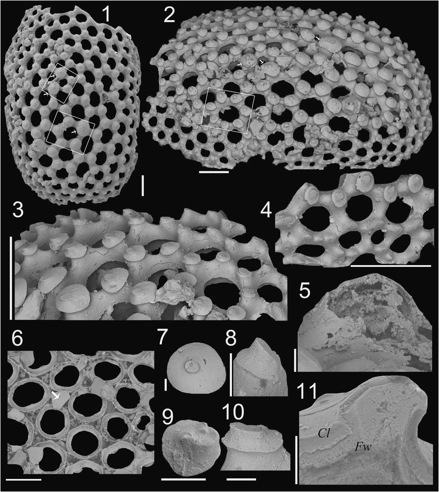
Microdictyon sp. from the Xinji Formation at Shuiyu section. (1–11) Plate S1, 166027; (1) plan external view; (2) oblique view; (3) magnification of the nodes at the margin of the plate; (4) magnification of the nodes to show the deformation of the nodes at the periphery of plate; (5) magnification of the hollow node; (6) internal view of the central region of the plate; (7–10) magnification of the nodes showing apex details; (11) magnification of the broken wall to show the microstructure of the plate. (1–4, 6) Scale bars=200 µm; (5, 7–11) scale bars=20 µm. Cl=capping layer; Fw=framework.

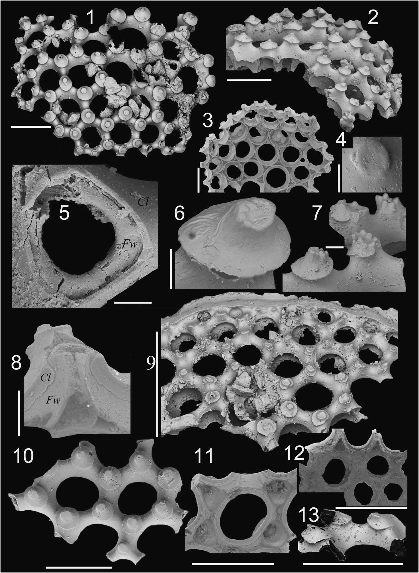
Microdictyon sp. from the Xinji Formation at Shuiyu section. (1–8) Plate S2, 166028; (1) plan external view; (2) oblique view; (3) internal view; (4) magnification of the apex of a node; (5) magnification of the broken base of a node; (6, 7) oblique view of the nodes with multiple tip apices; (8) oblique internal view of a broken node. (9, 12) Plate S5, 166031; (9) plan external view; (12) internal view. (10, 11, 13) Plate S4, 166030; (10) external view; (11) internal view; (13) lateral view of the nodes. (1–3, 9–13) Scale bars=200 µm; (4–8) scale bars=20 µm. Cl=capping layer; Fw=framework.

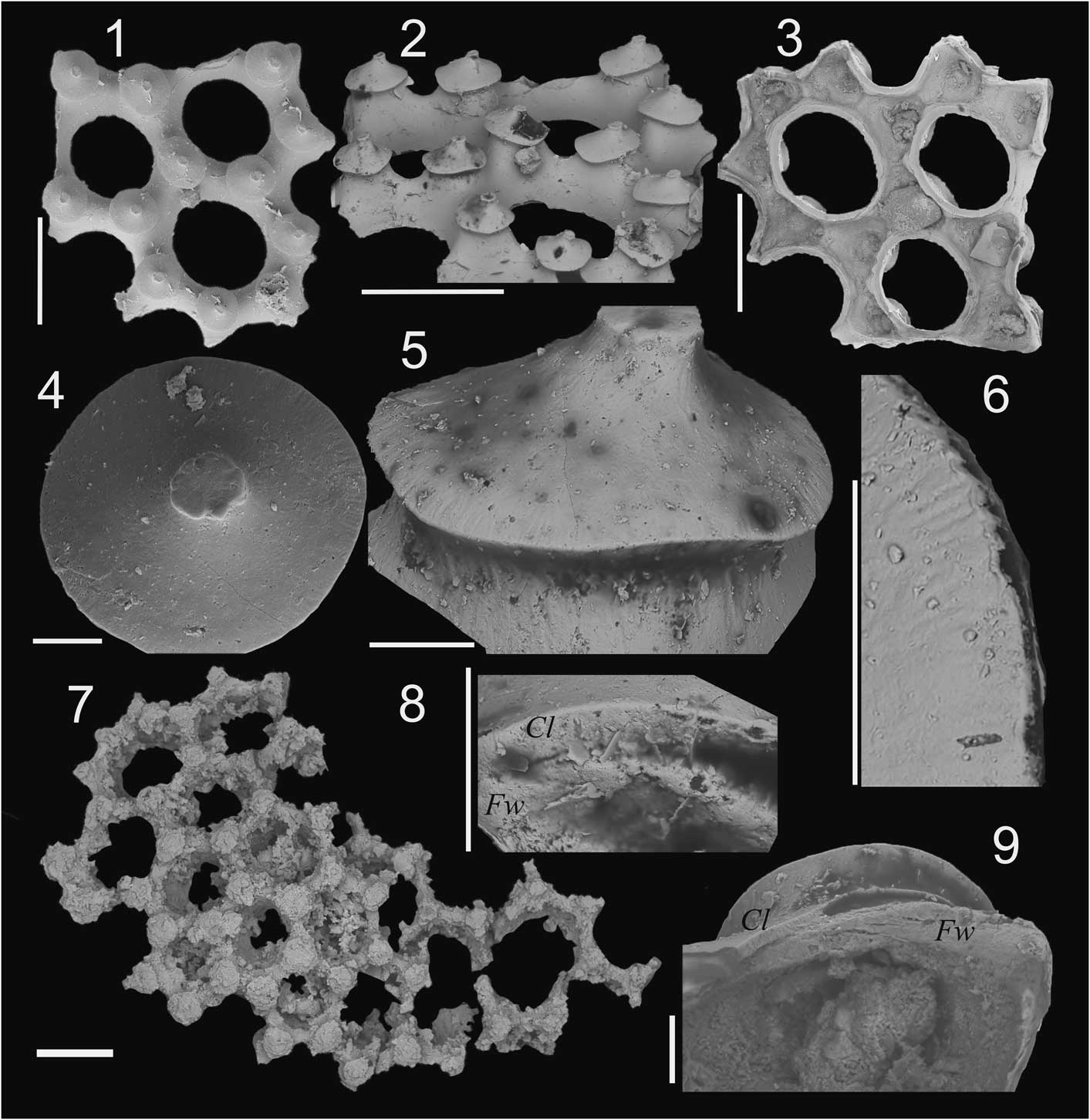
Microdictyon sp. from the Xinji Formation. (1–6, 8, 9) Plate S3, 166029, collected at Shuiyu section; (1) plan external view of the fragment; (2) oblique external view of the fragment; (3) internal view; (4) plan view of a node; (5) lateral view of a node; (6) magnification of the periphery of a node; (8) lateral view of a broken wall; (9) interval view of node. (7) Plate S6, 166032, collected at Shangwan section, plan external view. (4–6, 8, 9) Scale bars=20 µm; (1–3, 7) scale bars=200 µm. Cl=capping layer; Fw=framework.

Materials
One nearly complete specimen and four small fragmented specimens (described as S1, S2, S3, S4, and S5, respectively) were collected from the Xinji Formation at the Shuiyu section. One poorly preserved specimen (S6) was collected from the Xinji Formation at the Shangwan section.
Description
Plate S1 is ovoid and strongly convex, measuring 2,030 mm in length and 1,372 mm in maximum width, with a measured ratio of length to width at 1.48 (Fig. 2.1, 2.2). The holes are circular to ovoid in shape. The diameter (21.6 µm to 189 µm with the average of 115.35 µm) of the holes decreases toward the periphery of the plate (Fig. 2.1, 2.2, 2.4). Typically, each hole is surrounded by six hexagonally arranged nodes protruding from the junction of the walls. There is slight variation, with one hole surrounded by four nodes and 13 other holes respectively surrounded by five and seven nodes in plate S1 (square in Fig. 2.1, 2.2). In most cases, each node connects three walls; however, a few nodes can be observed connecting four walls in plate S1 (arrows in Fig. 2.1, 2.2, 2.6). Most of the nodes are low and mushroom-shaped, with an ovate to subtriangular outline. The length of the nodes ranges from 29.24 to 160.94 µm with the average of 77.26 µm and decreases toward the periphery of the plate. The majority of nodes show a single acentric low apex that protrudes from the surface. The apices roughly point in a single direction (Figs. 2.2, 2.3, 3.1, 3.10). It is noteworthy that many apices have a distinct circular or subcircular rim that forms a slightly concave platform at an oblique angle with the surface of the mushroom-shaped nodes (Fig. 2.3, 2.7–2.10). The apex of the node in plate S3 is quite distinct, with a relatively planar surface (Fig. 4.1, 4.2, 4.4, 4.5). The diameter of the apices ranges from 10.00 to 33.29 µm with the average of 21.52 µm. Toward the periphery of the plate, the mushroom-shaped nodes of plates S1 and S5 deform, giving the appearance of a single sharp tubercle with an indistinct rim (Figs. 2.2–2.4, 3.9), and some apices of nodes of plate S2 separate into multiple little tips (Fig. 3.1, 3.2, 3.7). The multiple tips only cover half of the apex of one node in plate S2 (Fig. 3.6). The upper surfaces of the nodes in plate S3 bear radial lines that originate from the apex and extend outward to the rim of the node (Fig. 4.4–4.6), possibly extending into the base of the nodes (Fig. 4.5). The mushroom-shaped nodes are hollow and sealed by one capping layer (Figs. 2.5, 3.8, 4.8, 4.9). On the inner surface, each hole has a cylindrical tubular wall with a basal opening (Figs. 2.6, 3.3). The peripheral girdle has a slightly inclined outer edge and an upright inner edge (Fig. 3.9). The plate microstructure consists of one basal framework layer (Fw) and one upper capping layer (Cl) attaching on the framework (Figs. 2.11, 3.5, 3.8, 4.8, 4.9). One poorly preserved fragment retrieved from the Shangwan section shows that holes are surrounded by regularly hexagonally arranged mushroom-shaped nodes (Fig. 4.7).
Remarks
The isolated plates of Microdictyon from the North China Platform show similar characteristics to previously reported isolated plates of Microdictyon. The ovoid, strongly convex plate and low mushroom-shaped nodes are quite similar to the type species, M. effusum (Bengtson et al., Reference Bengtson, Matthews and Missarzhevsky1986), but the single oblique platform-like or relatively protruding planar apices of the plates from North China contrast with the single spiny apices of M. effusum. Multi-tip apices have been described in Microdictyon chinense and M. jinshaense (Zhang and Aldridge, Reference Zhang and Aldridge2007; Topper et al., Reference Topper, Brock, Skovsted and Paterson2009), but this represents a rather pervasive character over the entire plate, rather than the rare peripheral occurrences observed in the North China material. The spiny nodes of the plates from North China occur in many species of Microdictyon, such as Microdictyon chinense, Microdictyon cf. M. effusum, Microdictyon jinshaense (Zhang and Aldridge, Reference Zhang and Aldridge2007), and Microdictyon cf. M. depressum (Skovsted, Reference Skovsted2006). But the plates described herein can be easily distinguished from the plates of these species by the combination of characters listed in the preceding. The outline and arrangement and microstructure of the nodes on the North China Microdictyon plates also show some similarities to the plates of M. depressum (Bengtson et al., Reference Bengtson, Conway Morris, Cooper, Jell and Runnegar1990), but the plates of M. depressum are quite flat and their single-tip apices are low. Topper et al. (Reference Topper, Brock, Skovsted and Paterson2011) reported 6 types of plates from the Ajax Limestone in South Australia. Like the specimens documented herein, some of them also bear multi-tip apices, but the shape and combination of the tips are obviously different. Both assemblages are relatively small (herein: five specimens; Topper et al., Reference Topper, Brock, Skovsted and Paterson2009: six specimens), impeding a detailed comparison. The radial line on the surface of the nodes is here reported for the first time on Microdictyon plates.
Discussion
Stratigraphical range and distribution of Microdictyon
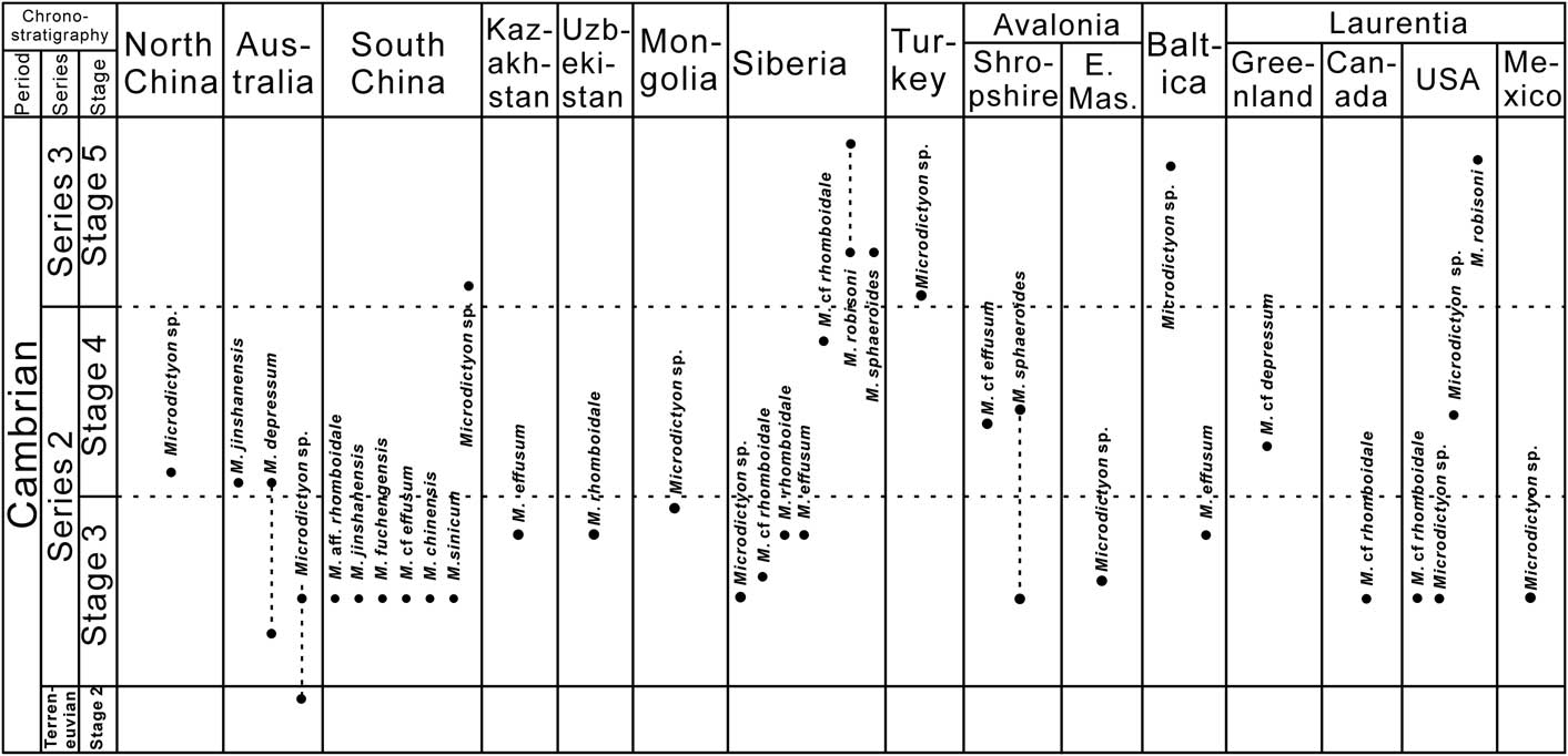
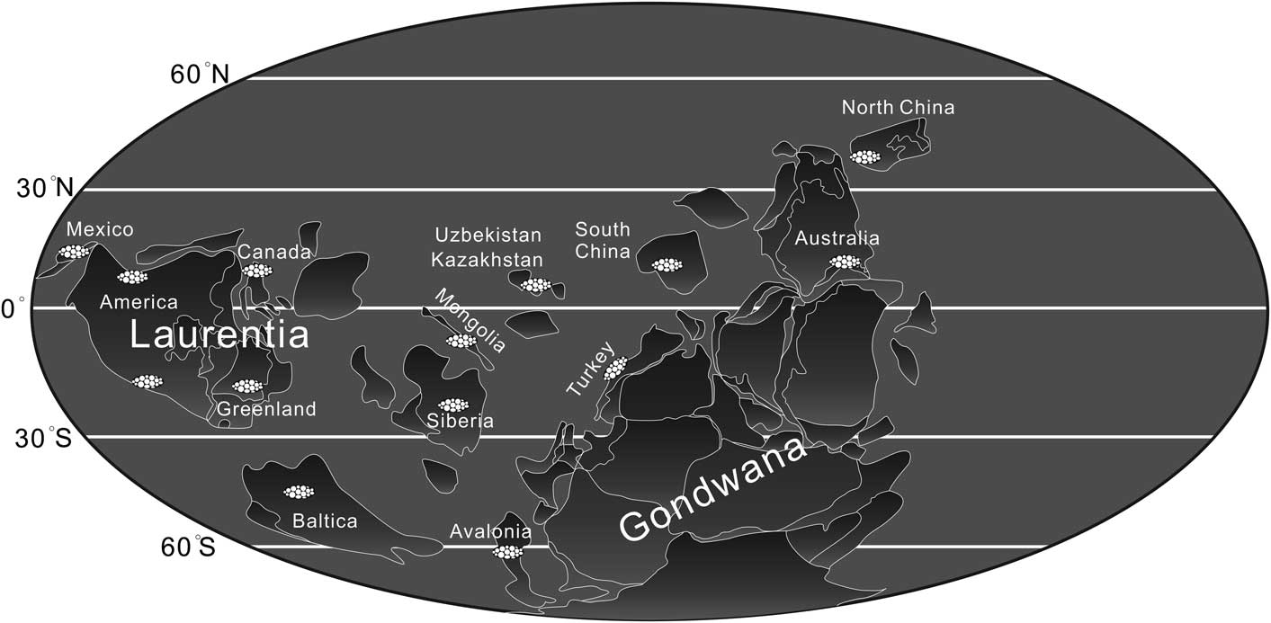
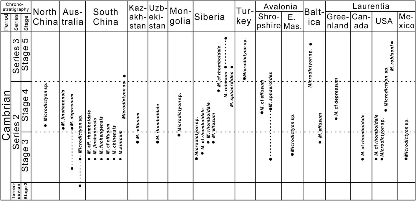
Microdictyon has a cosmopolitan geographic distribution from the Cambrian uppermost Stage 2 to uppermost Stage 5 (Figs. 5, 6). Complete soft-bodied specimens however have only been discovered from South China. Microdictyon is here documented from the lower Cambrian Xinji Formation of the North China Platform for the first time, extending the paleogeographic range of the genus in the late early Cambrian (Stage 4). The earliest occurrence of Microdictyon is from the lower Micrina etheridgei Zone (below the Abadiella huoi Zone) of the Ajax Limestone in section AJX-M from South Australia (Topper et al., Reference Topper, Brock, Skovsted and Paterson2011; Betts et al., Reference Betts, Paterson, Jago, Jacquet, Skovsted, Topper and Brock2016), which may represent the uppermost Cambrian Stage 2. Bengtson et al. (Reference Bengtson, Matthews and Missarzhevsky1986) also reported Microdictyon? tenuiporatum revised as Quadratapora tenuiporatum by Zhang and Aldridge (Reference Zhang and Aldridge2007) from the Tommotian Stage (pretrilobite stratum). In Stage 3, Microdictyon is reported from the Abadiella huoi Zone in Australia (Bengtson et al., Reference Bengtson, Conway Morris, Cooper, Jell and Runnegar1990; Topper et al., Reference Topper, Brock, Skovsted and Paterson2011) that correlates with the oldest trilobite zone, Parabadiella Zone in South China (Betts et al., Reference Betts, Paterson, Jago, Jacquet, Skovsted, Topper and Brock2016, Reference Betts, Paterson, Jago, Jacquet, Skovsted, Topper and Brock2017). This may be approximately coeval to the Eofallotaspis Zone in Morocco and the Profallotaspis jakutensis Zone in Siberia, as suggested by Yuan et al. (Reference Yuan, Zhu, Lin and Zhu2011). The genus then rapidly dispersed to many paleocontinents, with the majority of described species documented from the Eoredlichia-Wutingaspis Zone in South China (Chen et al., Reference Chen, Zhou and Ramsköld1995; Li and Zhu, Reference Li and Zhu2001; Zhang and Aldridge, Reference Zhang and Aldridge2007), the lower Nevadella Zone in Laurentia (British Columbia [Canada], New York, California, Nevada, Utah [United States], Mexico: Bengtson et al., Reference Bengtson, Matthews and Missarzhevsky1986; McMenamin, Reference McMenamin1984), the Callavia Zone in Avalonia (Avalonian part of eastern Massachusetts [United States]: Bengtson et al., Reference Bengtson, Matthews and Missarzhevsky1986; Landing, Reference Landing1988; Shropshire [England]: Hinz, Reference Hinz1987), and the upper Delgadella anabara Zone to the Judomia Zone in Siberia (Kouchinsky et al., Reference Kouchinsky, Bengtson, Clausen and Vendrasco2015). Some isolated plates of Microdictyon are also reported in the uppermost strata of Stage 3, in localities such as Kazakhstan (Bengtson et al., Reference Bengtson, Matthews and Missarzhevsky1986; Dzik, Reference Dzik2003), Uzbekistan (Bengtson et al., Reference Bengtson, Matthews and Missarzhevsky1986), Siberia (Bengtson et al., Reference Bengtson, Matthews and Missarzhevsky1986; Varlamov et al., Reference Varlamov, Rozanov, Khomentovsky, Shabanov and Abaimova2008), and Mongolia (Esakova and Zhegallo, Reference Esakova and Zhegallo1996). In Stage 4, Microdictyon has been documented from the Pararaia bunyerooensis Zone in Australia (Topper et al., Reference Topper, Brock, Skovsted and Paterson2009), the Lermontovia grandis Zone of Siberia (Demidenko, Reference Demidenko2006), the Bonnia-Olenellus Zone in Northeast Greenland (Skovsted, Reference Skovsted2006), the Elliptocephala asaphoides assemblage in the United States (Laurentia part of eastern New York; Bengtson et al., Reference Bengtson, Matthews and Missarzhevsky1986), the Strenuella Limestone and Protolenus Limestone in Shropshire (England/Avalonia; Bengtson et al., Reference Bengtson, Matthews and Missarzhevsky1986; Hinz, Reference Hinz1987), and North China (herein). There is a sharp decrease in distribution and diversity of Microdictyon in this period, with only M. sphaeroides, M. depressum, and M. jinshaense reported from Stage 3. In Stage 5, Microdictyon is reported from the Oryctocephalus indicus Zone in South China (Zhao et al., Reference Zhao, Yuan, Zhu, Yang, Guo, Qian, Huang and Pan1999, Reference Zhao, Yuan, Zhu, Yang, Guo, Peng and Yang2002, Reference Zhao, Zhu, Babcock, Yuan, Parsley, Peng and Wang2005), the Kuonamkites Biozone in Siberia (Kouchinsky et al., Reference Kouchinsky, Bengtson, Clausen, Gubanov, Malinky and Peel2011), the Acadoparadoxides mureroensis Zone in Turkey (Sarmiento et al., Reference Sarmiento, Fernández-Remolar and Göncüoglu2001; stratigraphy revised by Dean, Reference Dean2005), and the Ptychagnostus gibbus Zone (uppermost Stage 5) in North America and Baltica (Bengtson et al., Reference Bengtson, Matthews and Missarzhevsky1986). Species reported include M. robisoni (new occurrence), M. sphaeroides, and a few undetermined species.
Stratigraphic occurrences of Microdictyon species documented from different paleocontinents ranging from Cambrian Stage 2 to Stage 5. E. Mas.=eastern Massachusetts.
Paleogeographical distribution of Microdictyon from Cambrian Stage 2 to Stage 5 (paleogeographic map modified from Yang et al., Reference Yang, Steiner and Keupp2015).

The first appearance datum (FAD) of Microdictyon has been included in discussions as a potential marker for defining the global Stage 3 basal boundary (Peng et al., Reference Peng, Babcock and Cooper2012); however, the uncertainties regarding plate taxonomy and the sporadic appearance of Microdictyon specimens in Stage 2 in South Australia (Topper et al., Reference Topper, Brock, Skovsted and Paterson2011; Betts et al., Reference Betts, Paterson, Jago, Jacquet, Skovsted, Topper and Brock2016) hamper the biostratigraphic application of Microdictyon at the species level. There are morphological similarities between the species documented from China and Australia; for example, nodes with multiple-tip apices only occur in the plates of M. chinense and M. jinshaense from South China (Zhang and Aldridge, Reference Zhang and Aldridge2007) and M. jinshaense from South Australia (Topper et al., Reference Topper, Brock, Skovsted and Paterson2009, Reference Topper, Brock, Skovsted and Paterson2011) and the plates illustrated herein. This could indicate that North China had a close paleogeographic position with South Australia and South China in the early Cambrian. The early occurrence (Stage 2), wide distribution, and long stratigraphic range of Microdictyon suggest that the genus may have been a pioneering member of Cambrian communities displaying a strong adaptability to a variety of environments during the Cambrian explosion.
Morphological variation of the nodes of the isolated Microdictyon plates
It is obvious that some characters of the nodes of Microdictyon plates can occur in different Microdictyon species. For example, the short spiny nodes over the entire external surface of some medium-sized plates (possibly representing an intermediate ontogenetic growth stage) of Microdictyon chinense (Zhang and Aldridge, Reference Zhang and Aldridge2007) are also observed at the margin of the plates of several other species, such as Microdictyon cf. M. effusum, Microdictyon jinshaense (Zhang and Aldridge, Reference Zhang and Aldridge2007), Microdictyon cf. M. depressum (Skovsted, Reference Skovsted2006), and the plates herein. Thus, the short spiny node, which occurs in particular positions on plates or potentially in selective ontogenetic stages of some species, may be an intraspecific or ontogenetic variable character. The mushroom-shaped nodes are also quite common among isolated Microdictyon plates. Low mushroom-shaped nodes occur in nearly all the species of Microdictyon, but tall mushroom-shaped nodes only occur in the plates of M. robisoni (see Kouchinsky et al., Reference Kouchinsky, Bengtson, Clausen, Gubanov, Malinky and Peel2011). Because of the later occurrence (Cambrian, Stage 5) of M. robisoni with the tall mushroom-shaped nodes, this character might be considered a derived feature, and low mushroom-shaped nodes may be a primitive and conservative character. As discussed, the character of the node apices is critical to distinguishing species of Microdictyon when presented with only isolated plates; however, a cautious approach has been advocated (Zhang and Aldridge, Reference Zhang and Aldridge2007; Topper et al., Reference Topper, Brock, Skovsted and Paterson2009, Reference Topper, Brock, Skovsted and Paterson2011). As noted in the preceding, nodes with single-tip apices and multiple-tip apices can coexist in a single plate (Fig. 3.1, 3.2, 3.7) and possibly represent intraspecific or ontogenetic variation. Comparing with other types of single-tip apices documented in the literature, the oblique platform-shaped apices of the nodes figured herein are quite distinct but bear subtle similarities to the apices of the tall mushroom-shaped nodes of M. robisoni from Siberia described by Kouchinsky et al. (Reference Kouchinsky, Bengtson, Clausen, Gubanov, Malinky and Peel2011, figs. 28, 29). The apices of M. robisoni from Siberia, however, also typically bear several flattened tubercles that are not observed in the specimens herein. Due to the small sample set, it is not known whether the single oblique platform-shaped apices are a stable character of Microdictyon plates from North China, and the significance of this character for the taxonomy of the isolated Microdictyon plates is uncertain. The radial surface lines described herein on one specimen (Fig. 4.5, 4.6) may represent an aberrant form similar to the strong spine of Microdictyon cf. M. effusum and Microdictyon jinshaense (Zhang and Aldridge, Reference Zhang and Aldridge2007), or it may simply be a preservational artefact.
Conclusion
Microdictyon plates are reported for the first time from the lower Cambrian Xinji Formation (Stage 4, Series 2) of the North China Platform, extending the Cambrian paleogeographic range of Microdictyon. The plates described herein are characterized by low mushroom-shaped nodes and a single oblique platform-like apex. Some morphological variations do exist, with nodes in some plates characterized by multiple apices, and the upper surface of the nodes on another plate bears radial lines that extend from the apex to the rim of the node. These radial lines are a new nodular microstructure and may represent a case of intraspecific variation or a preservational artefact. Due to uncertainties regarding intraspecific and ontogenetic variation and the small sample size, the specimens herein have been left in open nomenclature. Microdictyon species described from isolated plates have in the past been established on the combination of several characters. However, studies dealing with the taxonomy of isolated plates should comprehensively analyze whether the morphological variability observed in the assemblage could represent intraspecific or ontogenetic variation. Much of this can only be done when examining large data sets. It appears possible that low mushroom-shaped nodes may represent a primitive character of the genus while the tall mushroom-shaped nodes may be a more derived character. It is likely that the minor morphological variations observed in this small assemblage represent intraspecific or ontogenetic variation; however, in the absence of additional material, this remains unresolved.
Acknowledgments
This work was supported by grants from the National Natural Science Foundation of China (41372021), the Chinese Academy of Sciences (XDB10010101), the Ministry of Science and Technology of China (2013CB835000), and a COFUND fellowship from Durham University. M. Zhu, F. Zhao (Nanjing), and L. Zhang (Xi’an) are thanked for the field guidance and assistance. G. Brock (Macquarie University), an anonymous reviewer, and the editors are also thanked for their careful work and constructive reviews that much improved the manuscript.