Introduction
Semicircular canal dehiscence syndrome is due to a pathological, mobile ‘third window’ of the inner ear caused by a defect in the semicircular canal. Patients with this syndrome present with various auditory and vestibular symptoms.Reference Minor, Solomon, Zinreich and Zee1 Most patients have superior semicircular canal dehiscence syndrome, first described in 1998, although some have posterior semicircular canal dehiscence syndrome.Reference Minor, Solomon, Zinreich and Zee1–Reference McCall, Curtin and McKenna4 Posterior semicircular canal dehiscence has been associated with a high-riding jugular bulb, impingement of the posterior cranial fossa, cholesteatoma, iatrogenic injury, and fibrous dysplasia of the temporal bone.Reference Brantberg, Bagger-Sjoback, Mathiesen, Witt and Pansell2–Reference Di Lella, Falcioni and Piazza5
In most patients with superior semicircular canal dehiscence, defect closure is accomplished by semicircular canal plugging or reroofing via a middle fossa approach.Reference Minor, Solomon, Zinreich and Zee1, Reference Minor6 Surgical treatments for posterior semicircular canal dehiscence include free abdominal fat obliteration of the mastoid cavity and posterior semicircular canal plugging without direct visualisation of the defect site.Reference Brantberg, Bagger-Sjoback, Mathiesen, Witt and Pansell2, Reference Mikulec and Poe7
Here, we describe a patient with posterior semicircular canal dehiscence due to a high-riding jugular bulb, who was surgically managed by resurfacing the defect site.
Case report
A 66-year-old man presented to the out-patient clinic with recently developed disequilibrium and vertigo induced by loud sounds such as shouts and motor horns, of two months' duration. His vertigo was aggravated when he lowered his head, such as when he bent forwards and downwards to tie his shoelaces. When he became dizzy while bending down, he noticed brief, pulsatile tinnitus and fullness of his right ear. Brief aggravation of his vertigo and pulsatile tinnitus could also be induced by the Valsalva manoeuvre. He had a 20-year history of hearing disturbance and chronic, nonpulsatile tinnitus in both ears, without any recent change in severity.
Physical examination revealed normal tympanic membranes bilaterally.
Videonystagmography showed no evidence of spontaneous or positional nystagmus, and the patient's caloric responses were within the normal range.
Pure tone audiometry detected a bilateral, downward-sloping, sensorineural hearing loss in the right ear with an air–bone gap only at 250 Hz.
Upon impedance audiogram, the patient's tympanometry was within the normal range, as was his stapedial reflex.
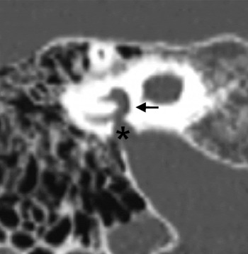
High resolution computed tomography revealed a high-riding jugular bulb impinging upon the right posterior semicircular canal, with a bony defect (Figure 1).
Coronal, high resolution computed tomography image showing the diverticulum of the high-riding jugular bulb (asterisk) impinging upon the right posterior semicircular canal (arrow).

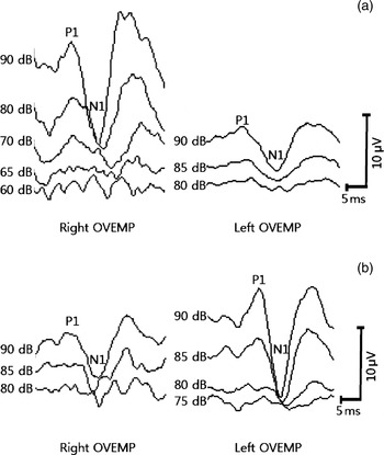
Ocular vestibular evoked myogenic potential testing, using a 500-Hz tone burst sound, demonstrated thresholds of 65 and 80 dBnHL in the right and left ears, respectively (Figure 2a). Cervical vestibular evoked myogenic potential testing demonstrated a threshold of 70 dBnHL in both ears. Application of a 500 Hz, 110 dB, pure tone stimulus to the patient's right ear with an insert earphone resulted in a sudden, brief movement of the trunk backward and to the right, along the plane of the right posterior semicircular canal. This brief postural sway movement was not observed with any pure tone stimulus below 110 dB in the right ear, or with any stimulus up to 120 dB in the left ear.
(a) Pre-operative ocular vestibular evoked myogenic potential (OVEMP), showing reduced thresholds in the right ear. (b) Post-operative OVEMP showing increased thresholds in the right ear.

No evidence of nystagmus was observed using Frenzel glasses during sound stimulus testing, or when pressure was applied through the right external ear canal using a Politzer bag. However, counter-clockwise (from the patient's perspective), down-beating nystagmus developed approximately 1 second after releasing positive pressure from the external ear canal, and continued for 4 to 5 seconds, suggesting inhibition of the right posterior semicircular canal. This negative pressure induced nystagmus was not observed in the left ear.
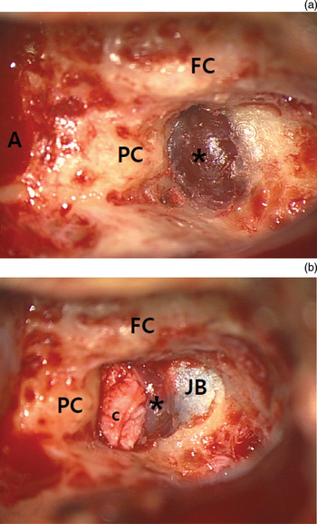
Due to continued daily discomfort, the patient elected to undergo surgical intervention. His right posterior semicircular canal was approached through the mastoid bone. The high-riding jugular bulb and the diverticulum adjacent to the inferior border of the right posterior semicircular canal were exposed (Figure 3a). After thinning, the bone covering the diverticulum of the jugular bulb was carefully removed with elevators, and the diverticulum was separated from the posterior semicircular canal inferiorly. A bony defect, about 2 mm in length, was found at the caudal aspect of the posterior semicircular canal just posterior to the ampulla; the inner endosteum could be observed through the defect. The dehiscent site was tightly covered with bone paté and reinforced with temporalis muscle fascia and conchal cartilage (Figure 3b). Fibrin glue was applied to secure the covering materials.
Intra-operative photographs showing findings during right transmastoid posterior semicircular canal dehiscence resurfacing. (a) The diverticulum (asterisk) of the right jugular bulb impinged upon the right posterior semicircular canal, and was detected after removal of the bony shell of the diverticulum. (b) The posterior semicircular canal dehiscence was obliterated by bone paté and temporalis muscle fascia covered by conchal cartilage. FC = facial canal; A = mastoid antrum; PC = posterior semicircular canal; c = conchal cartilage; JB = jugular bulb

The patient's disequilibrium improved gradually over the first two weeks after surgery, and no vestibular symptoms persisted.
At three-month follow up, the patient reported that he no longer experienced vertigo due to loud sounds or changes of position. Right-sided, intermittent, pulsatile tinnitus could not be elicited by either the Valsalva manoeuvre or a head-down position, and the previously observed abrupt body sway and vertigo could not be induced by a 500 Hz pure tone stimulus up to 120 dB bilaterally. The patient's ocular vestibular evoked myogenic potential thresholds were 85 and 75 dBnHL in his right and left ears, respectively, indicating the absence of a pathological third window (Figure 2b). Cervical vestibular evoked myogenic potential testing showed no post-operative threshold change in either ear.
Discussion
Although several case reports on posterior semicircular canal dehiscence syndrome have been published, including a series of 10 patients with non-iatrogenic posterior semicircular canal dehiscence, to date there have only been two reported cases of surgical management of posterior semicircular canal dehiscence.Reference Brantberg, Bagger-Sjoback, Mathiesen, Witt and Pansell2, Reference Gopen, Zhou, Poe, Kenna and Jones3, Reference Mikulec and Poe7
In one of these patients, dehiscence was caused by a high-riding jugular bulb.Reference Mikulec and Poe7 The natural posterior semicircular canal was plugged via a transmastoid approach, by opening the lateral surface, without attempting to directly identify the defect site by the jugular bulb.Reference Mikulec and Poe7 Although this patient showed partial improvement of symptoms and pure tone audiometry air–bone gap, the persistently reduced vestibular evoked myogenic potential thresholds and substantial remaining symptoms suggested that the defect site had been incompletely occluded.
In the second patient, posterior semicircular canal dehiscence was caused by a petrous apex cholesteatoma.Reference Brantberg, Bagger-Sjoback, Mathiesen, Witt and Pansell2 It was treated by packing free abdominal fat onto the defect site. However, the patient continued to experience vestibular symptoms and objective signs, indicating a persistent fistula.
In contrast, we were able to visually localise the defect site in our patient by separating the diverticulum of the jugular bulb from the dehiscent site. Since plugging of the semicircular canal may have changed our patient's auditory or vestibular function, due to the close proximity of the defect to the ampulla, we chose to resurface the defect site.Reference Carey, Migliaccio and Minor8, Reference Parnes9 Following surgery, our patient presented no further manifestations of posterior semicircular canal dehiscence, and his bilateral, moderate to severe, sensorineural hearing loss was not aggravated. To our knowledge, this is the first reported case of the direct and successful surgical management of a patient with posterior semicircular canal dehiscence, using a resurfacing manoeuvre.
The key features of semicircular canal dehiscence are vertigo induced by loud sounds (Tullio's phenomenon) and by pressure (Hennebert's sign).Reference Minor, Solomon, Zinreich and Zee1, Reference Minor6 Auditory presentations of posterior semicircular canal dehiscence include conductive or mixed hearing loss, ear fullness, and autophony, together with pulsatile tinnitus and auditory and vestibular features similar to those seen in superior semicircular canal dehiscence syndrome.Reference Gopen, Zhou, Poe, Kenna and Jones3 The manifestations of semicircular canal dehiscence may vary, and all features may not be present.Reference Minor, Solomon, Zinreich and Zee1, Reference Minor6 In symptomatic patients, the diagnosis of posterior semicircular canal dehiscence can be supported by a lowered vestibular evoked myogenic potential threshold and computed tomography findings.Reference Minor, Solomon, Zinreich and Zee1
Our patient showed fluctuations in vertigo and pulsatile tinnitus following head position changes and the Valsalva manoeuvre. His conductive hearing loss component was limited to 250 Hz (other low frequency regions were not affected). The positional and pressure-induced vertigo and the lowered ocular vestibular evoked myogenic potential threshold are considered to be due to defects in his bony canal.
Patients with canal dehiscence may show various sound- and/or pressure-induced signs, including head tilt, eye movement and postural sway.Reference Minor, Solomon, Zinreich and Zee1 Our patient showed several features which differed from those shown by patients with superior semicircular canal dehiscence syndrome, including eye movement induced by negative pressure in the right ear, compatible with inhibition of the right posterior semicircular canal. Negative pressure can result in ampullopetal flow brought about by bulging of the jugular diverticulum into the posterior canal space.
In addition, our patient displayed a sound-induced body sway in the plane of the right posterior canal. Body sway induced in patients with superior semicircular canal dehiscence has been reported to be in the plane of the involved superior semicircular canal.Reference Minor6
Another feature of our patient was his lowered pre-operative ocular vestibular evoked myogenic potential threshold, which returned to the level of the contralateral, healthy side post-operatively. However, his cervical vestibular evoked myogenic potential threshold was not lowered pre-operatively and did not change post-operatively. In contrast, most patients with superior semicircular canal dehiscence show lowered thresholds during both cervical and ocular vestibular evoked myogenic potential testing.Reference Welgampola, Migliaccio, Myrie, Minor and Carey10
• Posterior semicircular canal dehiscence due to jugular diverticulum is rare
• The condition creates a pathological ‘third window’ of the inner ear
• Surgical treatment of posterior semicircular canal dehiscence is rarely reported
• In this patient, the defect was successfully resurfaced under direct visualisation
The most frequent cause of posterior semicircular canal dehiscence is a high-riding jugular bulb, rising to the level of the basal turn of the cochlear or the inferior tympanic annulus.Reference Gopen, Zhou, Poe, Kenna and Jones3 A recent study showed that 6 of 11 patients with inner ear erosion caused by a jugular bulb had a diverticulum causing posterior semicircular canal dehiscence or vestibular aqueduct dehiscence. The pathophysiology of jugular bulb diverticulum formation is unclear, although the anomaly may be caused by venous hypertension and turbulent flow.Reference Otto, Hudgins, Abdelkafy and Mattox11 Other possible causes of posterior semicircular canal dehiscence include impingement of the posterior cranial fossa, cholesteatoma and fibrous dysplasia.Reference Brantberg, Bagger-Sjoback, Mathiesen, Witt and Pansell2–Reference McCall, Curtin and McKenna4, Reference Mikulec and Poe7
Conclusion
Posterior semicircular canal dehiscence syndrome caused by a diverticulum of the high jugular bulb is a rare condition. In the presented patient, the defect site was successfully covered by resurfacing the bony dehiscence with bone paté and reinforcing it with temporalis muscle fascia and conchal cartilage, under direct visualisation. Posterior semicircular canal dehiscence and superior semicircular canal dehiscence syndromes have different clinical symptoms and signs, which close examination of the patient may distinguish.