Case report
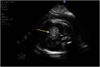
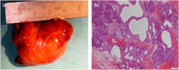
An intrapericardial vacuolated mass compressing and displacing the heart was diagnosed by echocardiography in a foetus of 22 weeks gestation. An intracardiac tumour was diagnosed by means of magnetic resonance imaging and foetal echography during foetal life (Fig 1). The mass was located laterally to the right atrium; it compressed the superior vena cava, leading to early signs of superior vena cava obstruction. Atrial outflows were free. Significant pericardial effusion without signs of cardiac tamponade was also present. Foetal echocardiography was carried out every other day. At 29 weeks of gestation, foetal lung maturity was stimulated by means of betamethasone and delivery was induced as a consequence of early signs of foetal distress and hydrops; a 1.55 kg male newborn was delivered by cesarean section. He underwent cardiopulmonary resuscitation as a result of acute cardiac failure and respiratory insufficiency that eventually required mechanical ventilation. Chest X-ray showed a left dislocation of an enlarged mediastinum. Echocardiography showed a vacuolated mass lying on the great vessels, partially compressing both the right atrium and superior vena cava. The heart was surrounded by a huge pericardial effusion; a patent ductus arteriosus was also present (Fig 1). Pericardial tapping was immediately performed and 20 ml of clear fluid was removed. The patient was eventually transferred to the neonatal intensive care unit as a consequence of worsened haemodynamic condition probably due to recurrent pericardial effusion. A pericardial drain was surgically inserted and a further 25 ml of blood-stained fluid was evacuated. After an initial improvement of clinical conditionsm progressive oedema of the upper body associated with systemic hypotension occurred. Therefore, surgical removal of the mass was planned. Surgery was performed by midline sternotomy: small thymus and intact pericardium were detected. At pericardial opening, a huge mass occupying the upper mediastinum and displacing the heart downwards and to the left was detected. The tumour was primarily connected to the aorta, the pulmonary trunk, the right pulmonary artery, and the right coronary artery, compressing the superior vena cava and displacing the right atrium posteriorly. The stalk of the mass arose from the ascending aorta just above the origin of the right coronary artery. During stalk dissection, the aorta was damaged and promptly repaired without complication. The tumour was resected en bloc and a 4.5 cm specimen was sent for histology (Fig 2a and b). Histology showed the features of a mixed germ cell tumour grade II according to the Gonzales-Crussi classification with 20% of immature component represented by neuroectodermal tissue and 30% of epithelial structures reminiscent of the lung and endometrial/intestinal tissues.Reference Gonzales-Crussi, Winkler and Mirkin 1 The superior caval syndrome progressively disappeared and the child experienced an uneventful post-operative course. Progressive weaning from the ventilator was performed in the neonatal intensive care unit and definitive extubation was achieved on the sixteenth post-operative day. The alpha-fetoprotein level progressively decreased from 70,885 ng/ml on the second post-operative day to 7053 ng/ml at hospital discharge.Reference Gonzales-Crussi, Winkler and Mirkin 1 Echocardiogram at discharge showed normal heart function. After 1 month, the child showed good clinical condition, regular growth (3.120 kg), and further reduction of alpha-fetoprotein (1743 ng/ml). At the age of 8 months, the child developed seizures as a result of West syndrome, which were controlled by adrenocorticotropic hormone administration.
Prenatal 2D-Echo: the arrow indicates the intrapericardial mass.

( a ) Tumour excised (length 4.5 cm). ( b ) Area of a well-differentiated teratoma consisting of cystic formations lined by monolayered columnar epithelium and lobular aggregations of exocrine glandular acini and ducts. Haematoxilin–eosin stain.

Discussion
Owing to the rarity of mixed germ cell tumours, their relative frequency is difficult to determine. It has been estimated that extragonadal germ cell tumours account for ~1–15% of the mediastinal tumour in adults and 25% in children.Reference Mullen and Richardson 2 Germ cell tumours of the mediastinum are classified in teratomatous and non-teratomatous lesions. Teratomas with malignant components are further subcategorised on the basis of the malignant elements present, sarcomatous, epithelial, or other germ cell malignant component, or a mixture of these types. The lesions are clinically classified in three stages (I, II, III) according to the involvement of the adjacent structures.Reference Moran and Suster 3
Diagnosis is mainly based on echocardiography; typical diagnostic ultrasonic findings are the multicystic nature, associated pericardial effusion due to obstruction of cardiac and pericardial lymphatics, and occasionally characterised by a rupture of cystic areas.Reference Rasmussen, Hwang, Harder, Nicholson, Davies and Nimrod 4 – Reference Reynolds, Donahue and Pearce 5 Magnetic resonance imaging is particularly helpful to define the relationship of the tumour with the surrounded structure, especially after delivery and during the follow-up period.
Prognostic factors are essentially represented by tumour stage and completeness of surgical resection.Reference Schneider, Calaminus and Reinhard 6 Other factors are anatomic site of the tumour, existence of metastasis, elevated β-HCG levels, and the amount of the immature tumour tissue.Reference Fritzsche, Kristiansen and Frauenfelder 7 The optimal approach to treatment is still controversial. The surgical excision is probably the most effective treatment for mixed germ cells tumours. Chemotherapy is sometimes added to surgery in case of additional malignant components, incomplete surgical resection, and in case of recurrent disease.Reference Marina, Cushing and Giller 8 Recent literature suggests that the treatment of mature teratoma is surgical resection and there is no role for chemotherapy unless elevated tumour markers are present.Reference Albany and Einhorn 9 The tumour we described was a grade II according to the Gonzales-Crussi classification with a well-differentiated yolk sac tumour component and a steady decrease in post-operative alpha-fetoprotein levels. Moreover, no adhesion to neighbouring tissues was evident at the time of surgery. Chemotherapy was therefore avoided. Further studies are needed to better describe symptoms associated with perinatal intrapericardial tumours and their appropriate clinical management.
Acknowledgements
A special thanks to Renata Boldrini, MD, for her fundamental contribution.