INTRODUCTION
The benthic fauna of Port Phillip Bay has been well studied including bay-wide surveys conducted as part of the Port Phillip Bay environmental studies (Poore et al., Reference Poore, Rainer, Spies and Ward1975; Wilson et al., Reference Wilson, Heislers and Poore1998). A thorough report and paper on marine biological invasions of Port Phillip Bay (Hewitt et al., Reference Hewitt, Campbell, Thresher and Martin1999; Reference Hewitt, Campbell, Thresher, Martin, Boyd, Cohen, Currie, Gomon, Keough, Lewis, Lockett, Mays, McArthur, O'Hara, Poore, Ross, Storey, Watson and Wilson2004) described the physical environment, species introductions and the history of shipping and trade. One chapter of the report listed introduced species of soft sediment crustaceans and discussed the potential for translocation of the major groups of taxa (Poore & Storey, Reference Poore and Storey1999). Collections from these surveys are housed in Museum Victoria and include three species of crangonid shrimp, Philocheras intermedius (Bate, Reference Bate1863), P. obliquus (Fulton & Grant, Reference Fulton and Grant1902), and P. victoriensis (Fulton & Grant, Reference Fulton and Grant1902). A fourth species P. flindersi (Fulton & Grant, Reference Fulton and Grant1902) is known from the geographically close Western Port and central and western Bass Strait.
Three specimens of an unfamiliar sand shrimp species were collected from the muddy intertidal zone of Newport, near the mouth of the Yarra River into Port Phillip Bay, in May and June 2008 by the Marine Research Group and brought to the attention of Museum Victoria. Newport is close to Melbourne's major international shipping port at the mouth of the Yarra River. The region is also close to the gas-fired Newport Power Station which discharges warmed cooling water through a channel within 400 m of the sand flats where these shrimps were taken. The cooling water channel is known as the ‘warmies’ by local fishers. The shrimps were distinct from other species of Crangonidae previously reported from Port Phillip Bay and after comparison with specimens lodged in the Natural History Museum and Institute, Chiba, Japan, have been determined as Crangon uritai Hayashi & Kim, Reference Hayashi and Kim1999.
The genus Crangon Fabricius, 1798, represented by 19 species (De Grave et al., Reference De Grave, Pentcheff, Ahyong, Chan, Crandall, Dworschak, Felder, Feldmann, Fransen, Goulding, Lemaitre, Low, Martin, Ng, Schweitzer, Tan, Tshudy and Wetzer2009), has not previously been recorded from the southern hemisphere and the known distribution of Crangon uritai is restricted to coastal and inshore waters of East Asia. Species of the genus Crangon commonly occur in littoral and sublittoral areas of cold and temperate regions of the northern hemisphere. Hayashi & Kim (Reference Hayashi and Kim1999) published a review of the East Asian species and believed them to be an abundant and important component of the coastal soft bottom communities as many Crangon species are described as prey or predators of flat fish (Seikai et al., Reference Seikai, Kinoshita and Tanaka1993; Minami, Reference Minami, Senta and Kinoshita1998; Mori, Reference Mori, Senta and Kinoshita1998).
Caridean shrimps have pelagic larval stages so there is the potential for them to be taken in with ships' ballast water and transported. Although Poore & Storey (Reference Poore and Storey1999) did not find any introduced caridean species in Port Phillip Bay at that time, they did suggest the possibility of this occurring citing that a species of Palaemonidae known from many coasts of the North Pacific, Palaemon macrodactylus Rathbun, 1902, has been reported from New South Wales (Pollard & Hutchings, Reference Pollard and Hutchings1990; Poore, Reference Poore2004).
MATERIALS AND METHODS
The examined material remains deposited in Museum Victoria (NMV) and the Natural History Museum and Institute, Chiba (CBM). The measurement provided is of the postorbital carapace length (cl) measured from the level of the posterior margin of the orbit to the midpoint of the posterodorsal margin of the carapace.
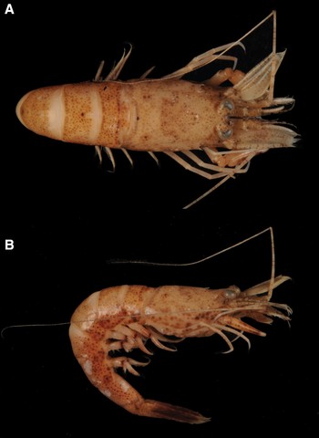
Crangon uritai Hayashi & Kim, Reference Hayashi and Kim1999, NMV J58308. (A) Dorsal view; (B) lateral view, taken of preserved specimen. Photographs: D. Staples, Museum Victoria.

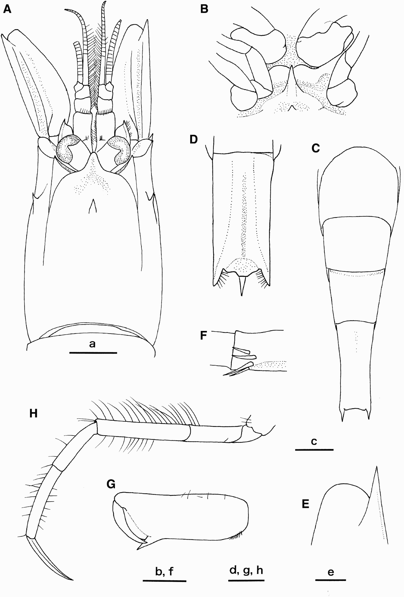
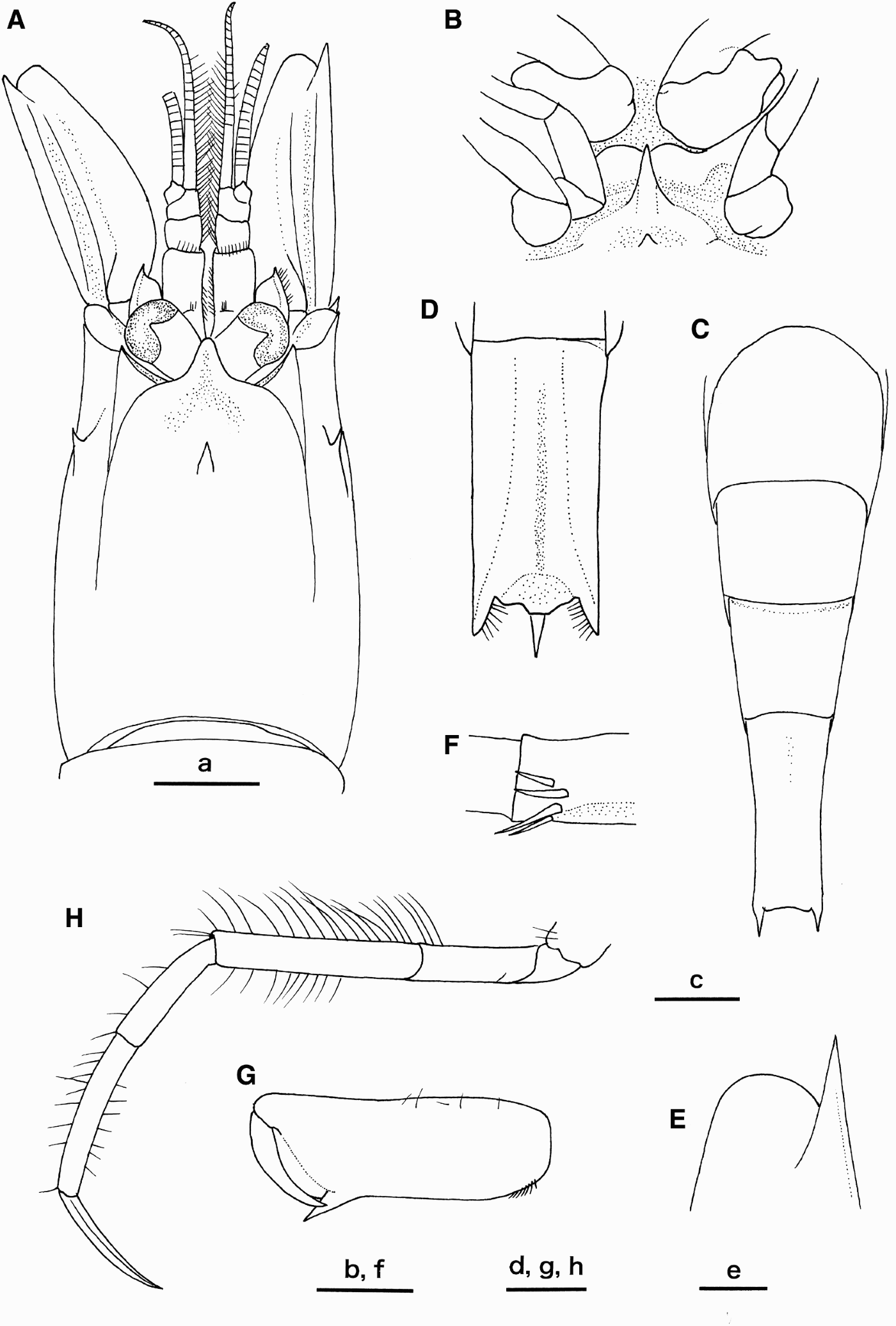
Crangon uritai Hayashi & Kim, Reference Hayashi and Kim1999, female (carapace length 6.3 mm), NMV J58308. (A) Carapace and cephalic appendages, dorsal view (setae partially omitted); (B) thoracic sternites 4 and 5, and coxae of pereopods 1–3, ventral view (setae omitted; left second pereopod detached); (C) abdominal somites 3–6, dorsal view; (D) abdominal somite 6, ventral view; (E) distal part of right antennal scale, dorsal view; (F) distal part of ventral surface of antepenultimate segment of maxilliped 3; (G) subchela of left pereopod 1, ventral view; (H) left pereopod 4, lateral view. Scale bars: 2 mm for a, c; 1 mm for b–d, g, h; 0.5 mm for e, f.
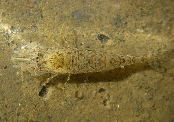
Crangon uritai Hayashi & Kim, Reference Hayashi and Kim1999, NMV J58308, image of live animal taken in situ. Photograph: John Eichler.

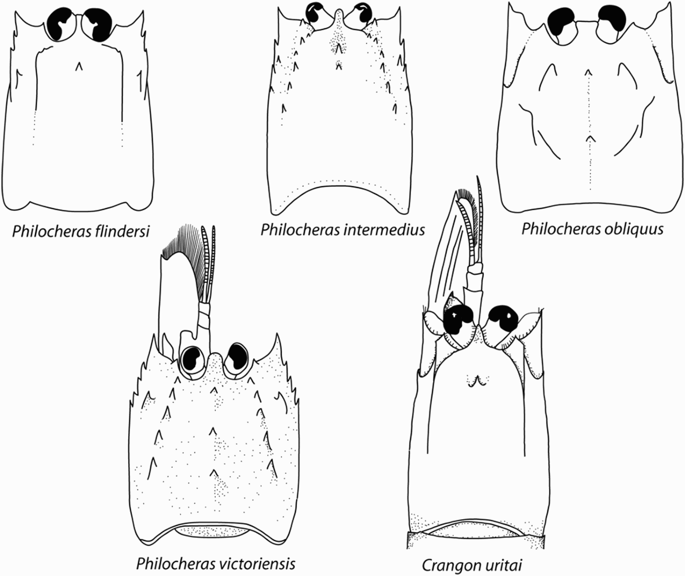
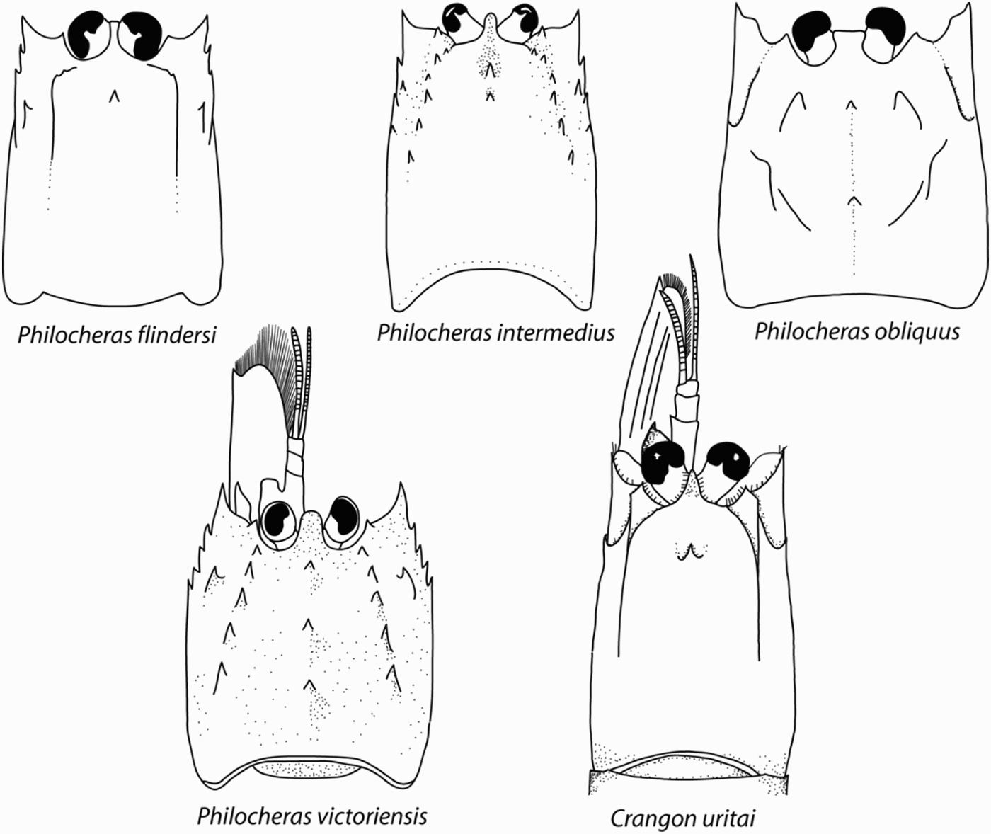
Carapace (dorsal view) of species of Crangonidae reported from Port Phillip Bay and nearby waters. Philocheras flindersi redrawn from Poore (Reference Poore2004: figure 36g); P. intermedius redrawn from Poore (Reference Poore2004: figure 36h); P. obliquus redrawn from Poore (Reference Poore2004: figure 36i); P. victoriensis redrawn from Poore (Reference Poore2004: figure 36c); Crangon uritai redrawn from Hayashi & Kim (Reference Hayashi and Kim1999: figure 13).
Crago sp. Urita, Reference Urita1926: 429.
Crangon crangon. Parisi, Reference Parisi1919: 90; Liu, Reference Liu1955: 58, pl. 21, figures 1–10. Not Crangon crangon Linnaeus, 1758.
Crangon affinis. Kubo, Reference Kubo, Okada, Uchida and Uchida1965: 622, figure 60, text figures 127 & 128 (in part); Miyake, Reference Miyake1982: 71, pl. 24, figure 4. Not Crangon affinis De Haan, 1849.
?Crango cassiope. Yasuda, Reference Yasuda1956: 8, 65, unnumbered figure, figure 29; Maekawa, Reference Maekawa1961: 172.
Crangon uritai. Hayashi & Kim, Reference Hayashi and Kim1999: 86, figures 13–26; Cha et al., Reference Cha, Lee, Park, Baik, Hong, Park, Lee, Choi, Hwang, Kim, Choi, Sohn, Sohn, Kim and Choi2001: 148–149, unnumbered figure.
MATERIAL EXAMINED
Australia: Victoria, Greenwich Bay, Newport (37°51′S 144°53′E), collected using hand sieve in muddy intertidal zone by B. Hall, 8 May 2008, NMV J46548 (1 female, cl 3.8 mm); same locality, collected by hand and sieved from among filamentous Chlorophyta, and Heterozostera nigricaulis Kuo, Reference Kuo2005 by V. Stajsic and J Eichlert, 8 June 2008, NMV J58308 (2 females, cl 6.1, 6.3 mm).
Japan: Iwate Prefecture, Ohtsuchi Bay, Shirahama, 2 m, sand bottom, sledge, coll. I. Takeuchi, 15 May 1995, CBM-ZC 5242 (1 female, cl 8.2 mm; 1 ovigerous female, cl 9.3 mm); Chiba Prefecture, Boso Peninsula, Katsuura, Yoshio, subtidal, sand bottom, beach seine, coll. E. Nishi, 29 July 1995, CBM-ZC 5246 (7 males, cl 4.5–6.3 mm; 10 ovigerous females, cl 7.0–10.8 mm).
COMPARATIVE MATERIAL EXAMINED
Crangon crangon (Linnaeus, 1758). England: Sussex, Orwell estuary, 51°59.043′N 01°16.004′E, subtidal, coll. S. De Grave and C. Asheley, CBM-ZC 9559 (8 males, cl 7.0–8.0 mm; 2 females, cl 9.0, 10.1 mm; 2 ovigerous females, cl 8.5, 11.0 mm).
TYPE LOCALITY
Dadaepo, Pusan, Korea, 1 m.
ABBREVIATED DESCRIPTION OF AUSTRALIAN SPECIMENS
Rostrum (Figure 2A) only reaching to mid-length of eyes, about 0.15 of carapace length, narrowing distally to rounded apex; carapace (Figure 2A) with median gastric spine arising from anterior about 0.20 of carapace length; hepatic spine arising slightly anterior to median gastric spine; hepatic groove distinct; antennal spine slightly falling short of or reaching rostral distal margin; branchiostegal spine reaching to level of distal corneal margin; orbital margin with distinct cleft; longitudinal suture extending from base of antennal spine to midlength of carapace.
Thoracic sternum is nearly flat, not depressed below; sternite 5 (Figure 2B) with short median spine only reaching anterior margin of transverse keel on sternite 4; sternite 6 with small median tubercle.
Abdominal somites 3–5 (Figure 2C) without median carina; abdominal somite 6 with shallow median groove ventrally (Figure 2D), without even trace of submedian carinae or median groove dorsally; pleura unarmed. Telson without median groove dorsally.
Antennular peduncle (Figure 2A) reaching mid-length of antennal scale; stylocerite abruptly tapering distolaterally in small acute spine. Antennal scale (Figure 2A) 0.7–0.8 of carapace length, about 2.6 times longer than wide; lateral margin nearly straight; distolateral spine distinctly overreaching rounded distal lamella (Figure 2E).
Maxilliped 3 reaching or slightly overreaching distal margin of lamella of antennal scale, bearing subdistal clump of 4 spines on ventral surface of antepenultimate segment (Figure 2F).
Pereopod 1 with palm about 3.0 times longer than wide; cutting edge moderately oblique (Figure 2G); merus with ventral spine arising at mid-length of ventral margin. Pereopod 2 chelate, subequal in length to pereopods 3–5, fully flexed at articulation between merus and carpus. Pereopod 3 slender, overeaching pereopod 1. Pereopod 4 (Figure 2H) with dactylus 0.8–0.9 times as long as propodus. Pereopod 5 similar to pereopod 4.
Colour in life: body light sandy-brown with darker brown speckled spots on dorsal surface of carapace, abdomen, chela and walking legs. Spots on pleura are darker and denser.
DISTRIBUTION
Natural geographical range includes the Yellow Sea, northern part of the East China Sea, central and southern part of the Sea of Japan, and Japanese mainland (southern Hokkaido to Kyushu); intertidal to 24 m (Hayashi & Kim, Reference Hayashi and Kim1999). The present discovery from Port Phillip Bay, Australia, is considered to be an artificial introduction from East Asian waters.
REMARKS
As noted by Hayashi & Kim (Reference Hayashi and Kim1999), Crangon uritai is morphologically very similar to C. crangon. Crangon septemspinosa Say, Reference Say, Cramer and Swann1818, known with certainty from the northern part of the Gulf of St Lawrence to east Florida, is also similar to these two species. Shared characters among the three species include: all abdominal somites are rounded dorsally, without trace of a mid-dorsal carina on the third to fifth somites or of submedian carinae or a mid-dorsal groove on the sixth somite; the sixth somite has a shallow median groove on the ventral surface. Crangon uritai can be distinguished from C. crangon and C. septemspinosa by the weak armature of the thoracic sternite 5. In non-spawning female of C. uritai, the median spine on the fifth thoracic sternite is relatively short, only reaching the anterior margin of the transverse keel on the fourth sternite; this sternal spine is reduced to a minute tubercle in spawning females. In C. crangon the median spine on the sternite 5 distinctly overreaches the transverse keel on the fourth sternite even in spawning females (this study). Squires (Reference Squires1990) noted that this sternite has a strong spine in C. septemspinosa, although details remain unclear. Subdistal spines on the ventral surface of the antepenultimate segment of maxilliped 3 are four or five in C. uritai, but two or three in C. crangon. Furthermore, C. septemspinosa appears different from C. uritai and C. crangon in the subtruncate, rather than rounded, distal lamella of the antennal scale (Squires, Reference Squires1965) and the much shorter maxilliped 3 that far falls short of the distal margin of the antennal scale (Williams, Reference Williams1965, Reference Williams1984; Squires, Reference Squires1990), rather than slightly falling short of or reaching beyond it in C. uritai and C. crangon.
The present three Australian specimens closely agree with Crangon uritai in the diagnostic features mentioned above, and are identified with the species with little hesitation.
Crangon uritai can be distinguished from other crangonid species known from intertidal sediments of Port Phillip Bay by traits that define the genus Crangon, such as pereopod 2 being chelate and subequal in length to other pereopods, a single arthrobranch on maxilliped 3 and one ventral spine on the merus of pereopod 1. It can further be distinguished from species of Philocheras by the armature of the dorsal carapace surface and rostrum shape (cf. Figure 4).
KEY TO SPECIES OF CRANGONIDAE OF PORT PHILLIP BAY AND WESTERN PORT, VICTORIA
1. Pereopods 2 subequal in length to other pereopods … … … Crangon uritai Hayashi & Kim, Reference Hayashi and Kim1999 [Port Phillip Bay]
— Pereopods 2 much shorter than other pereopods … … … … … … … … … … … …Philocheras 2
2. Carapace with 3 mid-dorsal teeth including epigastric tooth … … … … … … … … … … … … … P. victoriensis (Fulton & Grant, Reference Fulton and Grant1902) [Port Phillip Bay and Western Port]
— Carapace with 1 or 2 mid-dorsal teeth including epigastric tooth … … … 3
3. Carapace with 1 mid-dorsal tooth (epigastric tooth) … … … P. flindersi (Fulton & Grant, Reference Fulton and Grant1902) [Western Port]
— Carapace with 2 mid-dorsal teeth including epigastric tooth … … … 4
4. Carapace with longitudinal row of small teeth posterior to orbit … … P. intermedius (Bate, Reference Bate1863) [Port Phillip Bay]
— Carapace without longitudinal row of small teeth posterior to orbit … … … … … … … … … … … P. obliquus (Fulton & Grant, Reference Fulton and Grant1902) [Port Phillip Bay]
ACKNOWLEDGEMENTS
Thanks to the members of the Marine Research Group whose combined efforts in collecting, recognizing the species as ‘different’ and lodging material in Museum Victoria has instigated this report. Specimens were collected by Barbara Hall, Val Stajsic and John Eichler. Clarrie Handreck lodged the material at Museum Victoria alerting staff to his suspicion that it ‘wasn't one he'd seen before’. Collection management support was provided by Chris Rowley. David Staples kindly photographed the preserved specimens. The colour image was taken by John Eichler and reproduced with his permission. Special thanks to Gary Poore for his constructive comments after reading the manuscript draft. This paper is dedicated to the memory of Clarrie Handreck who was a wonderful educator and crusader of the importance of understanding the marine invertebrate fauna of Victoria.