Transcatheter closure of patent ductus arteriosus in extremely low-birth-weight infants using the Amplatzer Piccolo Occluder (APO; Abbott Structural Heart, Plymouth, MN, USA) has become widely accepted as a safe and efficient alternative to surgical ligation in infants of ≥ 700 grams, with a > 97% success rate and a low incidence of periprocedural complications. Reference Sathanandam, Gutfinger and O’Brien1–Reference Regan, Benbrik and Sharma3 Aortic obstruction due to device protrusion has been reported in 1.2% of cases. Reference Sathanandam, Gutfinger and Morray4 Late occurrence of device-induced aortic obstruction is exceptional.
Case description
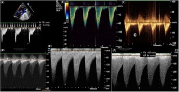
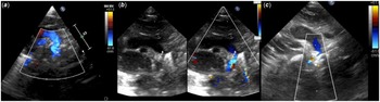
A 27-weeker premature infant was intubated at day 1 because of infant respiratory distress syndrome and remained ventilator-dependent. Echocardiography diagnosed a haemodynamically significant patent ductus arteriosus. After two unsuccessful courses of paracetamol (15mg/kg/day, 3 days), his clinical condition continued to deteriorate with necrotising enterocolitis and escalation in respiratory support with high-frequency oscillation ventilation. Transcatheter patent ductus arteriosus closure was performed at day 21 (procedural weight: 980 grams) under high-frequency oscillation. Last-minute echocardiography measured an 8.7-mm ductal length and a 2.6-mm minimal ductal diameter, leading to implant a 4/2 APO device through a 4-French femoral vein, under ultrasound guidance, with its aortic tip anterior to the orogastric tube on fluoroscopy. After device release, echocardiography checked co-axial, intraductal device positioning, with no peri-device residual shunt and peak velocities < 1.2 m/s in both left pulmonary artery and descending aorta. The baby was transferred back to the neonatal ICU Initial follow-up was uneventful with unchanged weekly ultrasound controls and de-escalation of respiratory support with extubation at day 7. From post-procedural week 4, echocardiography showed device protrusion to descending aorta, with descending aorta flow disturbances on colour Doppler imaging an increased Doppler velocity at 3.0 m/s and a discreet diastolic tail. At post-procedural week 8, aortic obstruction was more pronounced and looked solely related to late device protrusion, as no posterior shelf may evoke an associated native aortic coarctation. Close monitoring showed progressive aggravation of aortic obstruction (Fig 1, Supplementary materials S1), whilst the device looked as being pushed out of aortic end (Fig 2). At 4 months, the baby had gradually developed a symptomatic, acquired aortic coarctation. He underwent surgical coarctation repair through posterolateral thoracotomy, with end-to-end anastomosis without cardiopulmonary bypass. The aortic disk of the APO device was removed, and the aortic end of the ductus disconnected, with external ligation of the device into the duct in order to keep it firmly attached (Supplementary materials S2). After 5 years of follow-up, the child is alive and asymptomatic and required no re-intervention.
Doppler flow velocities in descending aorta demonstrating normal velocities at post-procedural day 1 ( a ), week 1 ( b ), and week 3 ( c ) with gradual increase from week 4 ( d ) and week 8 ( e ), towards severe aortic obstruction at week 16 ( f ).

Two-dimensional colour Doppler imaging showing an intraductally well-positioned device at post-procedural day 1 ( a ) with device protrusion at week 8 ( b ) and the device being pushed out of aortic end at week 16 ( c ).

Discussion
Device protrusion is a known complication following transcatheter patent ductus arteriosus closure, with a greatest potential for vascular obstruction in smaller infants. In extremely low-birth-weight infants, mild increase in descending aorta flow velocities in the early post-procedural period tends to improve spontaneously. Reference Markush, Tsing and Gupta5 Clinically significant device-induced aortic obstruction is rarer; its reported incidence was 1% (1/100) in the US multicenter premarket clinical trial (procedural weight: 1250 [700–2000] grams) and 1% (1/102) in the French multicenter study (procedural weight: 1543 ± 648 grams). Reference Sathanandam, Gutfinger and O’Brien1,Reference Malekzadeh-Milani, Akhavi and Douchin2 The time course for the development of post-procedure vessel obstruction is usually within 24 hours. Potential mechanisms for device protrusion have been proposed, most of which are related to technical issues or inadequate expertise leading to inappropriate device selection, inadequate imaging, manipulations errors, or device malposition. Reference Sathanandam, Gutfinger and Morray4 Device deformation has also been suggested, based on a small case series of 14 preterm infants reporting incredibly high immediate rates of aortic and severe left pulmonary artery obstruction (21.4% and 35.7%, respectively), Reference Chien, Wang and Lin6 but their device selection process was inappropriate as 12/14 infants received an oversized device. Reference Baruteau and Fraisse7 In another series, a 660-gram infant developed both left pulmonary artery and aortic obstructions following an oversized device implantation (ductal diameter/length: 2.9 mm/5.3 mm, device: 5/4 APO). Reference Wand, Lin, Hsieh, Wei, Ju and Wu8 Minocha et al. reported the aortic migration of a 5/2 APO device in a 2.4-kg, ex 23-weeker premature infant. Reference Minocha, Sutton and Crawford9 Not only the device was oversized (ductal diameter/length: 2.5 mm/8.1 mm) but also operator-related factors may be incriminated as device part of the aortic disc protruded into the aorta immediately after release, whilst it had been checked completely intraductally before release.
Delayed occurrence with late diagnosis of device protrusion is exceptional and may not be directly explained by operating factors. In our case, the device was (a) properly selected (ductal diameter/length: 3 mm/8 mm, device: 4/2 APO) and (b) properly positioned as it was placed intraductally with normal peak velocities in descending aorta including until the third week after device implantation. High-frequency oscillation ventilation should not compromise device placement. Reference Shibbani, Nijres and McLennan10
Hand injection of a small amount of contrast may have offered angiographic landmarks to accurately appreciate whether an anterior segment of the patent ductus arteriosus would have been let uncovered by the device at the pulmonic end. Reference Sathanandam, Gutfinger and Morray4 However, transcatheter patent ductus arteriosus closure in extremely low-birth-weight infants using no contrast injection has been reported by many groups including us, with similar success and complication rates as compared to those who use it. Reference Malekzadeh-Milani, Akhavi and Douchin2,Reference Regan, Benbrik and Sharma3 By the elimination of potential mechanisms, our understanding is that we may have let uncovered a segment of ductal tissue on the pulmonic end that could have led to post-procedure ductal vasoconstriction on pulmonic end, causing device to be pushed out of aortic end. This is in keeping with delayed gradual progression to aortic obstruction. To the best of our knowledge, this is the first report demonstrating that mechanism by serial ultrasound monitoring. This should encourage interventionists to let no uncovered segment at the pulmonic end when placing the APO device intraductally in < 2 kg infants, although it may increase the risk of device-related left pulmonary artery obstruction, which has to be carefully ruled out by echocardiography before device release. In our case, the device has been surgically removed, according to the management algorithm for device protrusion published in the consensus guidelines. Reference Sathanandam, Gutfinger and Morray4
Conclusion
Ductal vasoconstriction at the pulmonic end may lead to delayed device-induced, clinically significant aortic obstruction after transcatheter patent ductus arteriosus closure in extremely low-birth-weight infants. Being aware of that mechanism, clinicians should (a) strive to position the APO device intraductally with no uncovered patent ductus arteriosus segment at the pulmonic end in < 2 kg infants and (b) closely monitor the descending aorta velocities during at least the end of the first month after device implantation.
Supplementary material
To view supplementary material for this article, please visit https://doi.org/10.1017/S1047951123000938.
Acknowledgement
We gratefully acknowledge Pr Cyril Flamant, Pr Jean-Christophe Rozé, Dr Céline Grunenwald Gronier, Dr Mohamedou Ly, and the neonatal team for their help and support in developing the programme of transcatheter patent ductus arteriosus closure in premature infants at our institution. We also acknowledge GCS HUGO, AVIESAN, and FHU PRECICARE.
Financial support
A.-E.B is supported by the French Government as part of the “Investments of the future” programme managed by the National Research Agency (grant reference ANR-16-IDEX-0007); FHU PRECICARE.
Conflict of interest
A.-E.B. is consultant and proctor for Abbott. The other authors have no conflict of interest to declare.
Ethical standards
The authors assert that all procedures contributing to this work comply with the Helsinki Declaration of 1975, as revised in 2008.