Introduction
The World Health Organization (WHO) defines infertility as the failure to achieve clinical pregnancy within 12 months of regular unprotected sexual intercourses (Zegers-Hochschild et al., Reference Zegers-Hochschild, Adamson, de Mouzon, Ishihara, Mansour, Nygren, Sullivan and Vanderpoel2009). Male infertility accounts for ∼40% of cases among infertile couples (Simoni et al., Reference Simoni, Santi, Negri, Hoffmann, Muratori, Baldi, Cambi, Marcou, Greither, Baraldi, Tagliavini, Carra, Lombardo, Gandini, Pallotti, Krausz, Rastrelli, Ferlin and Menegazzo2016). This could be due to pre-testicular, testicular, or post-testicular defects (Condorelli et al., Reference Condorelli, La Vignera, Mongioì, Alamo and Calogero2018).
Testicular cells (e.g. Leydig and Sertoli cells) are under the control of autocrine, paracrine, and endocrine factors (Raad et al., Reference Raad, Hazzouri, Bottini, Trabucchi, Azoury and Grandjean2017). For instance, the hypothalamic–pituitary–gonadal (HPG) axis is the core regulator of testicular hormones and sperm production (Nicholls et al., Reference Nicholls, Harrison, Walton, Mclachlan, O’Donnell and Stanton2011). Notably, the hypothalamus releases gonadotropin-releasing hormone (GnRH) to stimulate the pituitary gland. The latter secretes luteinizing hormone (LH) and follicle-stimulating hormone (FSH) in the blood (Nicholls et al., Reference Nicholls, Harrison, Walton, Mclachlan, O’Donnell and Stanton2011). On the one hand, LH triggers testosterone synthesis by Leydig cells. Testosterone is a crucial hormone controlling reproductive physiology (Raad et al., Reference Raad, Hazzouri, Bottini, Trabucchi, Azoury and Grandjean2017). On the other hand, FSH regulates spermatogenesis by acting on the follicle-stimulating hormone receptor (FSHR) on Sertoli cells (SCs) (Nicholls et al., Reference Nicholls, Harrison, Walton, Mclachlan, O’Donnell and Stanton2011). This process occurs due to the attachment of the germ cells to SCs throughout gametogenesis (Nicholls et al., Reference Nicholls, Harrison, Walton, Mclachlan, O’Donnell and Stanton2011).
FSHR is a member of the G-protein coupled receptor (GPCR) superfamily (Gharesi-Fard et al., Reference Gharesi-Fard, Behrouz, Ghasemi, Shakeri, Behdin, Aghaei and Malek-Hosseini2015). It is made up of an amino-terminal extracellular domain (ECD), a transmembrane spanning polypeptide chain, and an intracellular carboxyl-terminal tail (C-terminus) (Ulloa-Aguirre et al., Reference Ulloa-Aguirre, Zariñán, Jardón-Valadez, Gutiérrez-Sagal and Dias2018). The FSHR gene is localized on chromosome 2p21-p16 (190 kb). It comprises 10 exons and nine introns (Gharesi-Fard et al., Reference Gharesi-Fard, Behrouz, Ghasemi, Shakeri, Behdin, Aghaei and Malek-Hosseini2015; Ulloa-Aguirre et al., Reference Ulloa-Aguirre, Zariñán, Jardón-Valadez, Gutiérrez-Sagal and Dias2018). Exons 1–9 encode the ECD. In parallel, exon 10 encodes the carboxyl-terminal of the ECD, the membrane-spanning protein domain, and the intracellular part (Ulloa-Aguirre et al., Reference Ulloa-Aguirre, Zariñán, Jardón-Valadez, Gutiérrez-Sagal and Dias2018). Upon the binding of FSH to FSHR, a conformational change is provoked (Ulloa-Aguirre et al., Reference Ulloa-Aguirre, Zariñán, Jardón-Valadez, Gutiérrez-Sagal and Dias2018). These conformational changes lead to the activation of a signal transduction that is transmitted to the intracellular milieu, leading to the transcription control of FSH-related genes in SCs (Gharesi-Fard et al., Reference Gharesi-Fard, Behrouz, Ghasemi, Shakeri, Behdin, Aghaei and Malek-Hosseini2015; Ulloa-Aguirre et al., Reference Ulloa-Aguirre, Zariñán, Jardón-Valadez, Gutiérrez-Sagal and Dias2018).
Several clinical studies have analyzed the effect of FSHR’s SNPs on gonad biology (Gharesi-Fard et al., Reference Gharesi-Fard, Behrouz, Ghasemi, Shakeri, Behdin, Aghaei and Malek-Hosseini2015; Ulloa-Aguirre et al., Reference Ulloa-Aguirre, Zariñán, Jardón-Valadez, Gutiérrez-Sagal and Dias2018). The two widely studied SNPs of the FSHR gene are present in exon 10. They correspond to amino acid positions 307 and 680, respectively. In these SNPs, the substitution of adenine (A) by guanine (G) results in an amino acid replacement. For instance, in codon 307, threonine (Thr, T) is replaced by alanine (Ala, A) and in codon 680, asparagine (Asn, N) is replaced by serine (Ser, S). These two polymorphisms may modify the FSHR biochemical properties (Zalata et al., Reference Zalata, Hassan, Nada, Bragais, Agarwal and Mostafa2008; Ulloa-Aguirre et al., Reference Ulloa-Aguirre, Zariñán, Jardón-Valadez, Gutiérrez-Sagal and Dias2018; Schubert et al., Reference Schubert, Pérez Lanuza and Gromoll2019). Of particular interest, p.Thr307Ala and p.Asn680Ser variants may remove a potential O-glycosylation site in the hinge region of the FSHR and create a phosphorylation site in the intracellular domain of the FSHR, respectively (Schubert et al., Reference Schubert, Pérez Lanuza and Gromoll2019). Curiously, Simoni and Casarini (Reference Simoni and Casarini2014) considered Ala307–Asn680 as the ancestral haplotype that is rarely found in modern humans (Simoni and Casarini, Reference Simoni and Casarini2014). In addition, the authors of this review hypothesized that two independent mutational events had occurred in these genomic regions (Simoni and Casarini, Reference Simoni and Casarini2014). For instance, in the introduced c.2039A>G (p.Asn680Ser), the Asn (ancient allele) is principally found in most populations. In parallel, in the introduced c.919A>G (p.Thr307Ala), the Thr (derived allele) is widely present in all ethnic groups except Africans (Simoni and Casarini Reference Simoni and Casarini2014; Conforti et al., Reference Conforti, Vaiarelli, Cimadomo, Bagnulo, Peluso, Carbone and Francesca Di Rella2019).
Different reports investigated the effect of these two FSHR gene SNPs on male infertility without yet reaching a firm consensus. This could be due to discrepancies detected among the published articles. The studies differ in the participants included (e.g. men with idiopathic infertility, with azoospermia, or with abnormal semen parameters), the selected control group (e.g. men with proven fertility or normozoospermic from infertile couples), and in the measured outcomes (e.g. frequencies of the genotypes in fertile and infertile men, semen parameters, and hormonal profiles) (Zalata et al., Reference Zalata, Hassan, Nada, Bragais, Agarwal and Mostafa2008; Lindgren et al., Reference Lindgren, Giwercman, Axelsson and Lundberg Giwercman2012; Grigorova et al., Reference Grigorova, Punab, Poolamets, Sõber, Vihljajev, Žilaitienė, Erenpreiss, Matulevičius, Tsarev and Laan2013; Gharesi-Fard et al., Reference Gharesi-Fard, Behrouz, Ghasemi, Shakeri, Behdin, Aghaei and Malek-Hosseini2015; Wu et al., Reference Wu, Xu, Wang, Li, Wang, Zhang and Shen2015; Tsitlakidis et al., Reference Tsitlakidis, Katopodi, Goulis, Papadimas and Kritis2017; Wu et al., Reference Wu, Zhang, Zhu, Jiang, Liu, Ni, Zhang, Li, Zhou, Cui and Xia2017). It is worth mentioning that a basic semen analysis is not sufficient to determine the quality of spermatozoa and their molecular composition. These two parameters were shown to be of paramount importance in regulating preimplantation embryo development (Avendaño and Oehninger, Reference Avendaño and Oehninger2011; Simon et al., Reference Simon, Murphy, Shamsi, Liu, Emery, Aston, Hotaling and Carrell2014; Castillo et al., Reference Castillo, Jodar and Oliva2018; Raad et al., Reference Raad, Azouri, Rizk, Zeidan, Azouri, Grandjean and Hazzouri2019a). As the quality of spermatogenesis is also under the control of FSH/FSHR (Muratori and Baldi, Reference Muratori and Baldi2018), it will be interesting to analyze the effect of FSHR SNPs on the sperm ability to fertilize oocytes and to maintain embryo development.
To the best of our knowledge, the influence of FSHR SNPs in infertile men on intracytoplasmic sperm injection (ICSI) outcomes was never fully investigated nor well understood. Within this framework, the primary objective of this prospective cohort study is to evaluate the effect of two FSHR SNPs p.Thr307Ala and p.Asn680Ser, in infertile men, on fertilization rates post-ICSI. As secondary objectives we aimed to assess the effect of these polymorphisms on sperm concentration, sperm motility, top embryo quality on day 3, top embryo quality on day 5, and clinical pregnancy rates.
Materials and methods
Study design, setting, participants
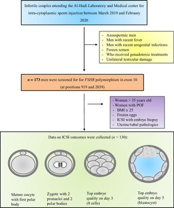
Infertile couples attending Al Hadi Laboratory and Medical Center for ICSI were invited to participate in this prospective cohort study. The study was performed between March 2019 and February 2020. A questionnaire about medical and fertility history was filled in after an in-person interview with each couple during the visit to the clinic for folliculometry. The couples were asked about their age, weight, height, body mass index (BMI), medications, infertility etiology, primary or secondary infertility, previous miscarriages, previous assisted reproductive technology (ART) attempts, previous surgeries, and history of chemotherapy. We only included men undergoing ICSI with their ejaculated fresh spermatozoa. All men suffering from recent fever, urogenital tract infection, unilateral testicular damage, or known genetic disease as well as the ones who had recently recovered from a surgery or gonadotoxic treatment, and those who were azoospermic were excluded from this study. Infertile men (n =173) were screened for FSHR polymorphisms at codons 307 and 680 (Figure 1). Thereafter, in order to analyze the effect of these FSHR gene variants in men on ICSI outcomes, we excluded couples in which the woman had a BMI ≥ 25 and/or who was more than 35 years old, women who had premature ovarian failure, uterine/tubal pathologies, undergoing ICSI with embryo biopsy, and/or using frozen eggs (Figure 1) (Bakos et al., Reference Bakos, Henshaw, Mitchell and Lane2011; Yamaguchi et al., Reference Yamaguchi, Ishikawa, Mizuta, Takeuchi, Matsubayashi, Kokeguchi, Habara, Ichioka, Ohashi, Okamoto, Kawamura, Kanto, Taniguchi, Tawara, Hara, Hibi, Masuda, Matsuyama and Yoshida2020).
Study design. Infertile couples attending Al Hadi Laboratory and Medical Center were invited to participate in this study (blue box). We excluded infertile men with: azoospermia, with recent fever, with recent urogenital infections, using frozen semen, men who received gonadotoxic treatments, or having unilateral testicular damage (yellow box). Infertile men were screened for FSHR polymorphism in exon 10 at positions 919 and 2039 (orange box). Data for ICSI were collected from couples (green boxes) for whom the female partners were not >35 years old, without premature ovarian failure (POF), with a body mass index (BMI) ≤ 25, and not using frozen eggs (purple boxes).

This study is reported according to the STrengthening the Reporting of OBservational studies in Epidemiology (STROBE) guidelines (Table S2).
Ethics statement and patient information
The enrolled participants received a full explanation of the study purpose and they signed a written consent form. The written consent form confirmed the participants’ approval of donating blood for research. In addition, it includes their acceptance to use their medical records anonymously. Ethical approval was obtained from Al Hayat Hospital, Lebanon (reference number ETC-02-2019).
Ovarian stimulation and oocytes culture
A gonadotropin-releasing hormone (GnRH) antagonist protocol was used for controlled ovarian stimulation as described elsewhere (Bakos et al., Reference Bakos, Henshaw, Mitchell and Lane2011). Briefly, daily injections of gonadotropins (Merional, IBSA, Lebanon) were administered starting the second day of the menstrual cycle. In addition, GnRH antagonist (Ovitrelle, Merck Serono, Lebanon) started to be administered from day 5 of the cycle. When at least three follicles reached 16 mm, human chorionic gonadotropin (hCG) (Ovitrelle, Merck Serono, Lebanon) was injected to trigger ovulation. After 36 h, cumulus–oocyte complexes (COCs) were retrieved using transvaginal ultrasound and cultured in fertilization medium (Life Global, Ibra Haddad, Lebanon). Oocyte denudation occurred after 3 h of the COCs culture by the removal of the cumulus and coronal cells using hyaluronidase (Life Global, Ibra Haddad, Lebanon). Then, the oocytes were transferred into culture medium (Total Global, Ibra Haddad, Lebanon) and incubated under 6% CO2 and 5% O2 at 37°C for 1 h (K-system incubator G185) (Raad et al., Reference Raad, Azouri, Rizk, Zeidan, Azouri, Grandjean and Hazzouri2019a).
Blood DNA extraction
On the day of oocytes pick-up, a blood sample (2 ml) was obtained from each man, anticoagulated with ethylenediaminetetraacetic acid (EDTA), and assigned for genomic DNA extraction. The extraction was performed according to the manufacturer’s instructions (Nucleospin Blood, Mini kit, for DNA from blood, Meato, Lebanon). For testing FSHR polymorphism, polymerase chain reactions (PCR) were performed in a thermocycler (BioRad CFX96, BioRad, Lebanon) with a 12.5 µl PCR Master Mix (REDTaq ReadyMix PCR reaction Mix, Sigma Aldrich, Lebanon), 20 pmol of each primer (Sigma Aldrich, Lebanon) and 100 ng of DNA in a final volume of 25 μl (Yan et al., Reference Yan, Gong, Zhang, Li, Li, Zhu and Sun2013).
p.Thr307Ala variant analysis
The polymorphism of the p.Thr307Ala variant was evaluated by nested PCR–restriction fragment length polymorphism (RFLP) as previously described (Yan et al., Reference Yan, Gong, Zhang, Li, Li, Zhu and Sun2013). At first, the FSHR gene was amplified by PCR from genomic DNA. The PCR was performed at an annealing temperature of 58°C with 35 cycles of 95°C (30 s), 58°C (30 s), and 72°C (45 s). The upstream primer used was 5′-TCTGAGCTTCATCCAATTTGCA-3′ and the downstream primer used was 5′-GGGAAAGAGGGCAGCTGCAA-3′ (Yan et al., Reference Yan, Gong, Zhang, Li, Li, Zhu and Sun2013).
Next, 5 µl of the amplicon was amplified by a second PCR using an upstream primer 5′-CAAATCTATTTTAAGGCAAGAAGTTGATTATATGCCTCAG-3′ and a downstream primer 5′-GTAGATTCCAATGCAGAGATCA-3′. Curiously, the A→G transition forms an endonuclease (Bsu36I) recognition site. Therefore, the second amplicon (364 bp fragment) was digested overnight using the Bsu36I restriction enzyme (Thermo Scientific, Ibra Haddad, Lebanon) at 37°C. The bands were visualized on a 2.5% agarose gel electrophoresis (100 mV, 30 min) stained with Novel Juice Stain (Novel Juice, GeneDireX, BioRad, Lebanon) (Yan et al., Reference Yan, Gong, Zhang, Li, Li, Zhu and Sun2013).
p.Asn680Ser variant analysis
In order to analyze the p.Asn680Ser variants, the FSHR gene was amplified by PCR using an upstream primer 5′-TTTGTGGTCATCTGTGGCTGC-3′ and a downstream primer 5′-CAAAGGCAAGACTGAATTATCATT-3′. Next, an amplification was performed with 35 cycles of 95°C (30 s) and 72°C (45 s) for a 519-bp DNA fragment. The PCR-RFLP fragment was assigned for a digestion by restriction enzyme (BsrI) (Thermo Scientific, Ibra Haddad, Lebanon). The bands were visualized on a 2.5% agarose gel electrophoresis (100 mV, 30 min) and stained with Novel Juice Stain (Novel Juice, GeneDireX, BioRad, Lebanon) (Yan et al., Reference Yan, Gong, Zhang, Li, Li, Zhu and Sun2013).
Outcomes measures
Semen analysis and motile sperm selection
A fresh semen sample was provided on the day of egg collection by masturbation into a sterile cup. After liquefaction, sperm concentration and motility were assessed according to the World Health Organization 2010 guidelines (WHO, 2010). Sperm concentration was evaluated using a haemocytometer counting chamber (reference value ≥ 15 × 106/ml). For sperm motility, 10 µl of semen were placed on a clean glass slide and covered with a 22 × 22 mm coverslip. Then, under ×40 magnification, at least 200 spermatozoa were classified as progressive motility, non-progressive motility, and non-motile [reference values: progressive motility ≥ 32%, total (progressive + non-progressive) motility ≥ 40%] (Menkveld, Reference Menkveld2010; Raad et al., Reference Raad, Mansour, Ibrahim, Azoury, Azoury, Mourad, Fakih and Azoury2019b).
Motile spermatozoa were selected after sperm processing using a density gradient technique. Briefly, in a 14-ml conical tube, 1.5 ml of a 40% layer was pipetted onto 1.5 ml of a 80% layer from the density gradient kit (Cook Medical, EMEC, Lebanon). Next, 1.5 ml of semen was pipetted onto these two layers and centrifuged for 10 min at 161 g. The obtained pellet was washed with Sperm Culture Medium (Cook Medical, EMEC, Lebanon) (followed by centrifugation for 5 min at 448 g) (Raad et al., Reference Raad, Azouri, Rizk, Zeidan, Azouri, Grandjean and Hazzouri2019a).
ICSI procedure and embryo culture
ICSI dishes were prepared prior to microinjection by placing two elongated droplets of PVP (Life Global, Ibra Haddad Lebanon), three droplets of HEPES medium (Life Global, Ibra Haddad, Lebanon), and covering them with culture oil (Life Global, Ibra Haddad, Lebanon). ICSI was performed under an inverted microscope (Leica DMi8) with a heating stage (37°C) using a micromanipulator (Eppendorf TransferMan 4r). Motile spermatozoa derived from a density gradient with good morphology were selected and immobilized in the PVP droplet with the microinjection pipette. Each mature oocyte was microinjected with a single sperm in the HEPES droplets. Following ICSI (day 0), oocytes were placed in Total Global medium (Life Global, Ibra Haddad, Lebanon) and incubated at a 37°C under 6% CO2 and 5% O2. On day 1, fertilization was assessed under an inverted microscope and abnormally fertilized zygotes were discarded. Next, embryos were cultured until day 3 or day 5 in Total Global medium (Life Global, Ibra Haddad, Lebanon) (Bellver et al., Reference Bellver, Mifsud, Grau, Privitera and Meseguer2013).
Morphological assessment of zygotes and embryos (day 3 and day 5)
Zygotes and embryos were classified according to the Istanbul consensus workshop on embryo assessment (ALPHA Scientists in Reproductive Medicine and ESHRE Special Interest Group Embryology, 2011). Between 16–18 h post-ICSI a normal zygote should have two polar bodies and two pronuclei. Fertilization rate is calculated as follows: number of normally fertilized eggs/number of injected mature eggs × 100. Cleavage embryos were morphologically scored based on blastomeres number, fragmentation rate, degree of blastomeres size equality, and multinucleation (ALPHA Scientists in Reproductive Medicine and ESHRE Special Interest Group Embryology, 2011). Top embryo quality on day 3 (TEQ day 3) was calculated by dividing the number of cleavage embryos on day 3 having eight even blastomeres (with no fragmentations, multinucleations, and vacuoles) by the number of normally fertilized eggs × 100. On day 5, blastocysts were scored based on the degree of the blastocoel expansion (1–6). Then, the quality of inner cell mass (A, B, C) and trophectoderm (A, B, C) were evaluated in blastocysts graded as 3–6. Top embryo quality on day 5 (TEQ day 5) was calculated by dividing the number of embryos scored ≥ 3AA on day 5 by the number of normally fertilized eggs × 100 (Gardner et al., Reference Gardner, Lane, Stevens, Schlenker and Schoolcraft2000).
Embryo transfer and pregnancy outcomes
Transfers were performed using a Tight Difficult Transfer (TDT) embryo transfer catheter (Omni Medical, Lebanon) under ultrasound guidance. For luteal support, a single injection of 500 IU hCG (Pregnyl, Organon) and daily vaginal progesterone (Crinone 8%, Merck Serono) were administrated. A clinical pregnancy was defined by the presence of a gestational sac on transvaginal ultrasound (Keltz et al., Reference Keltz, Zapantis, Jindal, Lieman, Santoro and Polotsky2015).
Statistical analysis
Statistical analysis was performed using IBM SPSS 23 software (IBM SPSS Statistics for Windows, Version 23.0. Armonk, NY: IBM Corp). Normal distribution of continuous variables was assessed using the Shapiro–Wilk test. Thereafter, the normally distributed continuous variables were compared using analysis of variance with post-hoc tests (e.g. Tukey–Kramer test). In parallel, non-normally distributed continuous variables were compared together using the Kruskal–Wallis test with post-hoc test Conover. For categorical data, the comparison was done using the chi-squared test. Results are expressed as median (minimum–maximum) for non-normally distributed continuous variables, as mean ± standard deviation (SD) for normally distributed continuous variables, and as percentages when applicable. A statistical significance was considered when a P-value was less than 0.05 (P < 0.05). Furthermore, the genotype distribution was assessed for Hardy–Weinberg equilibrium. In addition, the difference in genotype frequencies between the samples was examined using a chi-squared test for independence (Schmitz et al., Reference Schmitz, Bastos de Souza, Genro, Matte, de Conto and Sabino Cunha-Filho2015). Linear regression testing was conducted with adjustment for paternal age, maternal age, maternal BMI, number of previous attempts, sperm concentration, progressive motility, non-progressive motility, non-motile spermatozoa, number of collected eggs, number of mature eggs, and the polymorphisms at codons 307 and 680 (Grigorova et al., Reference Grigorova, Punab, Punab, Poolamets, Vihljajev, Žilaitiene, Erenpreiss, Matulevičius and Laan2014).
Results
FSHR gene variants frequencies
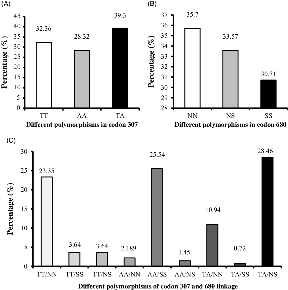
The three genotypes of codon 307 SNPs could be classified as homozygous genotypes (TT and AA) and heterozygous genotype (TA). Among the 173 infertile men, the frequencies of TT, AA, and TA were 32.6%, 28.32%, and 39.3%, respectively (Figure 2A). Concerning the codon 680 SNPs, they were categorized as homozygous genotypes (NN and SS) and heterozygous genotype (SN). The frequencies for NN, NS, and SS were 35.7%, 33.57%, and 30.71%, respectively (Figure 2B). Interestingly, allelic distributions for the two polymorphisms analyzed were in the Hardy–Weinberg equilibrium. Subsequently, the allelic variants combinations between the two codons 307 and 680 were analyzed. The frequencies of the possible combinations were 23.35% for TT–NN, 3.64% for TT–SS, 3.64% for TT–NS, 2.18% for AA–NN, 25.54% for AA–SS, 1.45% for AA–NS, 10.94% for TA–NN, 0.72% for TA–SS and 28.46% for TA–NS (Figure 2C). The following combinations: TT–SS (n = 5), TT–NS (n = 5), AA–NN (n = 3), AA–NS (n = 2), TA–NN (n = 15), and TA–SS (n = 1) had very low frequencies compared with other combinations. Thus, they will not be considered for further evaluation in this article.
The frequency of polymorphism in codon 307, codon 680, and both codons combined. (A) Percentages of the different polymorphisms in codon 307 in infertile men. (B) Percentages of the different polymorphisms in codon 680 in infertile men. (C) Percentages of different polymorphisms of codon 307 and 680 linkage. %, percentage.

Male fertility and the variants of the codon 307 of the FSHR gene
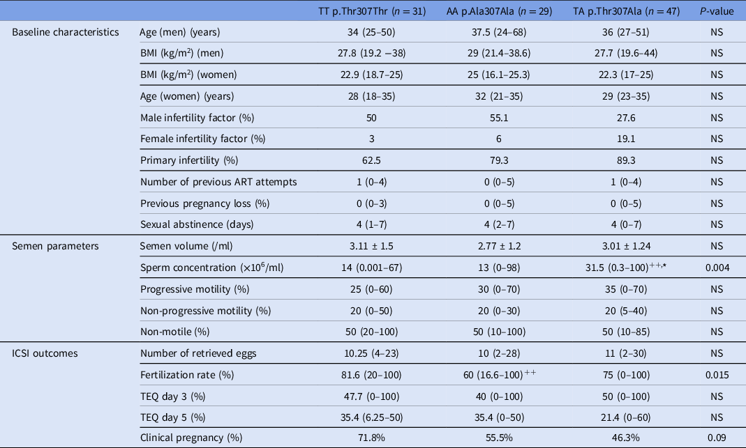
There were no statistically significant differences between the three genotypes TT, AA and TA regarding paternal age, paternal BMI, maternal age, percentage of male/female infertility factors, the percentage of couples with primary infertility, number of previous ART attempts, sexual abstinence days, semen volume and number of retrieved eggs (p>0.05). However, the percentage of couples with primary infertility was higher in the TA group compared with the TT genotype (P = 0.006). In comparison with the TA genotype, lower percentages of normal sperm concentration and motility samples (according to WHO, 2010) were found in the AA (P < 0.05) and TT (P < 0.01) genotypes. Interestingly, lower fertilization rates were found in the AA group than in TT group. This difference in fertilization rate remained significant after correcting the results for the paternal age as a confounding factor upon using a multiple linear regression analysis (Table S2). In contrast, there were no statistically significant differences between the studied codon 307 variants in terms of TEQ day 3, TEQ day 5 and clinical pregnancy rates (P > 0.05) (Table 1).
Male fertility status according to gene polymorphism of codon 307

Data expressed as median (minimum–maximum) for continuous variables with non-normal distribution, mean ± standard deviation for continuous variables with normal distribution, and as percentage where applicable. ++ P < 0.01 when compared with p.Thr307Thr group; *P < 0.05 when compared with p.Ala307Ala group. ART, assisted reproductive technology; TEQ day 3, top embryo quality on day 3; TEQ day 5, top embryo quality on day 5; NS, not significant.
Male fertility and the variants of the codon 680 of the FSHR gene
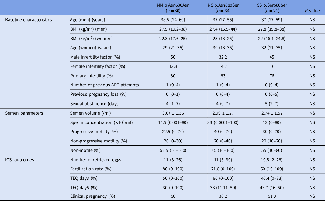
The baseline characteristics showed no significant differences between the three variants of the codon 608 concerning the paternal age, maternal age, paternal BMI, male/female infertility factors, percentage of couples with primary infertility, number of previous ART attempts, previous pregnancy loss and sexual abstinence (P > 0.05). In contrast, a lower percentage of couples with primary infertility was detected in the SS group than in the NN (P < 0.01) and NS (P < 0.001) groups. Moreover, there were no statistically significant differences between NN, NS, and SS for the semen volume, percentage of men with normal sperm concentration and motility (according to WHO, 2010), and ICSI outcomes (P > 0.05; Table 2).
Male fertility status according to gene polymorphism of codon 680

Data expressed as median (minimum–maximum) for continuous variables with non-normal distribution, mean ± standard deviation for continuous variables with normal distribution, and as percentage where applicable. ART, assisted reproductive technology; TEQ day 3, Top embryo quality on day 3; TEQ day 5, Top embryo quality on day 5; NS, not significant.
Male fertility and the most frequent allelic variants combinations TT–NN, AA–SS and TA–NS
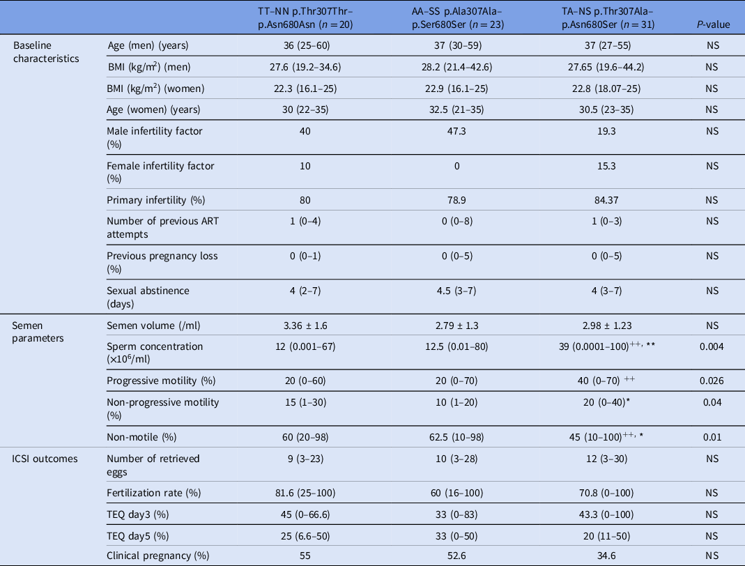
The TT–NN, AA–SS, and TA–NS combinations had the largest sample sizes and the highest frequencies among the different combinations of codons 307 and 680 variants (Figure 2C). Thereafter, these three genotypes will be considered to analyze their possible effect on male fertility potential. No significant differences were detected among the three genotypes in the baseline characteristics such as paternal age, maternal age, paternal BMI, male/female infertility factors, number of previous ART attempts, and sexual abstinence (P > 0.05). Regarding semen characteristics, higher sperm concentrations were found in the TA–NS group than in the TT–NN (P < 0.01) and AA–SS (P < 0.01) groups. Moreover, higher percentages of progressive sperm motility were found in the TA–NS group than in the TT–NN (P < 0.05) group. In addition, higher percentages of non-progressive sperm motility were detected in the TA–NS group than in the AA–SS (P < 0.05) group. Lower percentages of non-motile spermatozoa were identified in the TA–NS group than in the TT–NS (P < 0.01) and AA–SS (P < 0.05) groups. The analysis of ICSI outcomes showed a significant lower fertilization rate in the AA–SS group compared with the TT–NN group (P < 0.05). After conducting a multiple linear regression analysis, associations between fertilization rates and the studied allelic variants combinations were not detected (Table S1). In contrast, there were no significant differences in fertilization rates, TEQ day 3, TEQ day 5, and clinical pregnancy (P > 0.05) between the three groups (Table 3).
Male fertility status according to the most frequent allelic variants combinations between 307 and 680 codons

Data expressed as median (minimum–maximum) for continuous variables with non-normal distribution, mean ± standard deviation for continuous variables with normal distribution, and as percentage where applicable. + P < 0.05 when compared with p.Thr307Thr-Asn680Asn group; *P < 0.05 when compared with p.Ala307Ala-Ser680Ser group; ++ P < 0.01 when compared with the p.Thr307Thr–p.Asn680Asn group; **P < 0.01 when compared with p.Ala307Ala–p.Ser680Ser group. ART, assisted reproductive technology; TEQ day 3, top embryo quality on day 3; TEQ day 5, Top embryo quality on day 5; NS, not significant.
Discussion
The aim of this prospective study was to evaluate whether FSHR variants in infertile men could influence ICSI outcomes. The studied FSHR variants were at codon 307 (p.Thr307Ala) and codon 680 (p.Asn680Ser). In codon 307, we detected significant lower fertilization rates in men homozygous for the A-allele compared with those homozygous for the T-allele.
At first, we calculated the genotype frequencies in the selected population. Regarding the codon 307, the proportions of TT, AA, and TA were 32.36%, 28.32%, and 39.3%, respectively. In parallel, the analysis of the SNP in the codon 680 showed that the proportions of NN, NS, and SS were 35.7%, 33.57%, and 30.71%, respectively. Our results are in line with other reports in which the distribution of these variants between infertile men followed approximately the same trend (Zalata et al., Reference Zalata, Hassan, Nada, Bragais, Agarwal and Mostafa2008; Shimoda et al., Reference Shimoda, Koh, Yamamoto, Matsui, Sugimoto, Sin, Maeda, Kanaya, Yoshida and Namiki2009; Wu et al., Reference Wu, Zhang, Zhu, Jiang, Liu, Ni, Zhang, Li, Zhou, Cui and Xia2017). In parallel, we found that TT–NN, AA–SS, and TA–NS had higher frequencies compared with other combinations. This finding is in accordance with Lindgren et al. (who screened young men from the general population for FSHR SNPs in codons 307 and 680) (Lindgren et al., Reference Lindgren, Giwercman, Axelsson and Lundberg Giwercman2012) and with Shimoda et al. (Reference Shimoda, Koh, Yamamoto, Matsui, Sugimoto, Sin, Maeda, Kanaya, Yoshida and Namiki2009) who analyzed the codon 307 and 680 variants in fertile and infertile men) (Shimoda et al., Reference Shimoda, Koh, Yamamoto, Matsui, Sugimoto, Sin, Maeda, Kanaya, Yoshida and Namiki2009). In contrast, a case–control study in North China found a higher frequency of the TA–NN combination in infertile men (n = 212) compared with the control group (n = 164). The control group consisted of normozoospermic men from couples having female infertility factors (Wu et al., Reference Wu, Xu, Wang, Li, Wang, Zhang and Shen2015).
In parallel, we evaluated the effect of the SNPs on spermatozoa concentration and motility in the ejaculated semen. When the p.Thr307Ala change was analyzed, we detected a higher percentage of men with normal sperm concentration and sperm motility in the TA group compared with the TT and AA groups. Furthermore, men with the combined genotype TA–NS had significantly higher sperm concentration and motility when compared with the TT–NN and AA–SS genotypes. Hypothetically, the TA–NS genotype may influence serum FSH level and testicular response to it. Our results are in accordance with a recent meta-analysis performed on case–control studies. It indicated that FSHR polymorphisms p.Thr307Ala and p.Ans680Ser may increase the risk of male infertility (Wu et al., Reference Wu, Zhang, Zhu, Jiang, Liu, Ni, Zhang, Li, Zhou, Cui and Xia2017). In the same context, in 2015, a meta-analysis conducted on 21 papers published before February 2014 concluded that the combined genotype TT–NN had increased risk of male infertility (Wu et al., Reference Wu, Xu, Wang, Li, Wang, Zhang and Shen2015). It is worth mentioning that the authors highlighted some limitations of this meta-analysis such as small sample size, inappropriate control groups, and publication bias. In contrast, a meta-analysis performed on seven case–control studies published before July 2011 demonstrated that there was no association between these SNPs and male infertility. Among the limitations of this meta-analysis we can mention the small sample size and small number of papers included (Wu et al., Reference Wu, Cai, Sun, Lu, Zhao, Qin, Han, Niu, Lu, Xia, Wang, De Moor, Marchal and Wang2012).
FSHR SNPs could effect male fertility as FSH and FSHR play cardinal roles in spermatogenesis. FSHR is expressed on SCs, which are closely connected to germ cells in the seminiferous tubules (Raad et al., Reference Raad, Hazzouri, Bottini, Trabucchi, Azoury and Grandjean2017). Under the influence of FSH, FSHRs modulate the physiology of SCs to ensure spermatozoa survival and production (Raad et al., Reference Raad, Hazzouri, Bottini, Trabucchi, Azoury and Grandjean2017). For instance, seminiferous tubule fluid (STF) is secreted by SCs under the regulation of FSH. The STF creates a suitable environment for spermatogenesis and its dysregulation may cause infertility (Rato et al., Reference Rato, Socorro, Cavaco and Oliveira2010). In addition, SCs produce lactate from glycolysis that is the energy substrate of germ cells (Dias et al., Reference Dias, Alves, Silva and Oliveira2014). Alterations in carbohydrates metabolism may induce an imbalance between testicular reactive oxygen species and antioxidants leading to an oxidative stress. The latter may affect sperm physiology leading to infertility (Oliveira et al., Reference Oliveira, Alves, Rato, Laurentino, Silva, Sá, Barros, Sousa, Carvalho, Cavaco and Socorro2012; Rato et al., Reference Rato, Alves, Dias, Lopes, Cavaco, Socorro and Oliveira2013, Reference Rato, Alves, Cavaco and Oliveira2014). Moreover, the blood–testis barrier (BTB) is under the control of FSH. The BTB is made up of intercellular junctions between SCs. This barrier is important to create a suitable milieu for germ cells differentiation and immunoprotection. BTB breaching prompts spermatogenic arrest and infertility (Jiang et al., Reference Jiang, Bukhari, Zheng, Yin, Wang, Cooke and Shi2014). Interestingly, FSH may also control the detachment of mature spermatozoa from the seminiferous epithelium, a process known as spermiation. Abnormal spermiation could be the reason behind low sperm count and impaired motility in the ejaculated semen (Nicholls et al., Reference Nicholls, Harrison, Walton, Mclachlan, O’Donnell and Stanton2011; Stanton Reference Stanton2016). Furthermore, positive correlations were detected between sperm concentration and motility on the one hand and phospholipase C zeta (the oocyte activating factor) on the other hand. Lower concentrations of PLC zeta may lead to lower fertilization rates (Aghajanpour et al., Reference Aghajanpour, Ghaedi, Salamian, Deemeh, Tavalaee, Moshtaghian, Parrington and Nasr-Esfahani2011; Tavalaee et al., Reference Tavalaee, Kiani-Esfahani and Nasr-Esfahani2017). It will be interesting to assess whether a relationship exists between FSHR variants and PLC zeta expression levels.
Nonetheless, sperm concentration, motility, and morphology do not reflect the molecular composition of the spermatozoa (Raad et al., Reference Raad, Azouri, Rizk, Zeidan, Azouri, Grandjean and Hazzouri2019a). Curiously, morphologically normal motile spermatozoa may carry DNA breaks (Avendaño and Oehninger, Reference Avendaño and Oehninger2011). Sperm DNA fragmentation may occur in the seminiferous tubules, epididymis, semen, and during sperm manipulation in vitro (Muratori and Baldi, Reference Muratori and Baldi2018). This occurs due to abortive apoptosis in the germinal epithelium, alterations in the chromatin remodeling during spermiogenesis, or due to oxidative stress in the testicular/post-testicular milieu (Sakkas and Alvarez, Reference Sakkas and Alvarez2010). During zygote formation, the oocyte has the capacity to repair, to a certain extent, the sperm DNA fragmentation (Meseguer et al., Reference Meseguer, Santiso, Garrido, García-Herrero, Remohí and Fernandez2011). However, when it fails, fertilization and embryo development may be affected (Sarasa et al., Reference Sarasa, Jonás, Enciso, García, Leza, Steger and Aizpurua2020). As FSH is an anti-apoptotic endocrine factor and the crucial regulator of SCs, abnormal signalling via its receptor due to SNPs may lead to altered sperm nuclear quality (Sarasa et al., Reference Sarasa, Jonás, Enciso, García, Leza, Steger and Aizpurua2020). Spermatozoa carrying DNA breaks and/or high ROS levels may fail to fertilize the oocyte (Pregl Breznik et al., Reference Pregl Breznik, Kovačič and Vlaisavljević2013; Xue et al., Reference Xue, Wang, He, Mo, Huang, Wang, Mao, Cheng, Huang and Liu2016; Harchegani et al., Reference Harchegani, Rahmani, Tahmasbpour, Shahriary, Tahmasbpour and Shahriary2019; Kashir, Reference Kashir2020). In the present study we detected a higher fertilization rate in the TT–NN genotype than in the AA–SS genotype. In this context, Lindgren et al. (Reference Lindgren, Giwercman, Axelsson and Lundberg Giwercman2012) showed that the TT–NN combination had the lower FSH levels and higher estradiol and testosterone levels when compared with other combinations. This may reflect differential effects of FSHR polymorphisms on SCs and subsequently gamete quality. Further studies with larger sample sizes are needed to confirm the results of the present study.
It is worth mentioning that various studies have reported that FSHR may influence the ovarian response to exogenous FSH in women undergoing ART. For instance, it was shown that the number of collected oocytes was significantly higher in FSHR (rs6165) AA carriers and in FSHR (rs6166) AA carriers than in GG carriers. In addition, further studies are required to confirm the exact effect of FSHR polymorphisms on ovarian response and on live birth rates (Greb et al. Reference Greb, Behre and Simoni2005; Wunsch et al., Reference Wunsch, Sonntag and Simoni2007; Alviggi et al., Reference Alviggi, Conforti, Caprio, Gizzo, Noventa, Strina, Pagano, De Rosa, Carbone, Colacurci and De Placido2016, Reference Alviggi, Conforti, Santi, Esteves, Andersen, Humaidan, Chiodini, De Placido and Simoni2018).
In conclusion, the FSHR SNP p.Thr307Ala in infertile men undergoing ICSI may influence spermatozoa concentration, motility, and sperm fertilization ability. This could reflect the modulation of the FSHR activity by the SNPs. Thus, further studies using a larger sample size will be of paramount importance to confirm the results of the present scientific article.
Supplementary material
To view supplementary material for this article, please visit https://doi.org/10.1017/S0967199422000302
Acknowledgments
We would like to acknowledge the Al Hadi Laboratory and Medical Center team for their support throughout the project and Dr Janah Shaya for critical reading of the manuscript.
Author contribution
Chadi Fakih, Youmna Mourad, Fadi Fakih conceived and designed the project. Chadi Fakih, Mona Ghamloush, Neemtallah Safi, and Claira Costantine performed the experiments. Georges Raad and Chadi Fakih contributed to the data analysis. Georges Raad and Marwa Bazzi designed and wrote the manuscript. Georges Raad, Marwa Bazzi, Chadi Fakih, Youmna Mourad, Fadi Fakih, Mona Ghamloush edited the paper. All of the authors read and approved the final manuscript.
Funding
This work was funded by Al Hadi Laboratory and Medical Center.
Conflict of interest
The authors declare that there is no conflict of interest.