Introduction
The male reproductive system in amphibia is constituted by the testes, which are located in the body cavity at both sides of the median line (Hermosilla et al., Reference Hermosilla, Urbina and Cabrera1983; Oliveira & Vicentini, Reference Oliveira and Vicentini1998; Oliveira & Zieri, Reference Oliveira and Zieri2005; Santos & Oliveira, Reference Santos and Oliveira2008) and a series of ducts that transport the mature gametes to the external medium.
Although amphibia exhibit a wide variety of reproductive schemes, generally there are two types of gonadal cycles: (i) a continuous cycle in which sperm production is constant throughout the year; and (ii) a discontinuous cycle characterized by testes with spermatozoa present only during a particular season (van Oordt, Reference van Oordt1960; Paniagua et al., Reference Paniagua, Fraile and Sáez1990).
Spermatogenesis takes place during the testicular cycle. Sperm formation is a complex process that starts with the division of spermatogonia and ends with the transformation of spermatids into spermatozoa. This last process, called spermiogenesis, involves biochemical, cytochemical and morphological spermatid modifications (Phillips, Reference Phillips1974; Amaral et al., Reference Amaral, Fernandes, Báo and Recco-Pimentel1999).
Spermatogenesis in amphibians occurs in clusters called cysts, which in anurans are found inside the seminiferous tubules (Lofts, Reference Lofts and Lofts1974). Each cyst consists of single type of germ cell at the same stage of differentiation (Lofts, Reference Lofts and Lofts1974; Pudney, Reference Pudney1995) plus a Sertoli cell attached to the tubular basement membrane (Grier, Reference Grier1992).
There are few data concerning the morphological and functional characteristics of amphibian testes during the reproductive cycle (Sasso-Cerri et al., Reference Sasso-Cerri, de Faria, Freymüller and Miraglia2004). In Leptodactylus chaquensis males, knowledge of their reproductive biology is limited to a description of the sperm acrosomic reaction (Raisman et al., Reference Raisman, de Cunio, Cabada, del Pino and Mariano1980). This amphibian, which inhabits northern Argentina, has a relevant importance not only from an economic point of view as it provides meat for human consumption, but also because its population is perceptibly decreasing due to factors such as aggressive pesticides, diseases due to water pollution, predation, indiscriminate capture and/or occupation of their ecological niches by foreign species.
Taking into account that gametogenesis is an important aspect in the reproductive biology of any species and that morphological studies are required to establish the functional bases of the reproductive activity, the aim of the present work was to analyze the testicular organization and the histological aspects of the Leptodactylus chaquensis spermatogenesis. This study was undertaken during two periods of the reproductive cycle.
Materials and methods
Animals
Leptodactylus chaquensis adult males were collected from nearby areas of Tucumán, Argentina, during the December–March (reproductive) and May–July (postreproductive) periods. The animals (n = 6 for each period) were used immediately after capture or kept in captivity for 24 h in boxes with appropriate humidity at room temperature.
The protocols for this study followed approved guidelines for the ethical treatment of animals and all care was taken to prevent cruelty of any kind.
Gonads
After the animals were weighed, testes free of extraneous tissues were rapidly removed under anaesthesia and washed in amphibian Ringer's solution, pH 7.4.
Gonad samples were fixed in 10% formaldehyde for 24 h, included in Paraplast, embedded in paraffin and serially sectioned at 2–4 μm thickness. The sections were then stained with haematoxylin–eosin (H–E), routine staining, and toluidine blue (TB) pH 5.6, a dye that binds to molecules that contain acid groups and permits better visualization of the degree of condensation of the nuclear chromatin and of the acrosome. Finally, the samples were examined under an Axiolab (Zeiss) light microscope.
Morphometric parameters
The gonads were weighed for later calculation of the gonadosomatic index (GSI): gonad weight/100 g body weight. The length of the longer and shorter axes of the seminiferous tubules was determined using a micrometric ocular lens attached to an Axiolab (Zeiss) microscope under ×100 magnification. The axial length of 100 tubules was determined and the values were expressed as mean ± standard error (SEM) of the group from each period studied.
Statistical analysis
Statistical analysis was performed using the non-parametric Kruskal–Wallis test followed by a multiple comparison method (Dunn's method). All statistical comparisons were made above the 95% confidence level. Data are presented as mean ± SEM.
Results
Leptodactylus chaquensis male gonads are a pair of ovoid organs located in the abdominal body cavity that has a close relationship with the kidneys. The testes are surrounded by a thin capsule of connective tissue, the tunica albuginea, that gives them their white colour.
Gonad size and weight vary according to the period of the reproductive cycle. During the reproductive period (spring and summer), testis size was 1.78 ± 0.072 cm long, 1.06 ± 0.06 cm wide and 0.46 ± 0.04 cm thick, while during the postreproductive period (autumn and winter), values were 1.01 ± 0.039 cm, 0.36 ± 0.03 cm and 0.15 ± 0.01 cm respectively. Testicular weight was 0.720 ± 0.049 g during the reproductive period, decreasing significantly to 0.071 ± 0.007 g during the postreproductive period. Another morphometric parameter that showed marked differences between the two periods studied was GSI. In fact, during the reproductive period, the GSI was 2.50 ± 0.25, while in the postreproductive period it was 0.39 ± 0.03.
A relevant feature, intimately associated with the reproductive cycle, is the presence of fat bodies attached to the gonads. In the postreproductive period the fat bodies were well developed, with many lobes and with a strong orange colour, while in the breeding period they were smaller and pale coloured.
Testis histology study
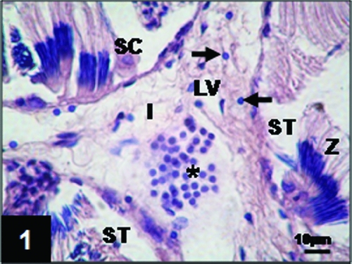
Leptodactylus chaquensis testes consist of a mass of convoluted seminiferous tubules between which is the interstitium, which contains connective tissue, blood and lymphatic vessels and Leydig cells either isolated or in small groups. These cells were characterized by their rounded nucleus that contained condensed chromatin with a uniform distribution in the nucleoplasm (Fig. 1).
Testis section of Leptodactylus chaquensis. In the interstitium (I), Leydig cells can be observed either isolated (arrows) or in groups (asterisk). LV: lymphatic vessel; SC: Sertoli cell; ST: seminiferous tubules; Z: spermatozoa. Haematoxylin–eosin.

The seminiferous tubules, surrounded by miode cells, are the structure where germ cells develop. In a transverse section of the tubule the size of their axis showed differences according to the period studied. In fact, during the reproductive period, the length of the long axis was 737 ± 71 μm and that of the short axis 395 ± 31 μm, while during the postreproductive period length were 421± 17 μm and 336 ± 16 μm respectively.
Reproductive period
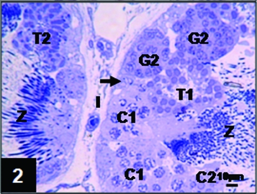
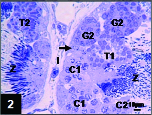
The testes from animals collected during the reproductive period presented seminiferous tubules with a wall that showed a marked thickness due to the presence of numerous cysts (Fig. 2).
Leptodactylus chaquensis testis architecture during the reproductive period. Germ cells are arranged in cysts constituted by: G2: secondary spermatogonia; C1: primary spermatocytes; C2: secondary spermatocytes; T1: primary spermatids; T2: secondary spermatids; Z: spermatozoa. I: interstitium; arrow: Sertoli cell. Toluidine blue.
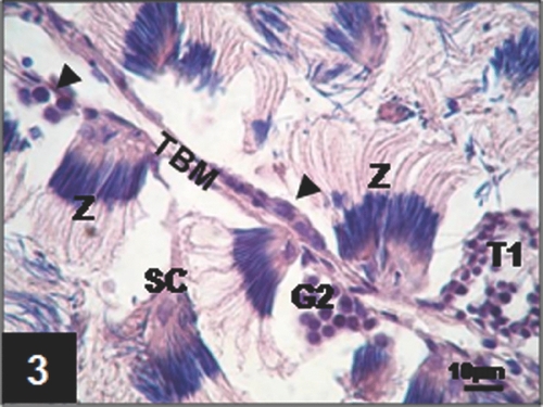
At the base of the tubular wall, primary spermatogonia, alone or in small groups of not more than four cells were seen (Fig. 3). These large cells, which do not form cysts, exhibited a dark cytoplasm and their nucleus showed marked indentations and homogeneously distributed chromatin granulations (see Fig. 9 later).
Seminiferous tubular wall showing a small group of primary spermatogonia (arrowhead). CS: Sertoli cell; G2: secondary spermatogonia cyst; T1: primary spermatids cyst; TBM: tubular basement membrane; Z: spermatozoa cyst. Haematoxylin–eosin.

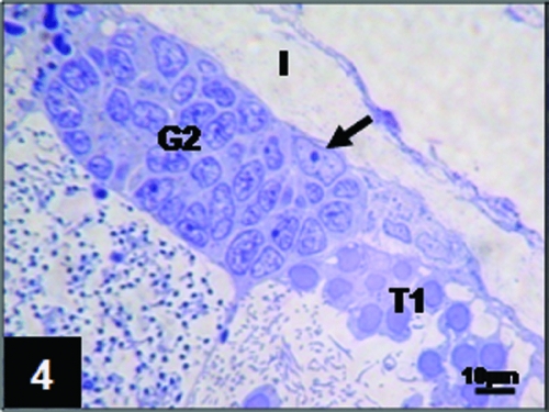
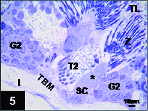
Primary spermatogonia divide mitotically into secondary spermatogonia. From this division on, all the cells of the germ line are grouped into cysts supported by the Sertoli cells whose cytoplasmic extensions constitute the cyst wall. Sertoli cells, which are characterized by a well defined nucleus with only one nucleolus, were in contact (Fig. 4), either directly or through cytoplasmic projections, with the tubular basement membrane (Fig. 5).
Seminiferous tubule section in which a secondary spermatogonia cyst (G2) associated with a Sertoli cell (arrow) can be observed. I: interstitium; T1: primary spermatids cyst. Toluidine blue.

Sertoli cell (SC) containing spermatozoa (Z) located close to the tubular lumen (TL). Note the cytoplasmic Sertoli cell projection (asterisk) to the tubular basal membrane (TBM). G2: secondary spermatogonia cysts; I: interstitium; T2: secondary spermatid cyst. Toluidine blue.

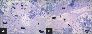
Secondary spermatogonia are smaller than primary spermatogonia. They are also usually located close to the basal wall. These cells showed scarce cytoplasm, a large nucleus with less indentation than primary spermatogonia and chromatin arranged in cumuli (Fig. 4). Mitosis of secondary spermatogonia generates cysts of primary spermatocytes, which represent a more advanced stage in spermatogenesis. These cells undergo the first meiotic division, so that different stages of this process can often be observed. Primary spermatocytes, whose size was smaller than those of their predecessors, presented a large nucleus with nuclear material with different compaction degrees according to the meiotic phase (Fig. 6 A, B).
Cyst containing primary spermatocytes at different stages of the first meiotic division. (A) Spermatocytes in prophase I (asterisk) and metaphase (arrowhead). C2: secondary spermatocytes cyst; SC: Sertoli cell. Toluidine blue. (B) Spermatocytes in metaphase (arrowhead) and anaphase (arrow). BV: blood vessel; G2: secondary spermatogonia cyst; I: interstitium. Toluidine blue.

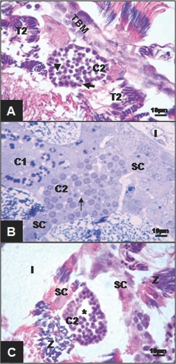
As spermatogenesis progresses, secondary spermatocytes are formed as a result of the first meiotic division; these spermatocytes are characteristically half the size of that of the primary spermatocytes. Many of these germ cells underwent the second meiotic division, so that different features of this process could be observed (Fig. 7 A). Secondary spermatocytes, which do not undergo meiosis, showed scarce cytoplasm while the nucleus, displaced at the cell periphery, had chromatin granulation close to the nuclear envelope (Fig. 7 B).
(A) Secondary spermatocytes (C2) in different stages of the second meiotic division. Arrowhead: metaphase II; arrow: telophase; T2: secondary spermatids cyst; TBM: tubular basement membrane. Haematoxylin–eosin. (B) Intercellular cytoplasmic bridges (thin arrow) connecting secondary spermatocytes (C2). C1: primary spermatocytes cyst; I: interstitium; SC: Sertoli cells. Toluidine blue. (C) Secondary spermatocytes cyst (C2) showing intercellular spaces (asterisks). I: interstitium; SC: Sertoli cells; Z: spermatozoa cyst. Haematoxylin–eosin.

In the cysts constituted by these cells one relevant feature was the presence of intercytoplasmic bridges between the germ cells (Fig. 7 B) and the appearance of intercellular spaces (Fig. 7 C).
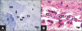
The secondary spermatocyte division generated primary spermatids with similar cytoplasmic and nuclear characteristics as in the previous stage. However, in this new population of cells, we could distinguish a structure, associated with the nucleus in a lateral position, that represented the preacrosomal vesicle (Fig. 8 A).
(A) Cysts of primary spermatids (T1). Note the presence of preacrosomal vesicles (arrow) adjacent to the nucleus of the cells. G2: secondary spermatogonia cyst; I: interstitium; SC: Sertoli cell; Z: spermatozoa cyst. Toluidine blue. ( B ) Intercellular spaces (asterisks) between primary spermatids (T1). SC: Sertoli cell; Z: spermatozoa. Haematoxylin–eosin.

Between these cells we found intercellular spaces (Fig. 8 B) that were more noticeable than the spaces between the secondary spermatocytes.
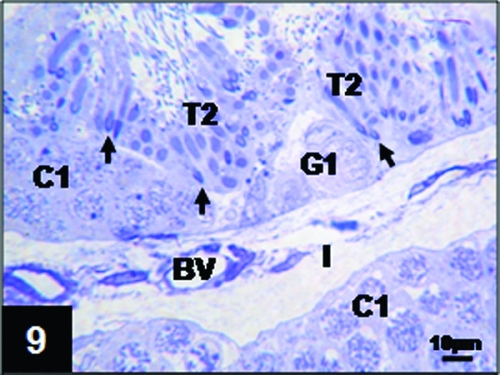
During spermiogenesis, primary spermatids continue their differentiation towards secondary spermatids. This process involves cellular and nuclear elongation, condensation of chromatin, which exhibited a uniform distribution, and progression of the preacrosomal vesicle to a well defined acrosome located at the apical side of the cell (Fig. 9).
Secondary spermatids (T2) exhibiting elongated shape with the nucleus surrounded by the preacrosomal vesicle (arrows). BV: blood vessel; C1: primary spermatocytes cyst; G1: primary spermatogonia; I: interstitium. Toluidine blue.

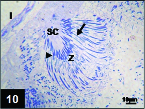
After a series of modifications that involved an important reduction in the cytoplasm, the secondary spermatids generated spermatozoa. These cells exhibited a head constituted by a highly compacted nucleus surrounded by the acrosome (Fig. 10). In this species, the acrosome had an important size and an arrowhead-like shape.
Spermatozoa (Z) anchored to the cytoplasm of the Sertoli cell (SC). Arrow: spermatozoa nucleus; arrowhead: acrosome; I: interstitium. Toluidine blue.

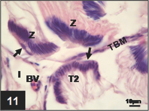
At the same time as flagellum formation, the opening of secondary spermatid cysts could be observed. The cells showed an arrangement in bundles anchored to the Sertoli cells (Fig. 11), which keep in contact with the tubular basement membrane through cytoplasmic projections as shown in Fig. 5.
Open cyst of secondary spermatids (T2) anchored to the Sertoli cell (arrow) adjacent to the tubular basement membrane (TBM). BV: blood vessel; I: interstitium; Z: spermatozoa cysts. Haematoxylin–eosin.

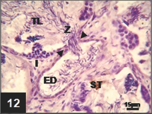
Later, a marked loss of association between the spermatozoa and the Sertoli cells took place, thus the majority of the gametes became free in the tubular lumen (Fig. 12).
In Leptodactylus chaquensis a noticeable feature during spermiation was the fusion between the seminiferous tubules and the efferent duct walls to form a path for spermatozoa release (Fig. 12).
Displacement of spermatozoa (Z) from the seminiferous tubule (ST) to the efferent duct (ED). Note the path formed by the fusion of the tubular wall with the efferent duct wall (arrowhead). I: interstitium; TL: tubular lumen. Haematoxylin.

Postreproductive period
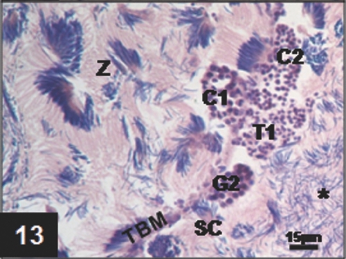
During this period all the cells of the germ line with the same morphological characteristics of the reproductive period could be observed. In general, the cysts also maintained their arrangement and location in the tubular wall (Fig. 13). However, the wall of the seminiferous tubules was thinner than that in the reproductive ones due to the presence of a reduced number of cysts.
Arrangement of the seminiferous tabules of L. chaquensis testis during the postreproductive period. The image shows: G2: secondary spermatogonia cyst; C1: primary spermatocytes cyst; C2: secondary spermatocytes cyst; SC: Sertoli cell; T1: primary spermatids cysts; TBM: tubular basement membrane; Z: spermatozoa cysts; asterisk: free spermatozoa. Haematoxylin–eosin.

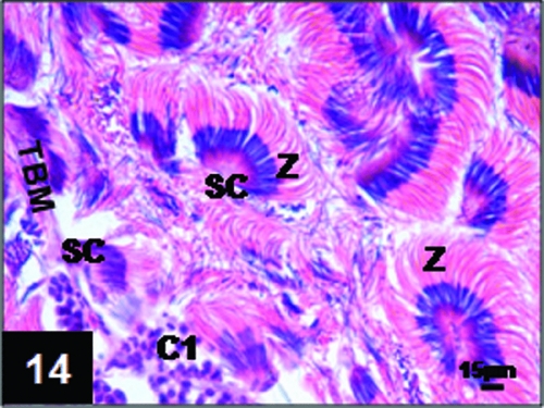
A characteristic to be noted is that during this period only a few free spermatozoa could be observed in the tubular lumen (Fig. 13), which was mostly filled with cysts of gametes that were still bound to the Sertoli cells (Fig 14).
L. chaquensis testis during the postreproductive period. Section of a seminiferous tubule showing C1: primary spermatocytes cyst; SC: Sertoli cells; TBM: tubular basement membrane; Z: spermatozoa cysts. Haematoxylin–eosin.

Discussion
In Leptodactylus chaquensis the gonads are covered by a white tunica albuginea. In contrast, the testes of other species of the Leptodactylidae family such as Physalaemus fuscomaculatus (Aoki et al., Reference Aoki, Vitale-Calpe and Pisanó1969), Physalaemus cuvieri (Oliveira et al., Reference Oliveira, Zanetoni and Zieri2002, Reference Oliveira, Sant'Anna, Omena, Santos and Zieri2003) and Physalaemus nattereri (Oliveira & Zieri, Reference Oliveira and Zieri2005) exhibit a dark brown colouration due to the presence of numerous pigmented cells located in the tunica albuginea and in the testicular interstitium. This finding demonstrates the variability of some morphological aspects between species of the same family (Oliveira et al., Reference Oliveira, Zanetoni and Zieri2002).
Fat bodies have been observed associated with the gonad that show pronounced cyclical changes with a maximal development in the postreproductive period and total regression during the reproductive one (Delgado et al., Reference Delgado, Gutiérrez and Alonso-Bedate1989). The inverse relationship between the development of the gonads and the fat bodies, considered to be a source of lipids necessary for sexual steroid synthesis, is supported by results obtained in some anuran species that show that total regression of the fat bodies is coincident with the high androgen levels (Itoh et al., Reference Itoh, Inoue and Ishii1990; Canosa & Ceballos, Reference Canosa and Ceballos2002) required for spermatogenesis.
Although in general the histomorphological organization of Leptodactylus chaquensis testis and the gametogenic process are similar to those described for other anuran amphibia such as Caudiverbera caudiverbera (Hermosilla et al., Reference Hermosilla, Urbina and Cabrera1983), Hyla japonica (Lee & Kwon, Reference Lee and Kwon1992 ), Scinax fuscovarius (Oliveira & Vicentini, Reference Oliveira and Vicentini1998), Physalaemus cuvieri (Oliveira et al., Reference Oliveira, Zanetoni and Zieri2002), Pseudis limellum (Ferreira et al., Reference Ferreira, Mehanna and Prado2008) and Dendropsophus minutus (Santos & Oliveira, Reference Santos and Oliveira2008), they also present characteristics of their own.
All cells of the germ line are present in the two periods studied. This fact allows us to suggest that Leptodactylus chaquensis could be an amphibian with a potentially continuous reproductive cycle similar to that of other species such as Telmatobius laticeps and Telmatobius pisanoi (Montero & Pisanó, Reference Montero and Pisanó1990), Hyla pulchella andina (Montero & Pisanó, Reference Montero and Pisanó1992), Pseudis limellum (Ferreira et al., Reference Ferreira, Mehanna and Prado2008), Dendropsophus minutus (Santos & Oliveira, Reference Santos and Oliveira2008). In fact, in Leptodactylus chaquensis it has been found that spermiation can be obtained under hormonal stimulation at any time of the year, a proposal that agrees with the results reported for Bufo arenarum (Canosa et al., Reference Canosa, Pozzi, Rosemblit and Ceballos2003). However, the gonads in the species studied show morphometric variations throughout the year. Not only the testis but also the seminiferous tubules are more developed in the reproductive period than in the postreproductive period, in which a reduced number of cysts and few free spermatozoa were observed in the tubular lumen.
The relationship between germ cells and Sertoli cells during spermatogenesis has a functional significance. Among other functions, these somatic cells are responsible for generating a microenvironment appropriate for germ cell development, as they constitute the hematotesticular barrier that enables the maintenance of the different composition of blood and testicular fluid (Santos & Oliveira, Reference Santos and Oliveira2008).
According to our observations, in Leptodactylus chaquensis the different types of cysts are not distributed randomly, as reported for Physalaemus cuvieri (Oliveira et al., Reference Oliveira, Zanetoni and Zieri2002). In fact, in the two periods studied, the different types of cysts showed a predominant localization in certain areas of the wall of the seminiferous tubules according to the cell developmental stage. Thus, cysts of the first stage of spermatogenesis are located in the basal zone while most of the cysts that contain cells at the last stages of differentiation are located towards the tubular lumen.
Primary spermatogonia, which are present in both periods studied, indicate the existence of a continuous source of germ cells. In contrast with the results reported for Rana esculenta (Rastogi et al., Reference Rastogi, Di Meglio, Di Matteo, Minucci and Iela1985) and Pachymedusa dacnicolor (Rastogi et al., Reference Rastogi, Bagnara, Iela and Krasovich1988), in which dark and clear primary spermatogonia were described, in Leptodactylus chaquensis only one class could be identified. Our observations are in agreement with those described for Bufo arenarum (Cavicchia & Moviglia, Reference Cavicchia and Moviglia1983), Hyla ranki (Taboga & Dolder, Reference Taboga and Dolder1991), Scinax fuscovarius (Oliveira & Vicentini, Reference Oliveira and Vicentini1998), Physalaemus cuvieri (Oliveira et al., Reference Oliveira, Zanetoni and Zieri2002), Dendropsophus minutus (Santos & Oliveira, Reference Santos and Oliveira2008) and Pseudis limellum (Ferreira et al., Reference Ferreira, Mehanna and Prado2008).
Secondary spermatogonia, cells that are already forming cysts, do not exhibit great morphological differences with their predecessors. Primary spermatocytes show all phases of the first meiotic division, even though a predominance of the prophase stage could be observed, as reported for Dendropsophus minutus (Santos & Oliveira, Reference Santos and Oliveira2008). With respect to secondary spermatocytes, although in the two periods studied stages of the second meiotic division could be seen, these cells were more often visible during the reproductive period, a finding that indicated greater gametogenic activity.
One remarkable feature, described as far as we know for the first time in amphibia, is the presence of intercellular bridges between the spermatocytes, in agreement with data reported for fish (Lo Nostro et al., Reference Lo Nostro, Grier, Meijide and Guerrero2003) and mammals (Guo & Zheng., Reference Guo and Zheng2004; Miething, Reference Miething2010). This intercellular communication between germ cells would allow molecular exchange (Gilbert, Reference Gilbert2000) that could be involved in the synchronization of the spermatogenic process. Our observations revealed the presence of intercellular spaces, which gradually appeared from spermatocyte stages and became more evident in those cysts constituted by spermatids as described for other amphibian species (Hermosilla et al., Reference Hermosilla, Urbina and Cabrera1983; Ferreira et al., Reference Ferreira, Mehanna and Prado2008). Even if the intercellular spaces increased in size as spermatogenesis progressed, in Leptodactylus chaquensis they did not become joined to form a central vacuole as reported by Lofts (Reference Lofts and Lofts1974) for Rana temporaria.
The transformations of spermatids into spermatozoa determined the formation of a gamete characterized by a large acrosome easily observed under the optical microscope. This particular characteristic is an advantage as it allows the following of the process of acrosome reaction after in vitro induction (Raisman et al., Reference Raisman, de Cunio, Cabada, del Pino and Mariano1980).
As in the case of most anamniotes, sperm release occurs after the rupture of the cyst wall (Pudney, Reference Pudney1995). One point to be emphasized, reported for the first time in amphibians, is the opening of a route resulting from the fusion of the seminiferous tubules with the efferent duct walls, which enables gamete transport during spermiation. Sperm progression toward the efferent duct would be favoured by the contraction of the miode cells present around the tubules.
In conclusion, although during the summer months (reproductive period) testicular activity is more remarkable, Leptodactylus chaquensis exhibits spermatogenic activity throughout the year, an observation that suggests that this species potentially has a continuous reproductive cycle.
The results described in this work would establish the bases for further molecular mechanism studies aimed at a better understanding of gonadal functions.
Acknowledgements
The present work was supported by a grant from PICT 2006 N° 2006 Agencia Nacional de Promoción Científica y Tecnológica (ANPCyT) and a grant 026/D425 Consejo de Investigaciones de la Universidad Nacional de Tucumán (CIUNT).